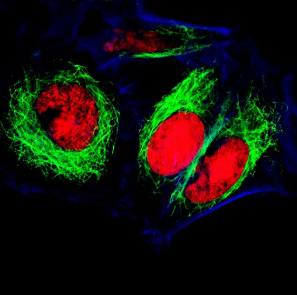
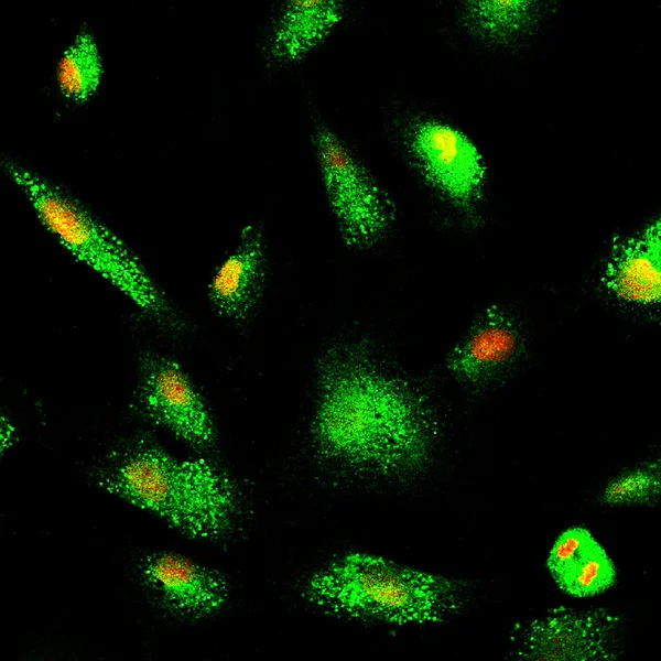
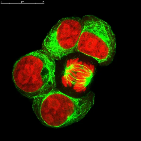
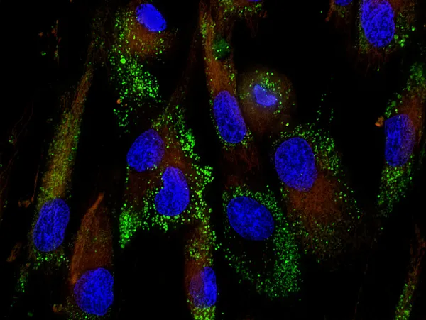
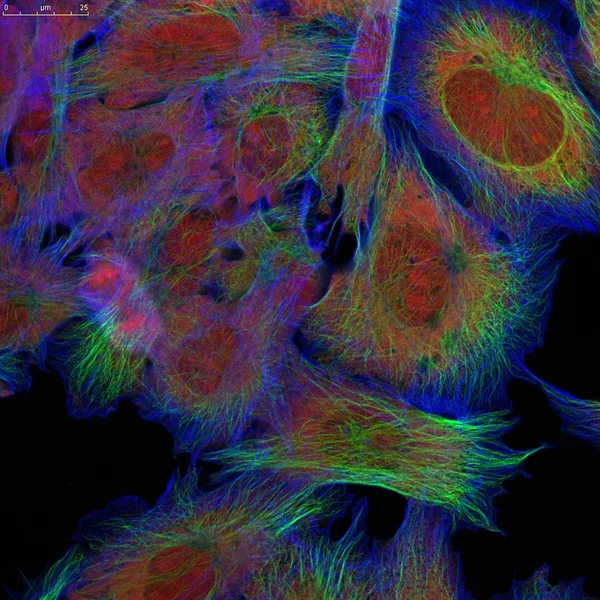
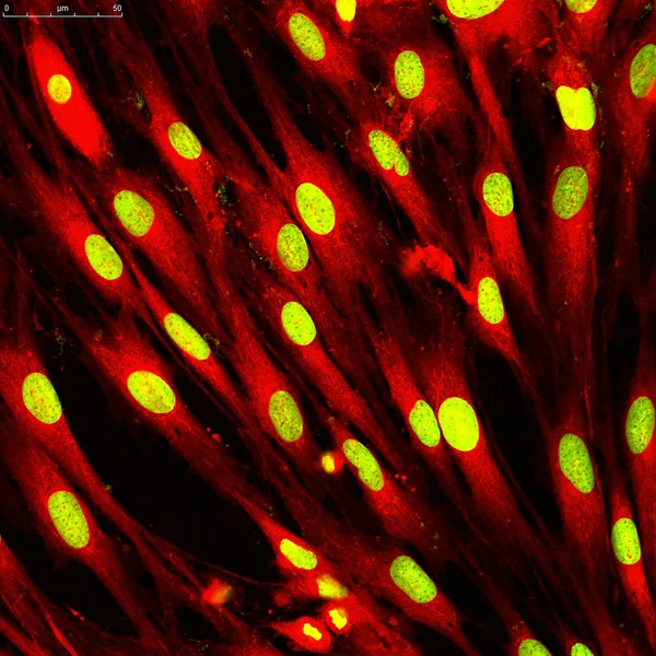
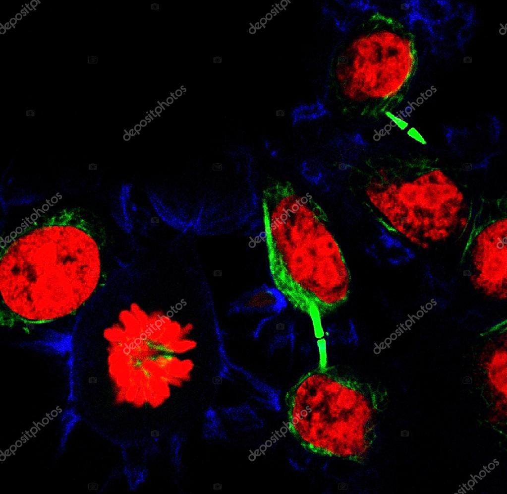
Epithelial tumor cells labeled with fluorescent molecules — Stock Photo, Image

Epithelial tumor cells labeled with fluorescent molecules — Photo

Tumor cells under microscope labeled with fluorescent molecules

 — Photo by vshivkova

Same Series:

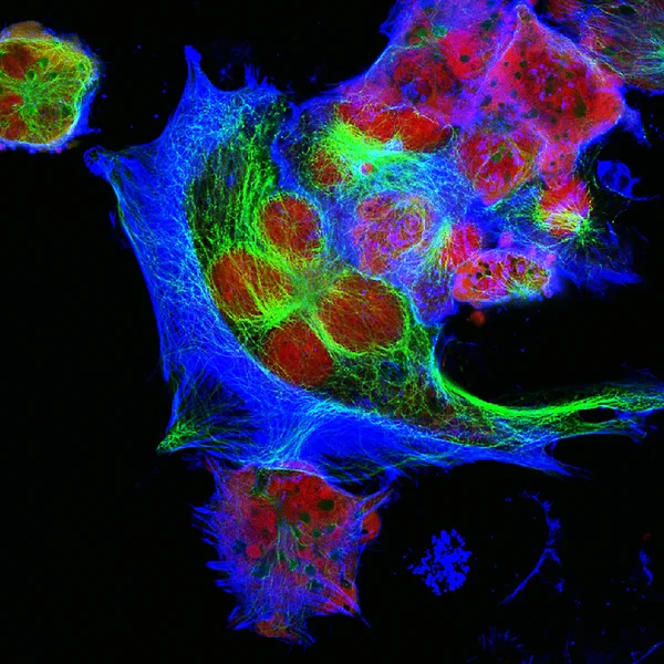
Tumor cells under microscope labeled with fluorescent molecules
Epithelial tumor cells labeled with fluorescent molecules — Stock Photo, Image
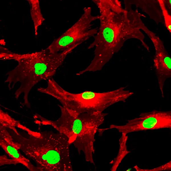
Mesenchymal stem cells labeled with fluorescent molecules
Mesenchymal stem cells labeled with fluorescent molecules — Stock Photo, Image
Real fluorescence microscopic view of human neuroblastoma cells
Human neuroblastoma cells — Stock Photo, Image
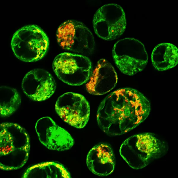
Real fluorescence microscopic view of human leukemic cells
Real fluorescence microscopic view of human leukemic cells — Stock Photo, Image
Mesenchymal stem cells labeled with fluorescent molecules
Mesenchymal stem cells — Stock Photo, Image
This image was taken with a digital camera on a confocal microscope.
Cells labeled with fluorescent dyes — Stock Photo, Image
Leukosis cell stained with monoclonal antibodies for different molecules
Leukosis cell line labeled with fluorescent dyes. One cell in division — Stock Photo, Image
Mesenchymal stem cells stained with monoclonal antibodies for different molecules
Mesenchymal stem cells labeled with fluorescent molecules — Stock Photo, Image
This image was taken with a digital camera on a confocal microscope. Here is real skin cells from patients, was grown in culture, then stained and photographed by myself.
Fibroblasts (skin  cells) labeled with fluorescent dyes — Stock Photo, Image
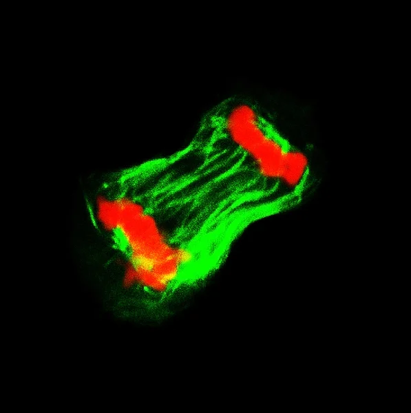
Leukosis cell line labeled with fluorescent dyes. One cell in division
Cell in division — Stock Photo, Image
This image was taken with a digital camera on a confocal microscope. Here is real skin cells from patients, was grown in culture, then stained and photographed by myself.
Fibroblasts (skin  cells) labeled with fluorescent dyes — Stock Photo, Image
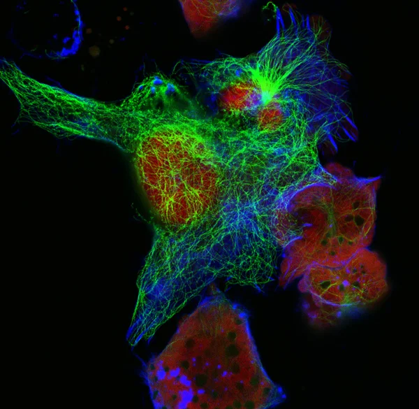
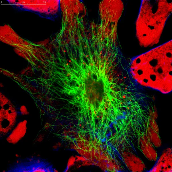
Neuroblastoma cells: nuclei are stained in red purple, microfilaments are in green and in blue
Real fluorescence microscopic view of mice neuroblastoma cell line — Stock Photo, Image
This image was taken with a digital camera on a confocal microscope. Here is real skin cells from patients, was grown in culture, then stained and photographed by myself.
Fibroblasts (skin  cells) labeled with fluorescent dyes — Stock Photo, Image
This image was taken with a digital camera on a confocal microscope. Here is real skin cells from patients, was grown in culture, then stained and photographed by myself.
Fibroblasts (skin  cells) labeled with fluorescent dyes — Stock Photo, Image
This image was taken with a digital camera on a confocal microscope. Here is real skin cells from patients, was grown in culture, then stained and photographed by myself.
Fibroblasts (skin  cells) labeled with fluorescent dyes — Stock Photo, Image
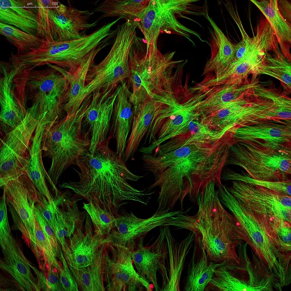
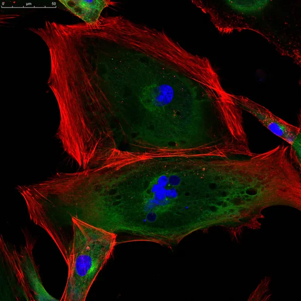
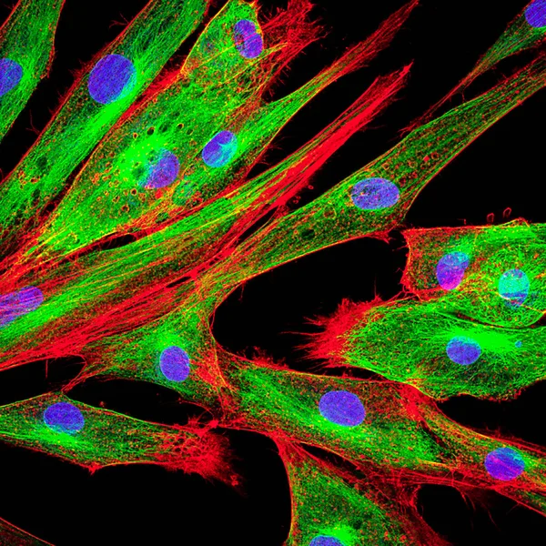
Real fluorescence microscopic view of human skin cells in culture. Nucleus are in blue, actin filaments are in red, tubulin was labeled with green
Real Fluorescence Microscopic View Human Skin Cells Culture Nucleus Blue — Stock Photo, Image

Similar Stock Videos:

Looped seamless footage for your event, concert, stage design, title, presentation, site, DVD, designers, editors and VJ s for led screens and projection mapping show
Looped Seamless Footage Your Event Concert Stage Design Title Presentation — Stock Video
Looped seamless footage for your event, concert, title, presentation, site, DVD, music videos, video art, holiday show, party, etc Also useful for motion designers, editors and VJ s for led screens.
Looped Seamless Footage Your Event Concert Title Presentation Site Dvd — Stock Video
Coronavirus, Covid-19 Vaccines. Coronavirus vaccine background footage. Disinfection Bubbles gel. Floating cells of covid 19 vaccine pills in organism of human. Coronavirus cell on black background.
Coronavirus Covid Vaccines Coronavirus Vaccine Background Footage Disinfection Bubbles Gel — Stock Video
Medical illustration 2d, 3d animation background of cells, bacteria viruses in world. Human risk virus. Coronavirus. Microscopic view. Macro cells models floating.
Medical Illustration Animation Background Cells Bacteria Viruses World Human Risk — Stock Video
Red Jellyfish in aquarium moving through the water in colorful neon light
Red Jellyfish Aquarium Moving Water Colorful Neon Light — Stock Video
Medical illustration 2d, 3d animation background of cells, bacteria viruses in world. Human risk virus. Coronavirus. Microscopic view. Macro cells models floating.
Medical Illustration Animation Background Cells Bacteria Viruses World Human Risk — Stock Video
Medical illustration 2d, 3d animation background of cells, bacteria viruses in world. Human risk virus. Coronavirus. Microscopic view. Macro cells models floating.
Medical Illustration Animation Background Cells Bacteria Viruses World Human Risk — Stock Video
Medical illustration 2d, 3d animation background of cells, bacteria viruses in world. Human risk virus. Coronavirus. Microscopic view. Macro cells models floating.
Medical Illustration Animation Background Cells Bacteria Viruses World Human Risk — Stock Video
Blooming marigold (tagetes) flowers, close up
Blooming marigold flowers — Stock Video
Computer generated abstract background of multicolor mosaic of various shapes, 3d rendering
Computer generated abstract background of colorful mosaic of various shapes, 3d rendering — Stock Video
Montage of opening colorful roses corners time-lapse in PNG+ format with ALPHA transparency channel isolated on black background
Montage Opening Colorful Roses Corners Time Lapse Png Format Alpha — Stock Video
Abstract animated twinging stained background seamless loop video - watercolor splotch effect - vibrant neon rainbow full color spectrum - magenta, pink, red, orange, yellow, green, blue, violet, purple
Abstract Animated Twinging Stained Background Seamless Loop Video Watercolor Splotch — Stock Video
Screen scanning - world map
Vector Boats - Worldwide - Trees growing - map of the world - black background - blue continent - Below View — Stock Video
Screen scanning - world map
Vector Boats - Worldwide - Trees growing - map of the world - black background - red continent - below View — Stock Video
Particle fluid collision overlayparticle fluid collision overlay
Particle Fluid Collision Overlayparticle Fluid Collision Overlay — Stock Video
4K. a group of fluorescent jellyfish swimming in an aquarium pool. transparent jellyfish underwater shots with a glowing jellyfish moving in the water.
Group Fluorescent Jellyfish Swimming Aquarium Pool Transparent Jellyfish Underwater Shots — Stock Video

Usage Information

You can use this royalty-free photo "Epithelial tumor cells labeled with fluorescent molecules" for personal and commercial purposes according to the Standard or Extended License. The Standard License covers most use cases, including advertising, UI designs, and product packaging, and allows up to 500,000 print copies. The Extended License permits all use cases under the Standard License with unlimited print rights and allows you to use the downloaded stock images for merchandise, product resale, or free distribution.

You can buy this stock photo and download it in high resolution up to 2463x2398. Upload Date: Mar 17, 2014