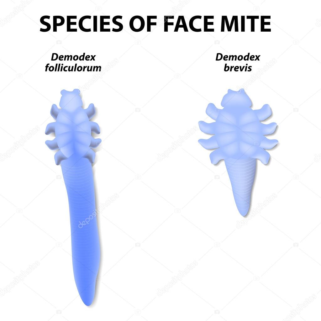
Species of face mite. — Stock Vector

Species of face mite. — Vector

Species of face mite. Demodex folliculorum and Demodex brevis. Demodex folliculorum lives in the hair follicles at the base of your eyelashes. Demodex brevis lives in oil glands connected to the hair follicles.

 — Vector by edesignua

Same Series:

Scabies. contagious skin infestation. Close-up of scabies mite and Human's skin with Magnified view of a burrowing trail of the mite.
Scabies Contagious Skin Infestation Close Scabies Mite Human Skin Magnified — Stock Vector
Echinococcus granulosus. causes cystic echinococcosis (hydatid disease). Vector illustration for biological, science, and medical use.
Echinococcus Granulosus Causes Cystic Echinococcosis Hydatid Disease Vector Illustration Biological — Stock Vector
Pityriasis rosea. close-up of skin rash. Signs and symptoms. The herald patch and Widespread rash. vector illustration
Pityriasis Rosea Close Skin Rash Signs Symptoms Herald Patch Widespread — Stock Vector
Microsporum, under microscope. Microscopic fungi that cause infections of scalp, skin of body and feet. vector illustration
Microsporum Microscope Microscopic Fungi Cause Infections Scalp Skin Body Feet — Stock Vector
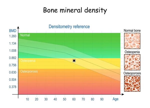
Bone mineral density (BMD). Densitometry reference (chart). close-up of condition of bone tissue from healthy to Osteopenia and osteoporosis. Aging process. vector illustration
Bone Mineral Density Bmd Densitometry Reference Chart Close Condition Bone — Stock Vector
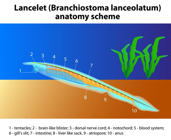
Lancelet. Branchiostoma lanceolatum anatomy scheme. Vector. The lancelet, is a member of Phylum Chordata and is important to the evolutionary view of the development
Branchiostoma lanceolatum anatomy scheme — Stock Vector
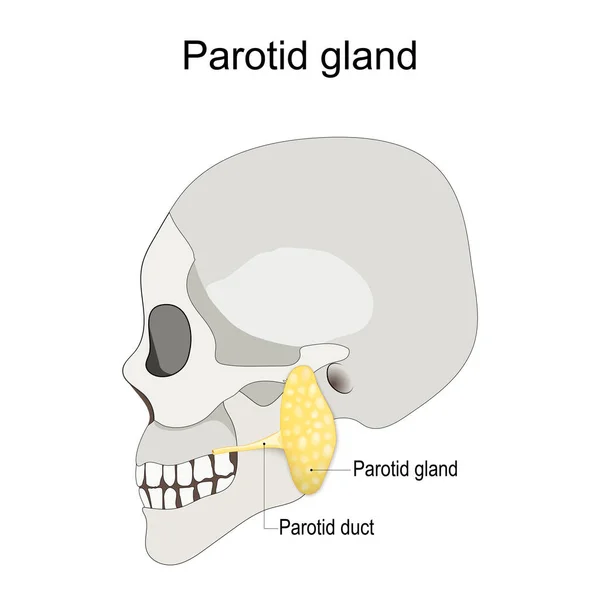
Location of the left parotid gland in humans. Humans skull with salivary gland and parotid duct. vector illustration
Location Left Parotid Gland Humans Humans Skull Salivary Gland Parotid — Stock Vector
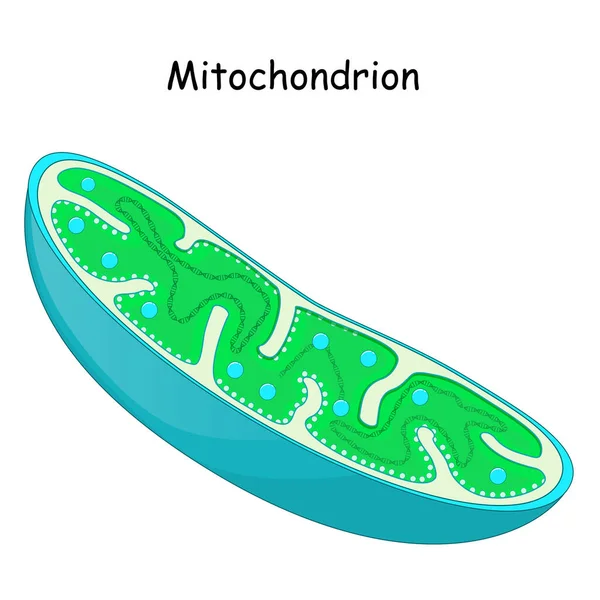
Mitochondria. structure and anatomy of a mitochondrion. vector icon
Mitochondria Structure Anatomy Mitochondrion Vector Icon — Stock Vector
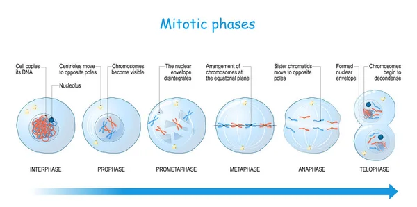
Mitosis stages from Interphase, Prophase, and Prometaphase to Metaphase, Anaphase, and Telophase. cell division. Vector illustration
Mitosis Stages Interphase Prophase Prometaphase Metaphase Anaphase Telophase Cell Division — Stock Vector
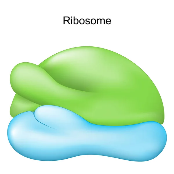
Ribosome. Cell organelle for Protein synthesis. Vector illustration
Ribosome Cell Organelle Protein Synthesis Vector Illustration — Stock Vector
Mendelian inheritance. model of Mendel's laws. biological inheritance for example pea plants.
Mendelian inheritance. model of  Mendel's laws. — Stock Vector
Epidermophyton floccosum. fungal infections of the skin. Vector illustration
Epidermophyton Floccosum Fungal Infections Skin Vector Illustration — Stock Vector
Ciliated columnar epithelium. epithelial cells forms the lining of the stomach and intestines, duodenum, fallopian tubes, uterus, central canal of the spinal cord, nose, ears and the taste buds.
Ciliated Columnar Epithelium Epithelial Cells Forms Lining Stomach Intestines Duodenum — Stock Vector
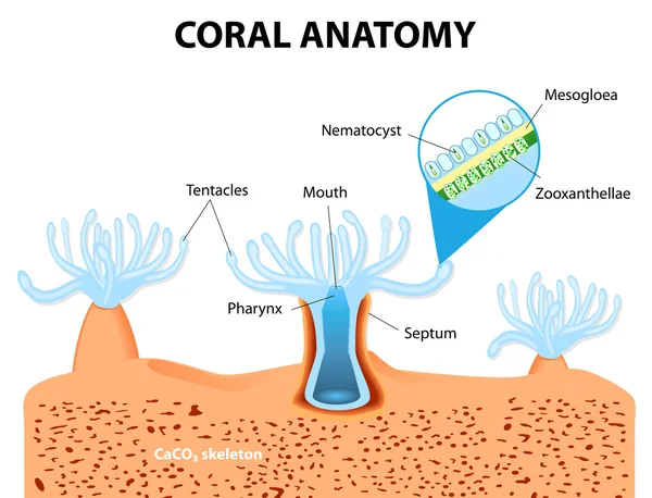
Structure coral polyp. Coral Anatomy. The coral polyps tend to live in colonies and form the building blocks of the reef.
Coral Anatomy. Vector diagram — Stock Vector
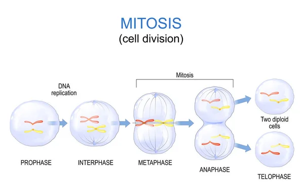
Mitosis cell division. Vector diagram. Poster for education
Mitosis Cell Division Vector Diagram Poster Education — Stock Vector
Auxins. plant hormones for plant body development.
Auxins plant hormones — Stock Vector

Usage Information

You can use this royalty-free vector image "Species of face mite." for personal and commercial purposes according to the Standard or Extended License. The Standard License covers most use cases, including advertising, UI designs, and product packaging, and allows up to 500,000 print copies. The Extended License permits all use cases under the Standard License with unlimited print rights and allows you to use the downloaded vector files for merchandise, product resale, or free distribution.

This stock vector image is scalable to any size. You can buy and download it in high resolution up to 4091x4091. Upload Date: Jan 12, 2015