Antibodies Binding Influenza Virus Surface Illustration — Stock Photo, Image

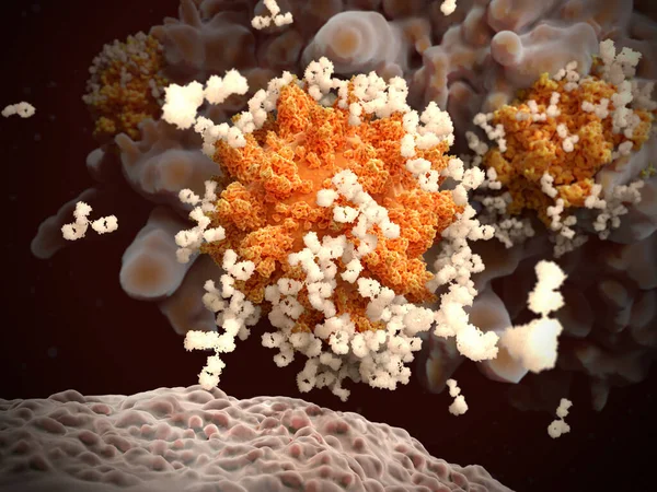
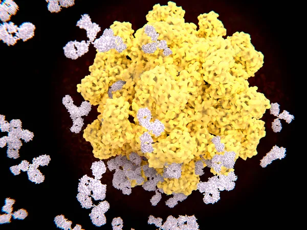
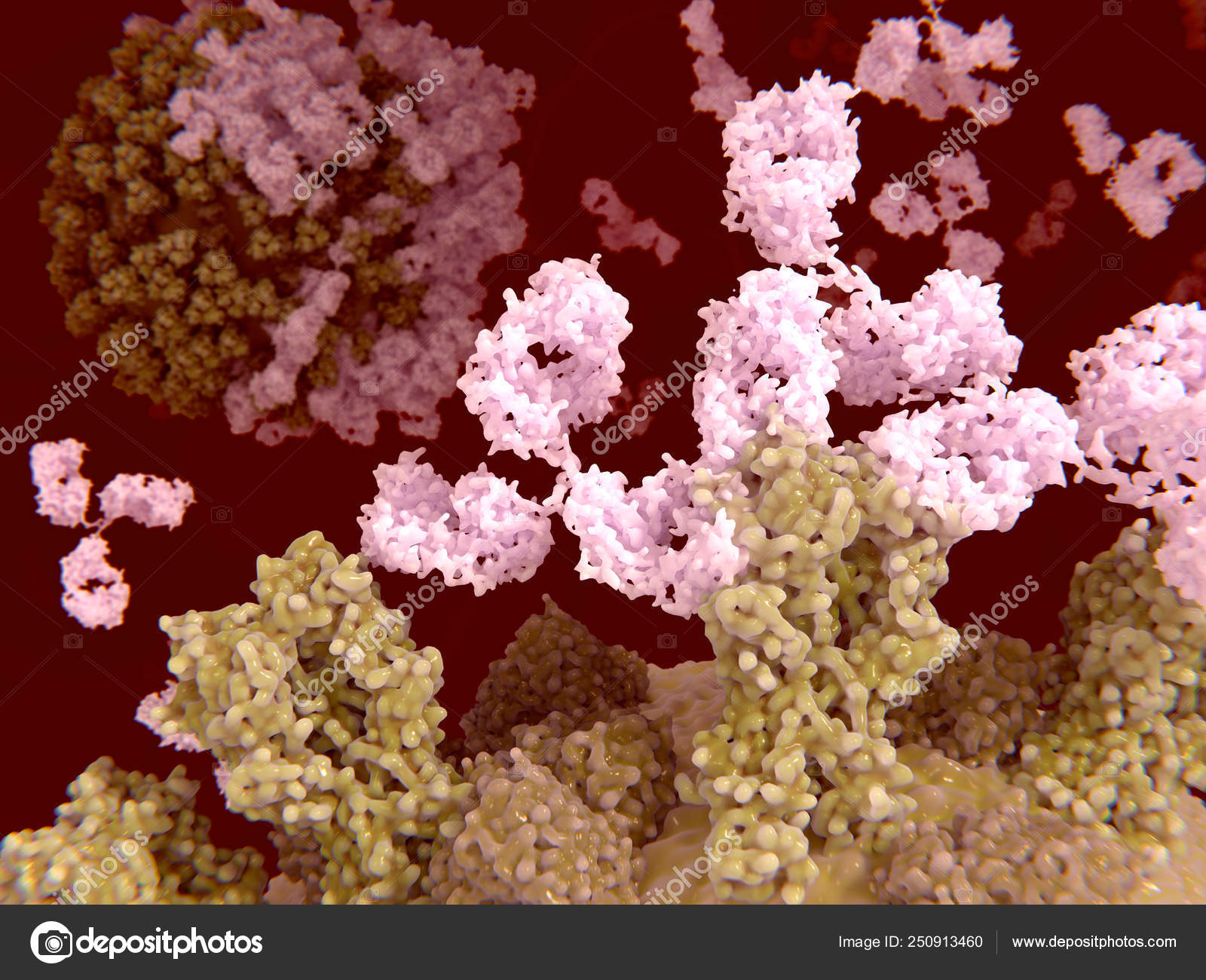
Antibodies binding to the influenza virus surface. Illustration — Photo

Antibodies binding to the influenza virus surface. Illustration

 — Photo by animaxx3d

Same Series:

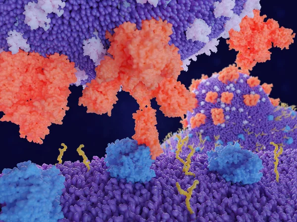
Antibody opsonization of coronaviruses SARS-CoV-2. The coated viruses can't penetrate into their target cells and are engulfed and destroyed by a macrophage.
Antibody Opsonization Coronaviruses Sars Cov Coated Viruses Can Penetrate Target — Stock Photo, Image
Immune response to SARS-CoV-2: B-lymphocyte releases antibodies that bind to and mark coronaviruses. Hence, the viruses can't penetrate into their target cells and are engulfed and destroyed by a macrophage (cell in the background).
Immune Response Sars Cov Lymphocyte Releases Antibodies Bind Mark Coronaviruses — Stock Photo, Image
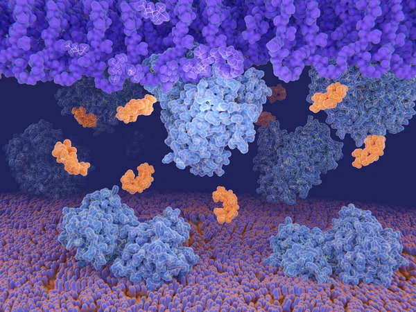
D-alanyl-D-lactate ligase (blue) synthesizes cell-wall precursors in bacteria, that are resistant to the antibiotic vancomycin (yellow), so that it cannot bind to the cell-wall. Source: PDB entry 1e4e.
Bacterial resistance to the antibiotic vancomycin — Stock Photo, Image
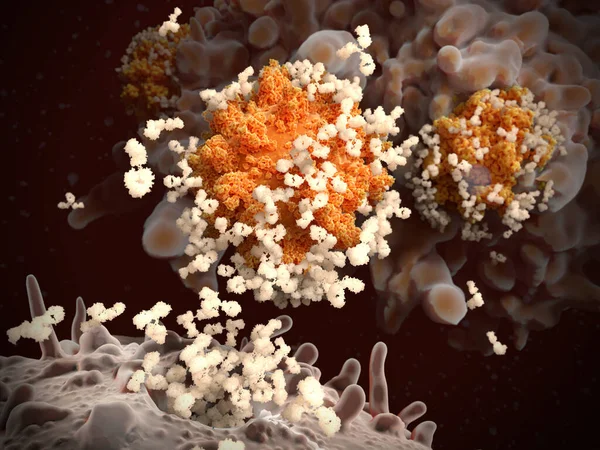
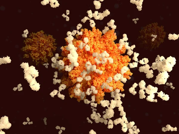
Antibody opsonization of influenza viruses. 3D rendering. Viruses coated with antibodies can't penetrate into their target cells and are engulfed and destroyed by a macrophage. Background cell. Illustration
Antibody Opsonization Influenza Viruses Rendering Viruses Coated Antibodies Can Penetrate — Stock Photo, Image
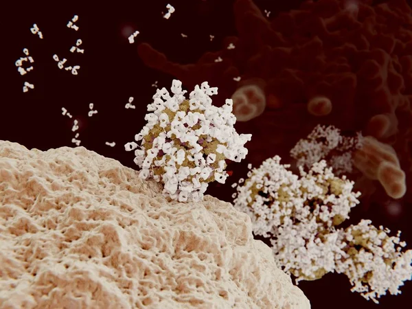
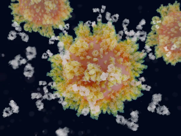
Antibodies binding to a virus. Antibodies identify and neutralize pathogens such as viri. 3D Rendering. Illustration
Antibodies Binding Virus Antibodies Identify Neutralize Pathogens Viri Rendering Illustration — Stock Photo, Image
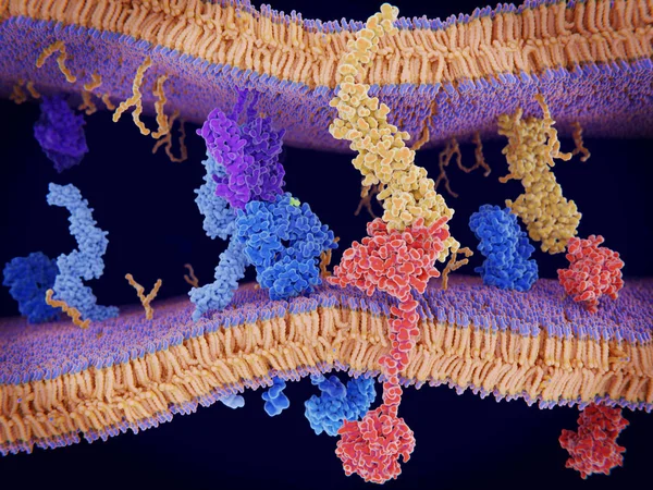
Immunologically active proteins on a T-cell. TCR (blue), CD-4 (light blue), CD-28 (dark blue), PD-1 (magenta), CTLA-4 (violet), Ca-channel (dark violet). The T-cell receptor, CD-4 and CD-28 activate T-cells, while PD-1 and CTLA-4 inhibit the activat
Immunologically Active Proteins Cell Tcr Blue Light Blue Dark Blue — Stock Photo, Image
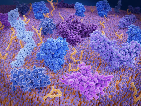
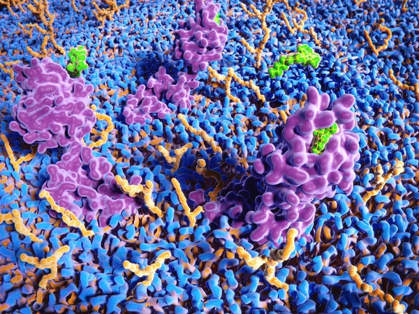
Membrane proteins (violett), glycolipids (yellow) and several ligands of the proteins
Membrane Proteins Violett Glycolipids Yellow Several Ligands Proteins — Stock Photo, Image
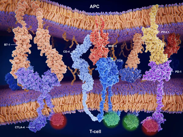
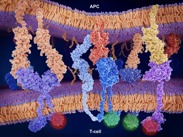
Interactions of MHC-II with the T-cell receptor and CD4 and B7-1 with CD-28 activates T-cells while the interactions of P7-1 with CTLA-4 and PD-L1 with PD-1 deactivates T-cells.
Interactions Mhc Cell Receptor Cd4 Activates Cells While Interactions Ctla — Stock Photo, Image
PD-1 (red) extends from the surface of a T-cell and interacts with the ligand protein PD-L1 (yellow) from a antigen presenting cell. Although the T-cell has been activated through the interaction of a T-cell receptor (blue) and a MHC protein (viole
Red Extends Surface Cell Interacts Ligand Protein Yellow Antigen Presenting — Stock Photo, Image
Interaction of MHC-II (red) with the T-cell receptor (blue) and CD4 (light blue) and B7-1 (orange) with CD-28 (dark blue) activates T-cells while the interaction of P7-1 with CTLA-4 (violett) and PD-L1 (yellow) with PD-1 deactivates T-cells
Interaction Mhc Red Cell Receptor Blue Cd4 Light Blue Orange — Stock Photo, Image
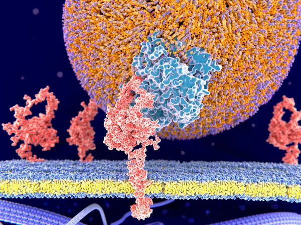
LDL particlel binding to the LDL receptor. It binds the Apoprotein B100 (blue) from LDL-particles and mediates their internalization (endocytosis) in the cell. Illustration
Ldl Particlel Binding Ldl Receptor Binds Apoprotein B100 Blue Ldl — Stock Photo, Image
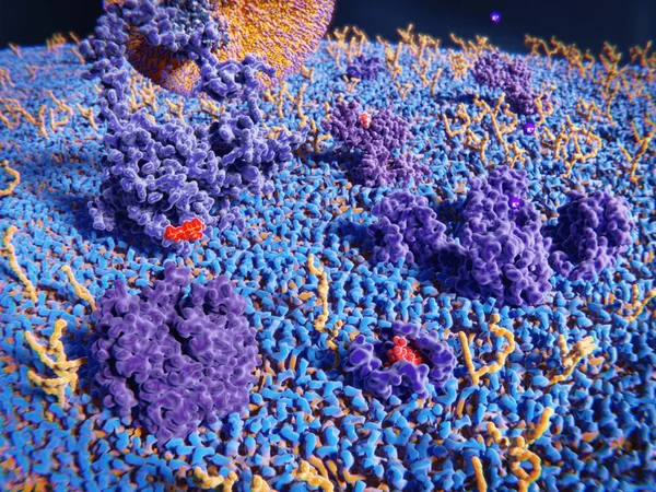
The cannabinoid receptor appears mainly in the nervous system and is involved in appetite, pain-sensation, mood and memory.
Cannabinoid Receptor Appears Mainly Nervous System Involved Appetite Pain Sensation — Stock Photo, Image
Binding of the coronavirus spike protein(red) to an ACE2 receptor (blue) on a human cell leads to the penetration of the virus in the cell, as depicted in the background.
Binding Coronavirus Spike Protein Red Ace2 Receptor Blue Human Cell — Stock Photo, Image
Antibodies binding to a coronavirus. Binding of antibodies to the spike (S)-protein of the SARS-CoV-2 virus is an essential step for developing immunity to the coronavirus.
Antibodies Binding Coronavirus Binding Antibodies Spike Protein Sars Cov Virus — Stock Photo, Image
Binding of antibodies to the spike (S)-protein of the SARS-CoV-2 virus is an essential step for developing immunity to the coronavirus.
Binding Antibodies Spike Protein Sars Cov Virus Essential Step Developing — Stock Photo, Image

Usage Information

You can use this royalty-free photo "Antibodies binding to the influenza virus surface. Illustration" for personal and commercial purposes according to the Standard or Extended License. The Standard License covers most use cases, including advertising, UI designs, and product packaging, and allows up to 500,000 print copies. The Extended License permits all use cases under the Standard License with unlimited print rights and allows you to use the downloaded stock images for merchandise, product resale, or free distribution.

You can buy this stock photo and download it in high resolution up to 8000x6000. Upload Date: Mar 14, 2019