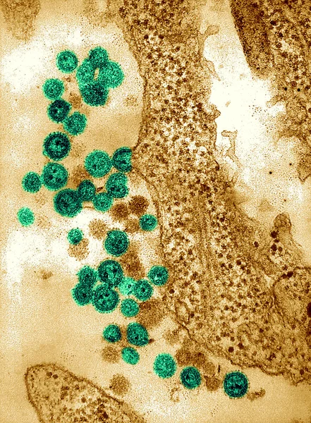
Virus Used Preparation Smallpox Vaccine Electron Micrograph Vaccinia Virus Vaccinia — 스톡 사진

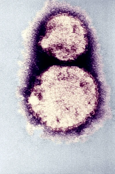
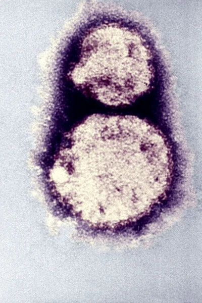
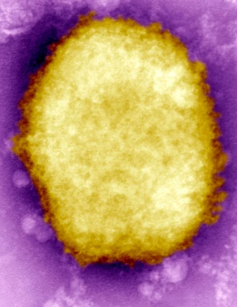
Virus used in the preparation of the smallpox vaccine. An electron micrograph of a Vaccinia virus. Vaccinia virus is normally confined to cattle, but is conveyed to humans through vaccination, thereby imparting immunity to the smallpox virus. — 스톡 이미지

Virus used in the preparation of the smallpox vaccine. An electron micrograph of a Vaccinia virus. Vaccinia virus is normally confined to cattle, but is conveyed to humans through vaccination, thereby imparting immunity to the smallpox virus.

 — 님의 사진 imagepointfr

동일 시리즈:

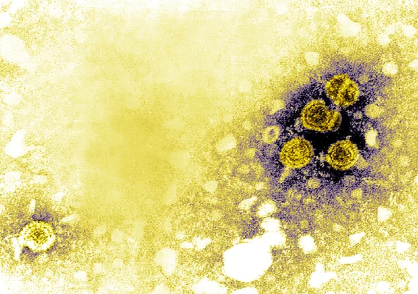
Hantavirus, the causal agent of hantavirus pulmonary syndrome (HPS).
Hantavirus Causal Agent Hantavirus Pulmonary Syndrome Hps — 스톡 사진
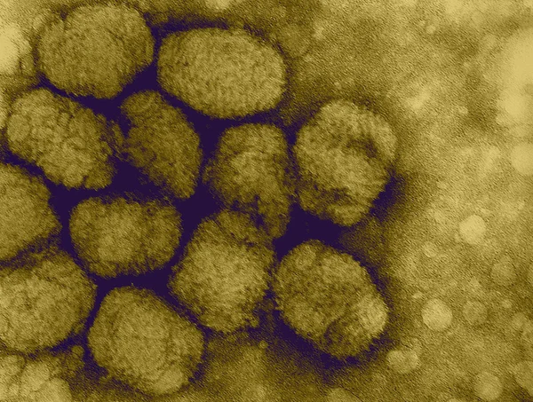
현미경 천연두 바이러스
천연두 바이러스 — 스톡 사진
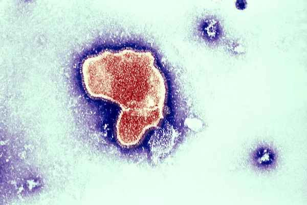
Thic electron micrograph reveals the morphologic traits of the Respiratory Syncytial Virus (RSV). The virion is variable in shape, and size (average diameter of between 120-300nm). RSV is the most common cause of bronchiolitis and pneumonia among inf
Thic Electron Micrograph Reveals Morphologic Traits Respiratory Syncytial Virus Rsv — 스톡 사진
웨스트 나 일 바이러스는 모기에 물린 상처를 통해 인 간에 게 전달
웨스트 나 일 바이러스 — 스톡 사진
Hantavirus hantavirus 폐 증후군 (hps의 인과 에이전트)
Hantavirus 폐 증후군 — 스톡 사진
천연두 바이러스의 전송 전자 현미경 사진
천연두 바이러스 — 스톡 사진
고양이 calicivirus
고양이 calicivirus — 스톡 사진
모양의 icosahedral herpesvirus virions
모양의 icosahedral herpesvirus virions — 스톡 사진
B 형 간염의 전송 전자 현미경 사진
B 형 간염 — 스톡 사진
Monkeypox virus present in human vesicular fluid. Here a negative stain electron micrograph reveals a "M" (mulberry type) monkeypox virus virion in human vesicular fluid. The surface of "M", or "mulberry" virions are covered with short, whorled filam
Monkeypox Virus Present Human Vesicular Fluid Here Negative Stain Electron — 스톡 사진
현미경에서 바이러스
현미경에서 바이러스 — 스톡 사진
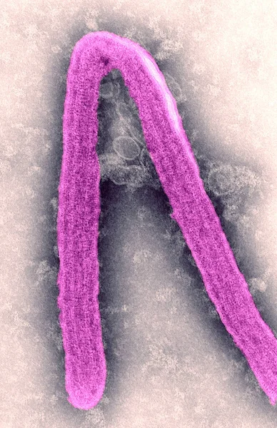
This negative stained transmission electron micrograph (TEM), captured by F.A. Murphy in 1968, depicts a Marburg virus virion, which had been grown in an environment of tissue culture cells. Marburg hemorrhagic fever is a rare, severe type of hemorrh
Negative Stained Transmission Electron Micrograph Tem Captured Murphy 1968 Depicts — 스톡 사진
This transmission electron micrograph from a pelleted specimen depicts three icosahedral-shaped herpes virus virions. The staining technique used to process this specimen revealed the following findings: virions with a dark center, where the stain ha
Transmission Electron Micrograph Pelleted Specimen Depicts Three Icosahedral Shaped Herpes — 스톡 사진
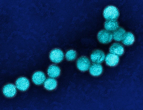
Electron micrograph of the poliovirus. Poliovirus is a species of Enterovirus, which is a genus in the family of Picornaviridae, and is an RNA virus.
Electron Micrograph Poliovirus Poliovirus Species Enterovirus Which Genus Family Picornaviridae — 스톡 사진
Machupo 바이러스의 전자 photomicrograph
Machupo 바이러스 — 스톡 사진
인간의 기공을 액체에서 존재 monkeypox 바이러스
Monkeypox 바이러스 — 스톡 사진

사용 정보

표준 또는 확장 라이센스에 따라 이 로열티-프리 "Virus used in the preparation of the smallpox vaccine. An electron micrograph of a Vaccinia virus. Vaccinia virus is normally confined to cattle, but is conveyed to humans through vaccination, thereby imparting immunity to the smallpox virus." 사진을 개인적 및 상업적 목적으로 사용할 수 있습니다. 표준 라이센스는 광고, UI 디자인, 제품 패키지를 비롯한 대부분의 사용을 커버하며, 최대 500,000부까지 인쇄할 수 있습니다. 확장 라이센스는 무제한 인쇄 권한과 함께 표준 라이센스 하의 모든 사용을 커버하며 다운로드한 스톡 이미지를 상품, 제품 재판매 또는 무료 배포에 사용할 수 있도록 합니다.

이 스톡 사진 구입 시 최대 3630x2794 의 고해상도로 다운로드할 수 있습니다. 업데이트 날짜: 2022년 8월 11일