• Home
  • Photos
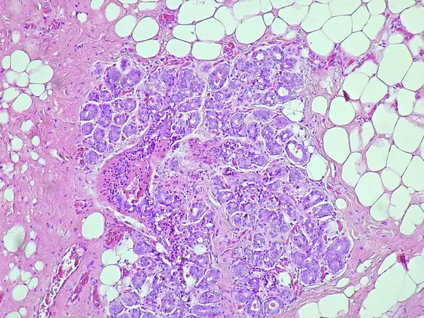
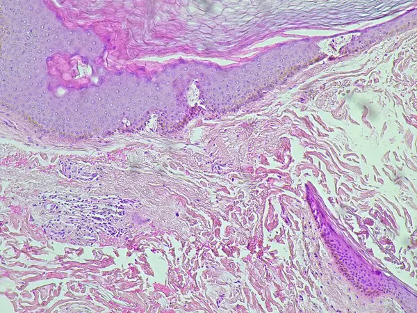
  • Human cell tissue under microscope in pathology laboratory. Microscopic picture of chronic gastric disease showing Helicobacter pylori bacterial infection, hematoxylin eosin staining
Human Cell Tissue Microscope Pathology Laboratory Microscopic Picture Chronic Gastric Disease Showing Helicobacter Pylori Bacterial Infection Hematoxylin Eosin Staining

Human cell tissue under microscope in pathology laboratory. Microscopic picture of chronic gastric disease showing Helicobacter pylori bacterial infection, hematoxylin eosin staining — Photo

Human cell tissue under microscope in pathology laboratory. Microscopic picture of chronic gastric disease showing Helicobacter pylori bacterial infection, hematoxylin eosin staining

 — Photo by DidinMH

Same Series:

Human breast cell tissue under a microscope in a pathology laboratory. Microscopic view of fibrosing mammary adenosis, hematoxylin eosin staining
Human Breast Cell Tissue Microscope Pathology Laboratory Microscopic View Fibrosing Mammary Adenosis Hematoxylin Eosin Staining
Photomicrograph showing histology of a atheroma cyst on the lobulus auricula in the anatomical pathology laboratory
Photomicrograph Showing Histology Atheroma Cyst Lobulus Auricula Anatomical Pathology Laboratory
Human cervical cell tissue under microscope in pathology laboratory. Microscopic view of invasive carcinoma, adenocarcinoma of human breast cancer, hematoxylin eosin staining
Human Cervical Cell Tissue Microscope Pathology Laboratory Microscopic View Invasive Carcinoma Adenocarcinoma Human Breast Cancer Hematoxylin Eosin Staining
Background of human ovarian cyst cell tissue under microscope in pathology laboratory. View microscopically positive cervix adenocarcinoma, tissue sections by H and E
Background Human Ovarian Cyst Cell Tissue Microscope Pathology Laboratory View Microscopically Positive Cervix Adenocarcinoma Tissue Sections
Human cervical cell tissue under microscope in pathology laboratory. Microscopic view of invasive carcinoma, adenocarcinoma of human breast cancer, hematoxylin eosin staining
Human Cervical Cell Tissue Microscope Pathology Laboratory Microscopic View Invasive Carcinoma Adenocarcinoma Human Breast Cancer Hematoxylin Eosin Staining
Ulcerative colitis photomicrograph in anatomical pathology laboratory
Ulcerative Colitis Photomicrograph Anatomical Pathology Laboratory
Background of human ovarian cyst cell tissue under microscope in pathology laboratory. View microscopically positive cervix adenocarcinoma, tissue sections by H and E
Background Human Ovarian Cyst Cell Tissue Microscope Pathology Laboratory View Microscopically Positive Cervix Adenocarcinoma Tissue Sections
Human breast cell tissue under a microscope in a pathology laboratory. Microscopic view of fibrosing mammary adenosis, hematoxylin eosin staining
Human Breast Cell Tissue Microscope Pathology Laboratory Microscopic View Fibrosing Mammary Adenosis Hematoxylin Eosin Staining
Photomicrograph of pyonephrosis + angiomyolipoma in a patient diagnosed with pyonephrosis sinistra in the anatomical pathology laboratory
Photomicrograph Pyonephrosis Angiomyolipoma Patient Diagnosed Pyonephrosis Sinistra Anatomical Pathology Laboratory
Photomicrograph Endometrial hyperplasia, photo under microscope in the anatomical pathology laboratory
Photomicrograph Endometrial Hyperplasia Photo Microscope Anatomical Pathology Laboratory
Human cell tissue under microscope in pathology laboratory. Microscopic picture of chronic gastric disease showing Helicobacter pylori bacterial infection, hematoxylin eosin staining
Human Cell Tissue Microscope Pathology Laboratory Microscopic Picture Chronic Gastric Disease Showing Helicobacter Pylori Bacterial Infection Hematoxylin Eosin Staining
Photomicrograph of pyonephrosis + angiomyolipoma in a patient diagnosed with pyonephrosis sinistra in the anatomical pathology laboratory
Photomicrograph Pyonephrosis Angiomyolipoma Patient Diagnosed Pyonephrosis Sinistra Anatomical Pathology Laboratory
Photomicrograph of pyonephrosis + angiomyolipoma in a patient diagnosed with pyonephrosis sinistra in the anatomical pathology laboratory
Photomicrograph Pyonephrosis Angiomyolipoma Patient Diagnosed Pyonephrosis Sinistra Anatomical Pathology Laboratory
Photomicrograph showing histology of a atheroma cyst on the thorax in the anatomical pathology laboratory
Photomicrograph Showing Histology Atheroma Cyst Thorax Anatomical Pathology Laboratory
Photomicrograph of soft tissue tumor of colli area with microscopic results of specific chronic inflammation in anatomical pathology laboratory
Photomicrograph Soft Tissue Tumor Colli Area Microscopic Results Specific Chronic Inflammation Anatomical Pathology Laboratory
Photomicrograph of Hashimoto's thyroiditis (chronic lymphocytic thyroiditis) in pathology anatomy laboratory
Photomicrograph Hashimoto Thyroiditis Chronic Lymphocytic Thyroiditis Pathology Anatomy Laboratory

Usage Information

You can use this royalty-free photo "Human cell tissue under microscope in pathology laboratory. Microscopic picture of chronic gastric disease showing Helicobacter pylori bacterial infection, hematoxylin eosin staining" for personal and commercial purposes according to the Standard or Extended License. The Standard License covers most use cases, including advertising, UI designs, and product packaging, and allows up to 500,000 print copies. The Extended License permits all use cases under the Standard License with unlimited print rights and allows you to use the downloaded stock images for merchandise, product resale, or free distribution.

You can buy this stock photo and download it in high resolution up to 3840x2160. Upload Date: Apr 1, 2025