Complex adaptive system Stock Photos

31 Complex adaptive system pictures are available under a royalty-free license

Machine Learning
Machine Learning
Machine Learning AI Artificial Intelligence Abstract Concept
Machine Learning Artificial Intelligence Abstract Concept
Machine Learning AI Artificial Intelligence Abstract Concept
Machine Learning Artificial Intelligence Abstract Concept
Machine Learning
Machine Learning
Machine Learning
Machine Learning
Machine Learning AI Artificial Intelligence Abstract Concept
Machine Learning Artificial Intelligence Abstract Concept
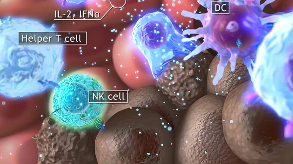
3D medical illustration cytotoxic T cell and Helper T cell destroys the stress-related molecule
Medical Illustration Cytotoxic Cell Helper Cell Destroys Stress Related Molecule
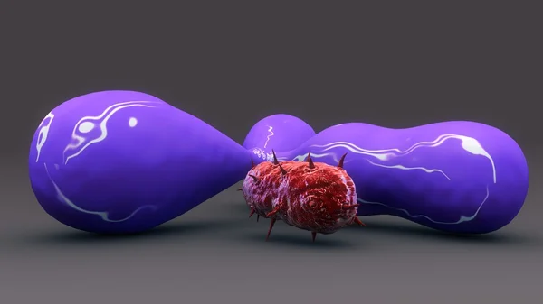
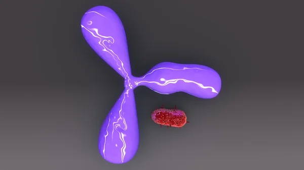
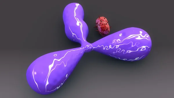
Human Antigene and bacteria
3D medical illustration cytotoxic T cell and Helper T cell destroys the stress-related molecule
Medical Illustration Cytotoxic Cell Helper Cell Destroys Stress Related Molecule
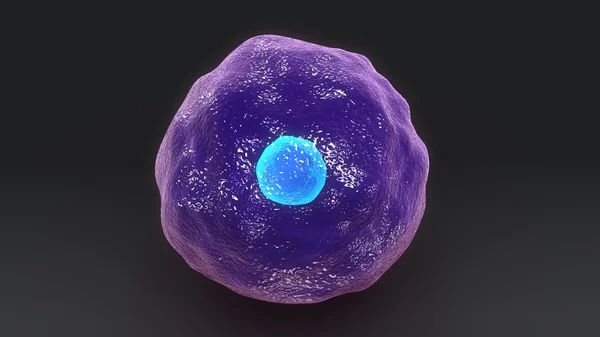
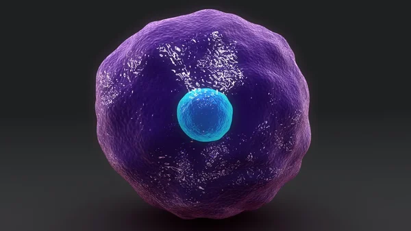
Human Cytotoxic T cell
Human Antigene and bacteria
Human Cytotoxic T cell
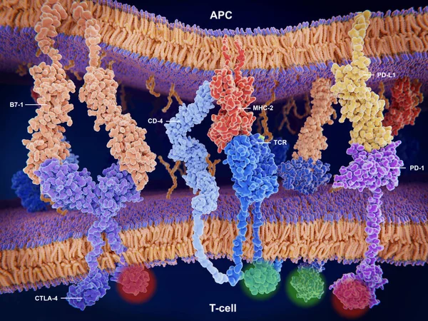
Interactions of MHC-II with the T-cell receptor and CD4 and B7-1 with CD-28 activates T-cells while the interactions of P7-1 with CTLA-4 and PD-L1 with PD-1 deactivates T-cells.
Interactions Mhc Cell Receptor Cd4 Activates Cells While Interactions Ctla Deactivates Cells
Microscopic metastasis of cancer
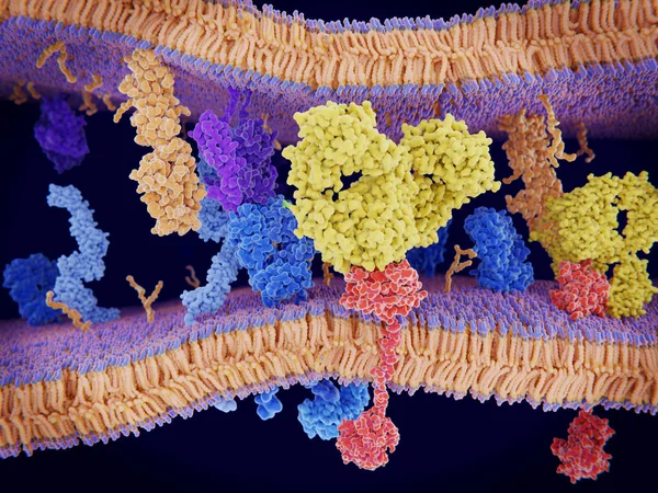
Cancer cells express PD-L1 (orange) proteins on their surface to trick the immune system. The interaction of PD-L1 with PD-1 of T-cells leads to a down-regulation of T-cells. The antibody (yellow) blocks this interaction.
Cancer Cells Express Orange Proteins Surface Trick Immune System Interaction Cells Leads Regulation Cells Antibody Yellow Blocks Interaction
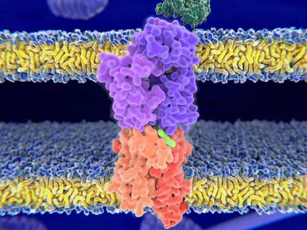
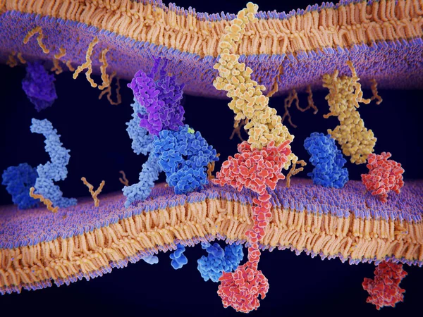
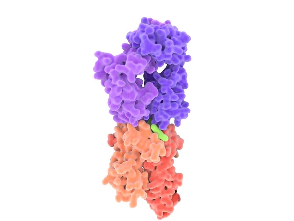
T-cell receptor in complex with the MHC class II-peptide complex. The antigen (light green) is a peptide from a tumor cell, bacteria or virus. Complex embedded in the membranes. 3D-Rendering. Illustration
Cell Receptor Complex Mhc Class Peptide Complex Antigen Light Green Peptide Tumor Cell Bacteria Virus Complex Embedded Membranes Rendering Illustration
Interaction of MHC-II (red) with the T-cell receptor (blue) and CD4 (light blue) and B7-1 (orange) with CD-28 (dark blue) activates T-cells while the interaction of P7-1 with CTLA-4 (violett) and PD-L1 (yellow) with PD-1 deactivates T-cells
Interaction Mhc Red Cell Receptor Blue Cd4 Light Blue Orange Dark Blue Activates Cells While Interaction Ctla Violett Yellow Deactivates Cells
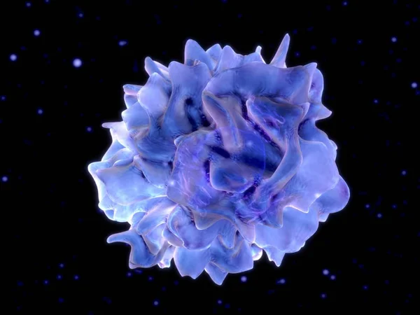
Dendritic cell. 3D-rendering. Dendritic cells are antigen-presenting cells of the immune system. Illustration
Dendritic Cell Rendering Dendritic Cells Antigen Presenting Cells Immune System Illustration
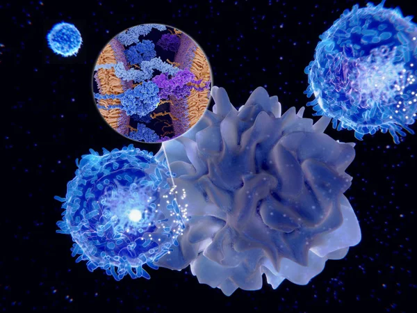
Dendritic cells present antigens (green) to lymphocytes through their membran bound MHC-molecules (violet). CD4 molecules (light blue) bind to other portions of the MHC, strengthening the interaction.
Dendritic Cells Present Antigens Green Lymphocytes Membran Bound Mhc Molecules Violet Cd4 Molecules Light Blue Bind Other Portions Mhc Strengthening Interaction
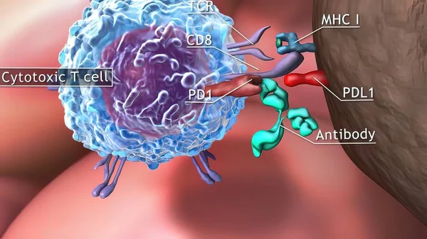
PD-1 (red) extends from the surface of a T-cell and interacts with the ligand protein PD-L1 (yellow) from a antigen presenting cell. Although the T-cell has been activated through the interaction of a T-cell receptor (blue) and a MHC protein (viole
Red Extends Surface Cell Interacts Ligand Protein Yellow Antigen Presenting Cell Although Cell Has Been Activated Interaction Cell Receptor Blue Mhc Protein Viole
Dendritic cell. Dendritic cells are antigen-presenting cells of the immune system. They process antigen material and present it on the cell surface to the T cells of the immune system. 3D Rendering. Illustration
Dendritic Cell Dendritic Cells Antigen Presenting Cells Immune System Process Antigen Material Present Cell Surface Cells Immune System Rendering Illustration
3d computer illustration of a dendritic cell. They areantigen-presenting cells of the immune system. Their main function is to process antigen material and present it on the cell surface to the T cells of the immune system. They are messengers betwe
Computer Illustration Dendritic Cell Areantigen Presenting Cells Immune System Main Function Process Antigen Material Present Cell Surface Cells Immune System Messengers Betwe
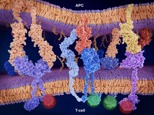
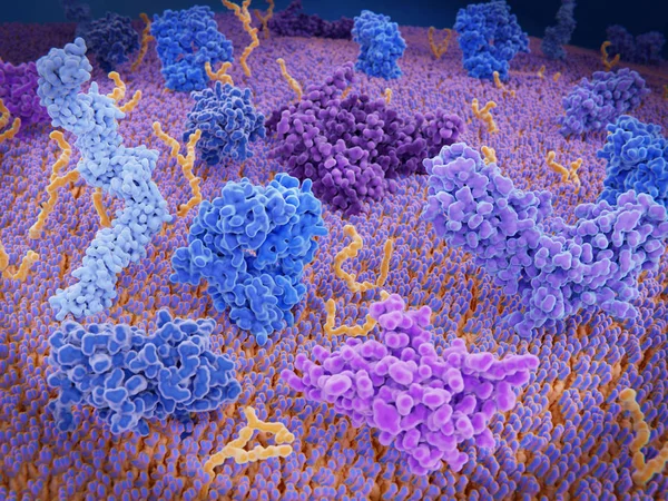
Immunologically active proteins on a T-cell. TCR (blue), CD-4 (light blue), CD-28 (dark blue), PD-1 (magenta), CTLA-4 (violet), Ca-channel (dark violet). The T-cell receptor, CD-4 and CD-28 activate T-cells, while PD-1 and CTLA-4 inhibit the activat
Immunologically Active Proteins Cell Tcr Blue Light Blue Dark Blue Magenta Ctla Violet Channel Dark Violet Cell Receptor Activate Cells While Ctla Inhibit Activat
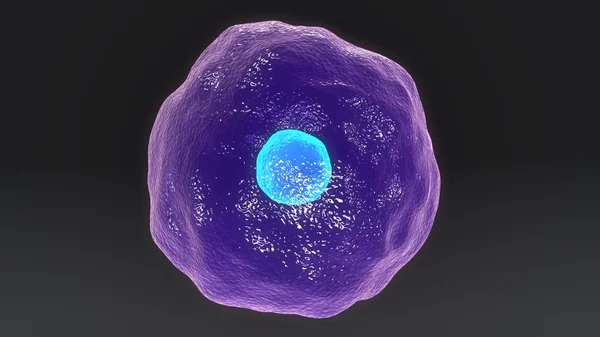
Human Cytotoxic T cell
T-cell receptor in complex with the MHC class II-peptide complex. The antigen (light green) is a peptide from a tumor cell, bacteria or virus. Different stages of the interaction. 3D-Rendering. Illustration
Cell Receptor Complex Mhc Class Peptide Complex Antigen Light Green Peptide Tumor Cell Bacteria Virus Different Stages Interaction Rendering Illustration
3D medical illustration cytotoxic T cell and Helper T cell destroys the stress-related molecule
Medical Illustration Cytotoxic Cell Helper Cell Destroys Stress Related Molecule
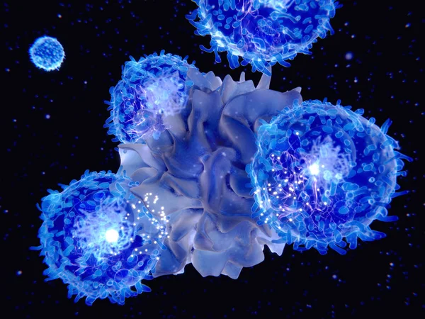
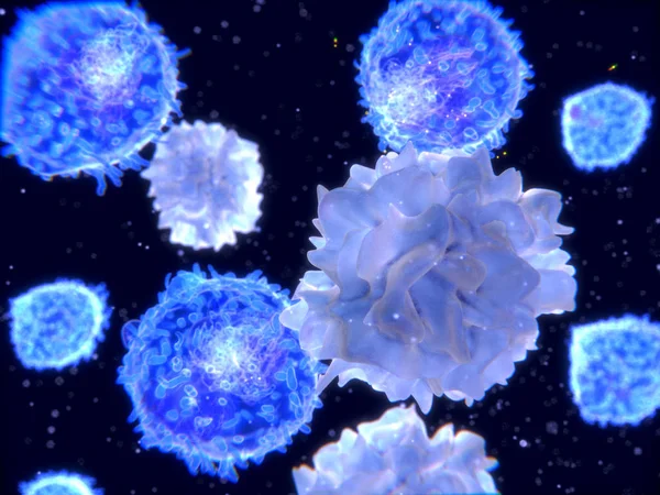
T-lymphocytes and dendritic cells, 3D-rendering; Dendritic cells are antigen-presenting cells of the immune system. They process antigen material and present it on the cell surface to the T-cells. Illustration
Lymphocytes Dendritic Cells Rendering Dendritic Cells Antigen Presenting Cells Immune System Process Antigen Material Present Cell Surface Cells Illustration
Human Cytotoxic T cell