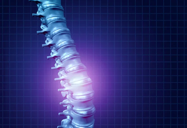
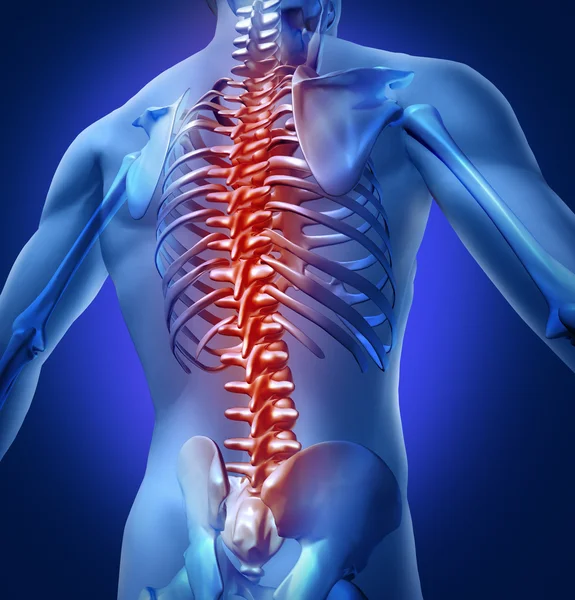
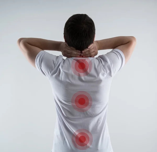
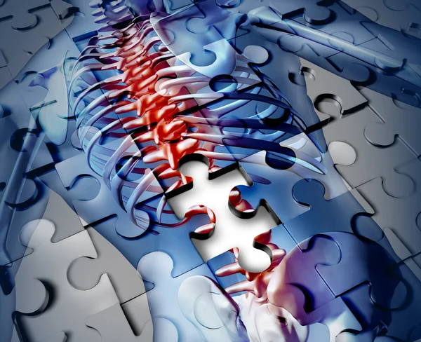
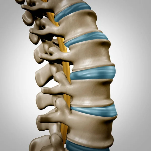
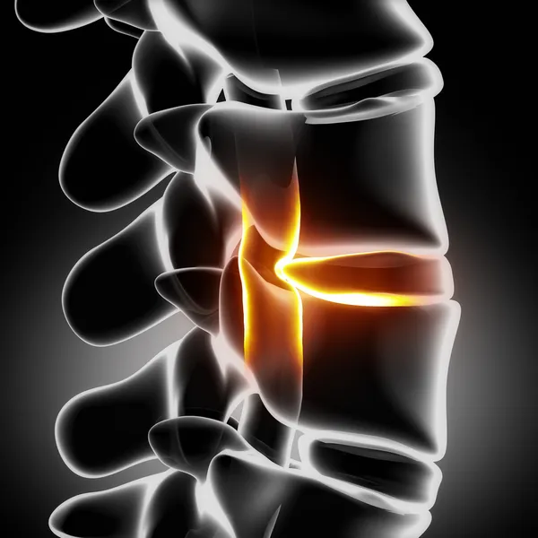
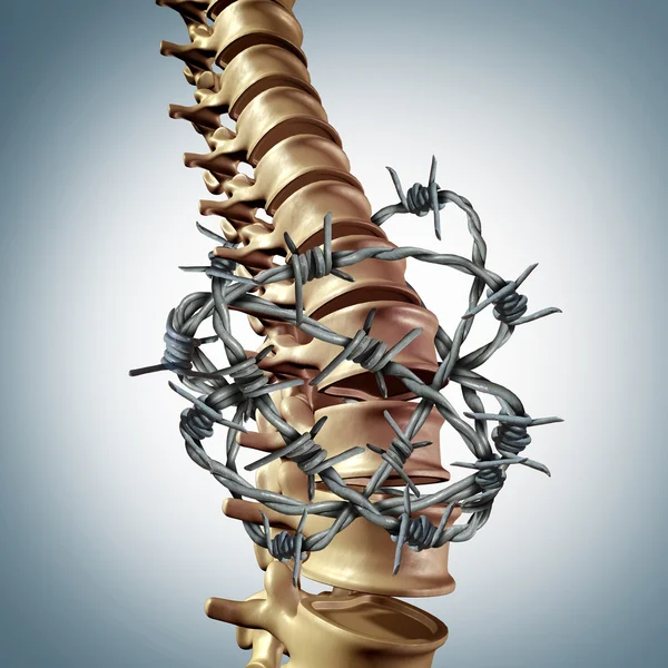
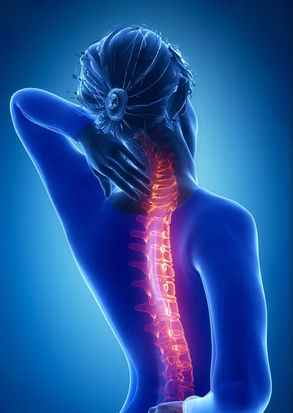
Back Spine Pain
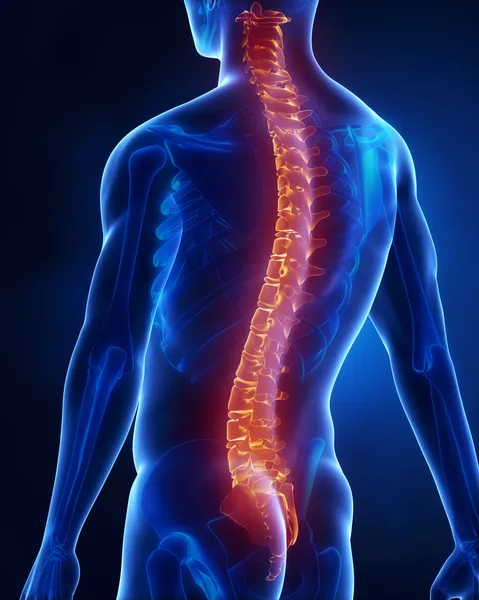
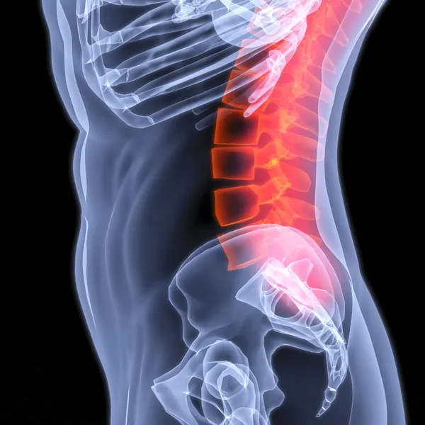
Back Spine Pain — Stock Photo, Image
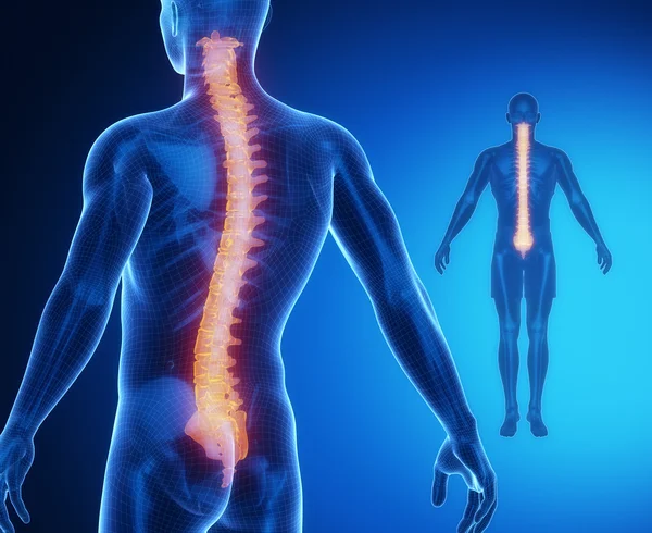
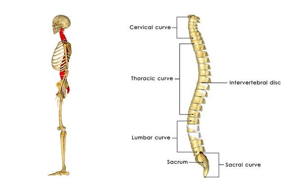
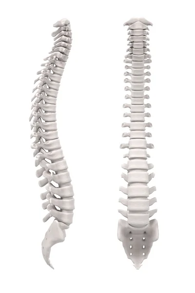
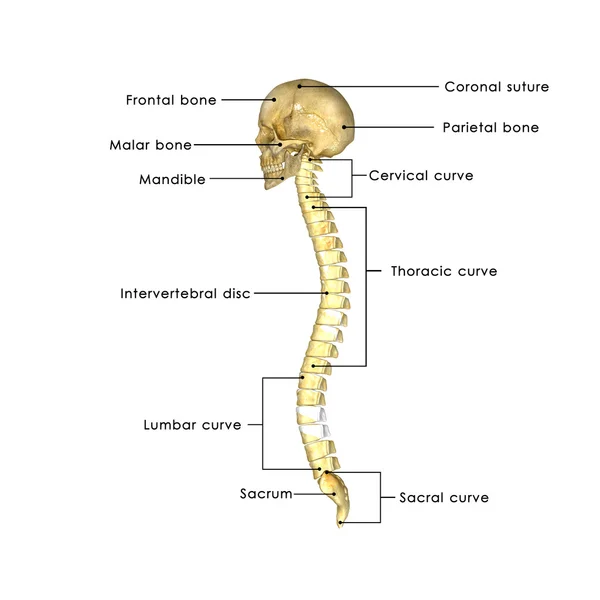
Human spine in details
Human spine in details — Stock Photo, Image
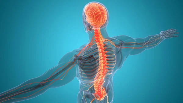
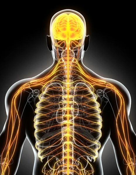
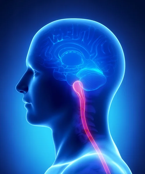
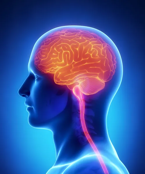
Central Organ of Human Nervous System Brain Anatomy. 3D
Central Organ Human Nervous System Brain Anatomy — Stock Photo, Image
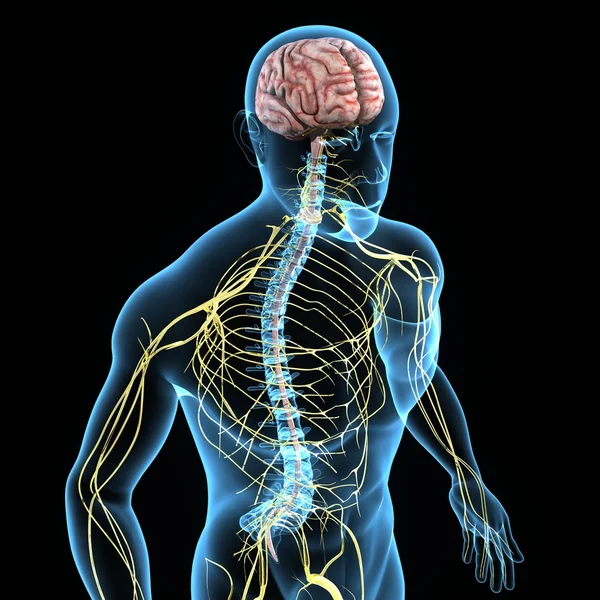
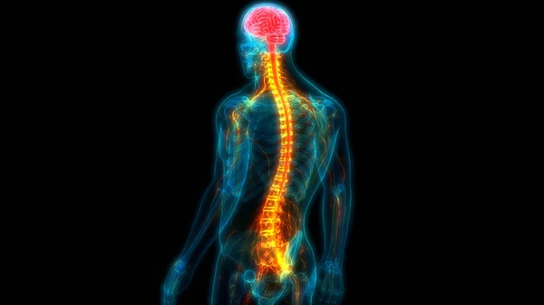
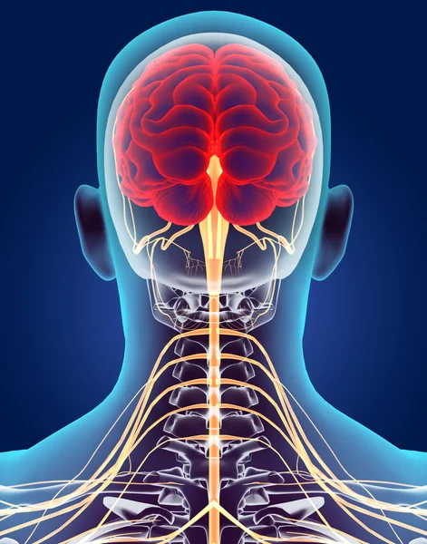
Nervous system
Nervous system — Stock Photo, Image
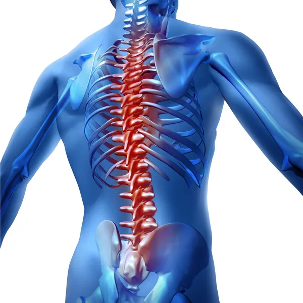
Human-Back-Pain
Human-Back-Pain — Stock Photo, Image
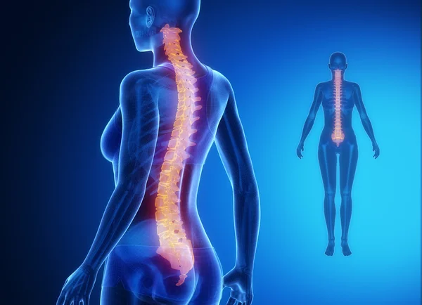
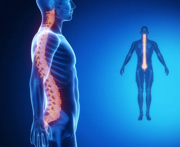
SPINE blue x--ray bone scan
SPINE blue x--ray bone scan — Stock Photo, Image
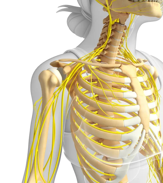
Female ribcage and nervous system artwork
Female ribcage and nervous system artwork — Stock Photo, Image
Back Pain In Human Body
Back Pain In Human Body — Stock Photo, Image
Backbone disease
Backbone disease — Stock Photo, Image
SPINE bones anatomy scan
SPINE bones anatomy scan — Stock Photo, Image
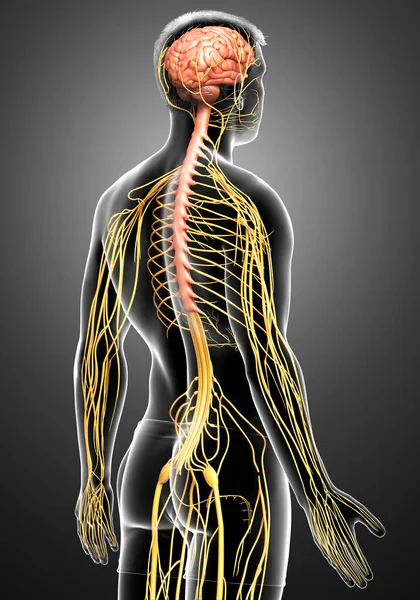
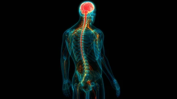
3D illustration male nervous system.
3D illustration male nervous system. — Stock Photo, Image
Spinal Stenosis
Spinal Stenosis — Stock Photo, Image
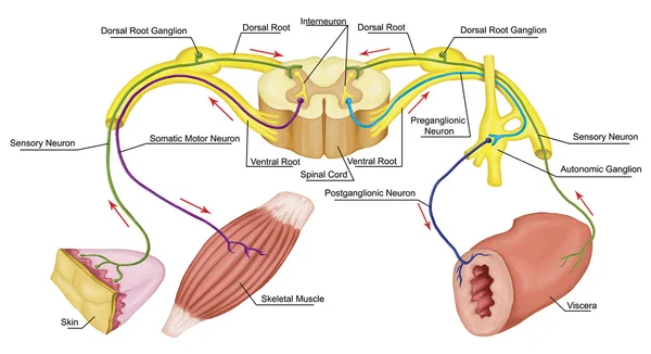
Somatic and Automatic motor reflex, somatic and Automatic nervous system, peripheral and visceral nervous system, voluntary and involuntary control of body and visceral functions
Somatic and Automatic motor reflex, somatic and Automatic nervous system,  peripheral and visceral nervous system, voluntary and involuntary control of body and visceral functions — Stock Photo, Image
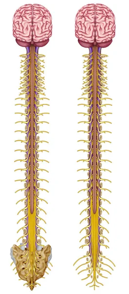
Spinal Cord
Spinal Cord — Stock Photo, Image
This system includes all the nervous structures that leave the central nervous system to the rest of our body.
System Includes All Nervous Structures Leave Central Nervous System Rest — Stock Photo, Image
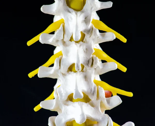
Spine vertebra model
Spine vertebra model — Stock Photo, Image
Somatic and Automatic motor reflex, somatic and Automatic nervous system, peripheral and visceral nervous system, voluntary and involuntary control of body and visceral functions
Somatic and Automatic motor reflex, somatic and Automatic nervous system, peripheral and visceral nervous system, voluntary and involuntary control of body and visceral functions — Stock Photo, Image
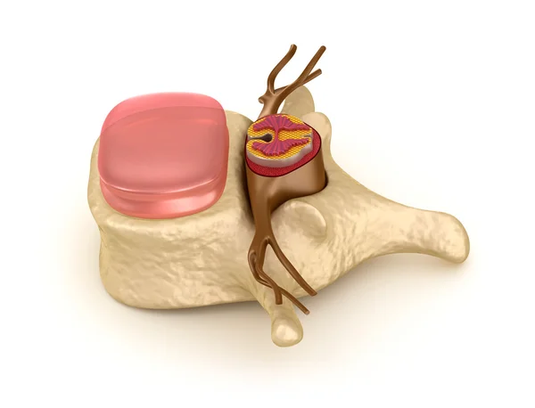
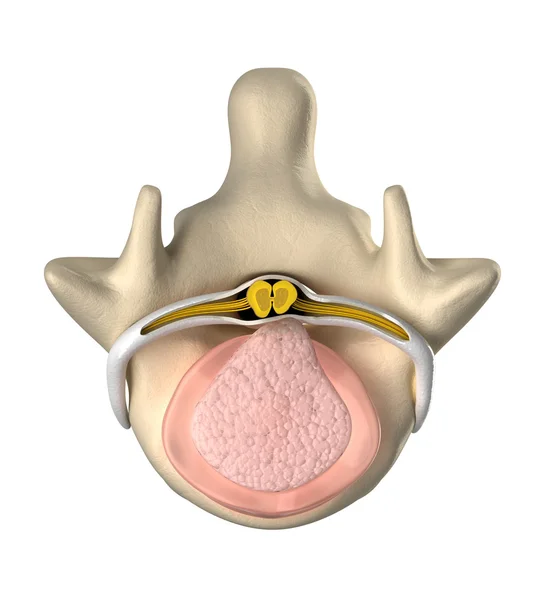
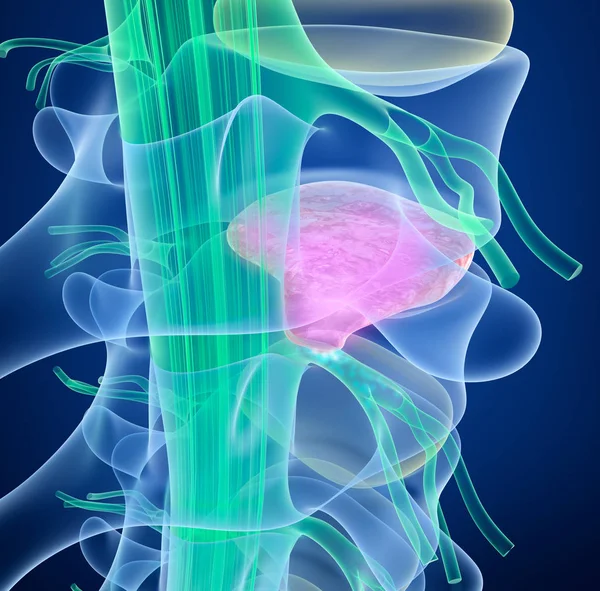
Normal intervertebral disc, spinal cord and nucleus pulposus - top view
Normal intervertebral disc, spinal cord and nucleus pulposus - top view — Stock Photo, Image
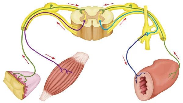
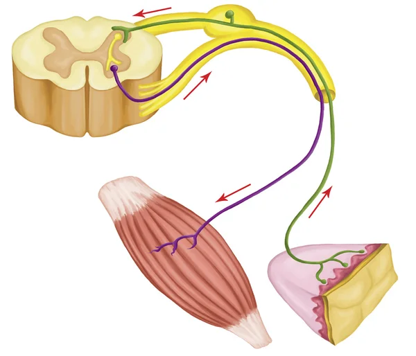
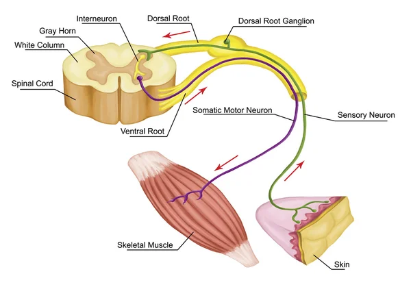
Somatic motor reflex, somatic nervous system, peripheral nervous system, voluntary control of body movements via skeletal muscles, afferent and efferent nerves
Somatic motor reflex, somatic nervous system, peripheral nervous system, voluntary control of body movements via skeletal muscles, afferent and efferent nerves — Stock Photo, Image
Skull with spinal cord
Skull with spinal cord — Stock Photo, Image
SPINE bones anatomy scan
SPINE bones anatomy scan — Stock Photo, Image
Human Back Disease
Human Back Disease — Stock Photo, Image
Jogging man with visible spine
Jogging man  with visible spine — Stock Photo, Image
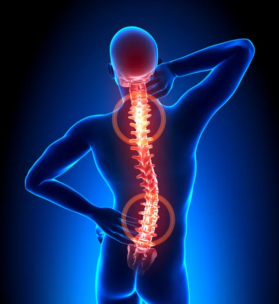
Man has pain in back and neck
Man has pain in back and neck — Stock Photo, Image
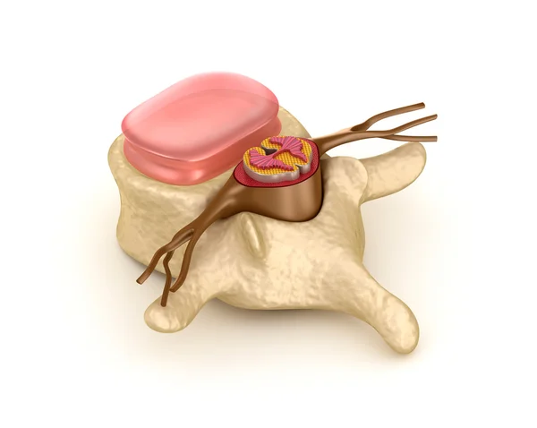
Vertebra, intervertebral disc and spinal cord
Vertebra, intervertebral disc and spinal cord — Stock Photo, Image
Patient and doctor looking at x-ray image and discussing problem with spinal cord
Patient Doctor Looking Ray Image Discussing Problem Spinal Cord — Stock Photo, Image
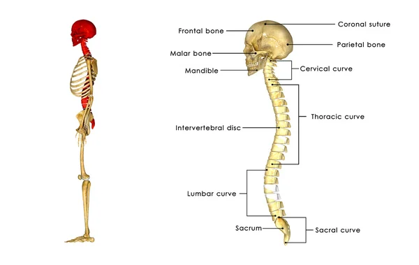
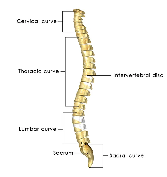
Vertebral Column Lateral view
Vertebral Column Lateral view — Stock Photo, Image
Vertebrae illness
Vertebrae illness — Stock Photo, Image
Human Spine Anatomy Section
Human Spine Anatomy Section — Stock Photo, Image
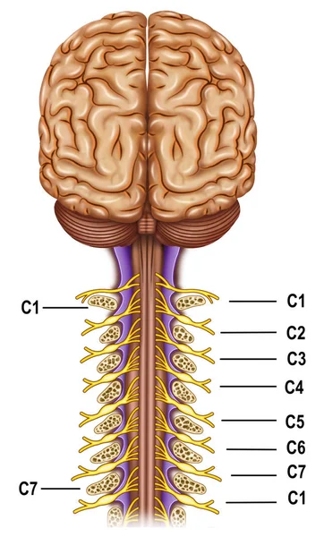
Vertebrae and nerves cervical plexus
Vertebrae and nerves cervical plexus — Stock Photo, Image
Human Central Nervous System Brain Anatomy. 3D
Human Central Nervous System Brain Anatomy — Stock Photo, Image
Human Circulatory System Heart Anatomy. 3D
Human Circulatory System Heart Anatomy — Stock Photo, Image
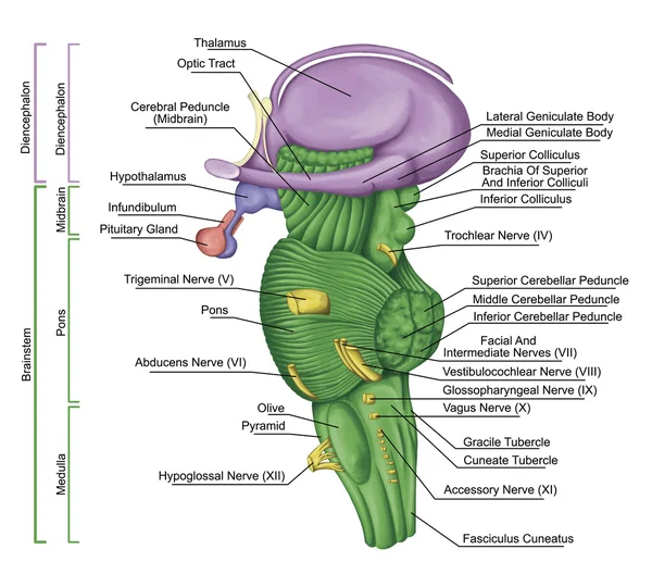
Brainstem, brain stem, lateral view, adjoining and structurally continuous with the spinal cord, parts of the diencephalon, motor and sensory innervation to the face and neck via thecranial nerves
Brainstem, brain stem, lateral view, adjoining and structurally continuous with the spinal cord, parts of the diencephalon, motor and sensory innervation to the face and neck via thecranial nerves — Stock Photo, Image
SPINE bones anatomy scan
SPINE bones anatomy  scan — Stock Photo, Image
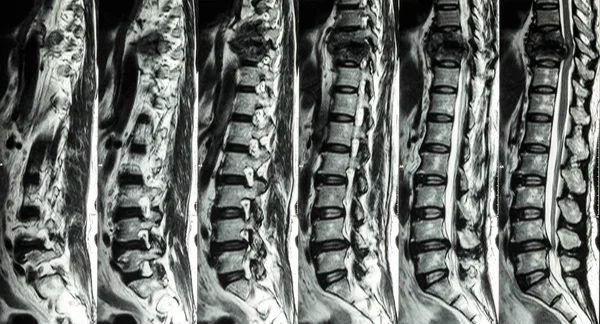
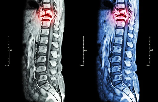
MRI of Lumbar & Thoracic spine : show fracture of thoracic spine and compress spinal cord ( Myelopathy )
MRI of Lumbar & Thoracic spine : show fracture of thoracic spine and compress spinal cord ( Myelopathy ) — Stock Photo, Image
Spinal Cord
Spinal Cord — Stock Photo, Image
Somatic motor reflex, somatic nervous system, peripheral nervous system, voluntary control of body movements via skeletal muscles, afferent and efferent nerves
Somatic motor reflex, somatic nervous system,  peripheral nervous system, voluntary control of body movements via skeletal muscles, afferent and efferent nerves — Stock Photo, Image
Spine metastasis ( cancer spread to thoracic spine ) ( MRI of thoracic and lumbar spine : show thoracic spine metastasis and compress spinal cord ( Myelopathy ) ) ( sagittal plane )
Spine metastasis ( cancer spread to thoracic spine ) ( MRI of thoracic and lumbar spine : show thoracic spine metastasis and compress spinal cord ( Myelopathy ) ) ( sagittal plane ) — Stock Photo, Image
Brain and spinal cord medical x-ray scan
Brain and spinal cord medical x-ray scan — Stock Photo, Image
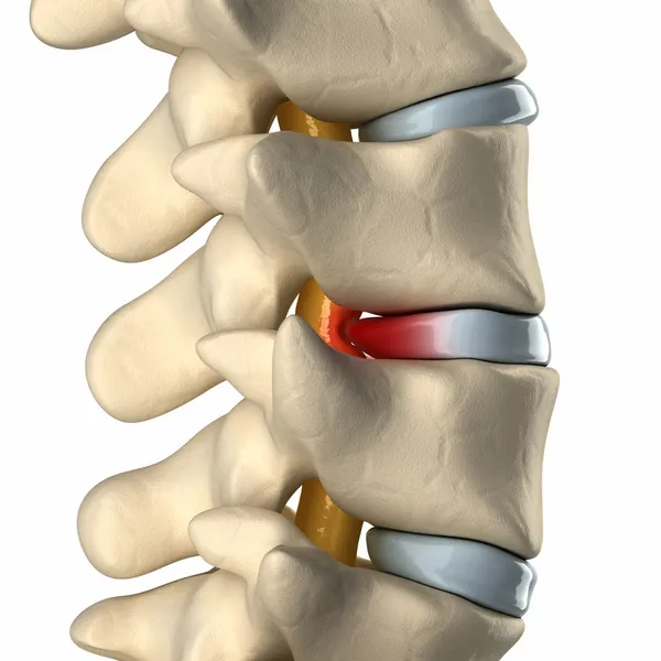
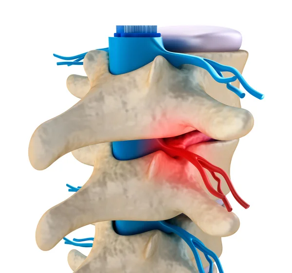
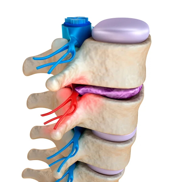
Spinal cord under pressure of bulging disc
Spinal cord under pressure of bulging disc — Stock Photo, Image
Brain Anatomy - Spinal cord Nerves
Brain Anatomy - Spinal cord Nerves — Stock Photo, Image
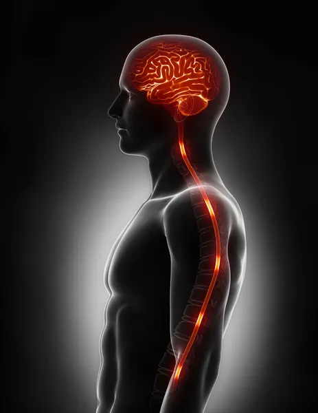
Anatomy of brain with backbone
Anatomy of brain with backbone — Stock Photo, Image
Herniated disc with pressure on spinal cord close-up
Herniated disc with pressure on spinal cord close-up — Stock Photo, Image
Spinal cord under pressure of bulging disc
Spinal cord under pressure of bulging disc — Stock Photo, Image
Doctor Explaining The Problems About Spinal Cord To Patient
Doctor Explaining The Problems About Spinal Cord To Patient — Stock Photo, Image
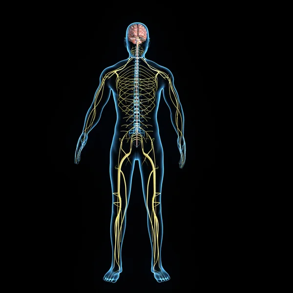
3D illustration male nervous system.
3D illustration male nervous system. — Stock Photo, Image
SPINE bones scan
SPINE  bones scan — Stock Photo, Image
3d renderings of spinal cord
3d renderings of spinal cord — Stock Photo, Image
SPINE bones anatomy scan
SPINE bones anatomy  scan — Stock Photo, Image
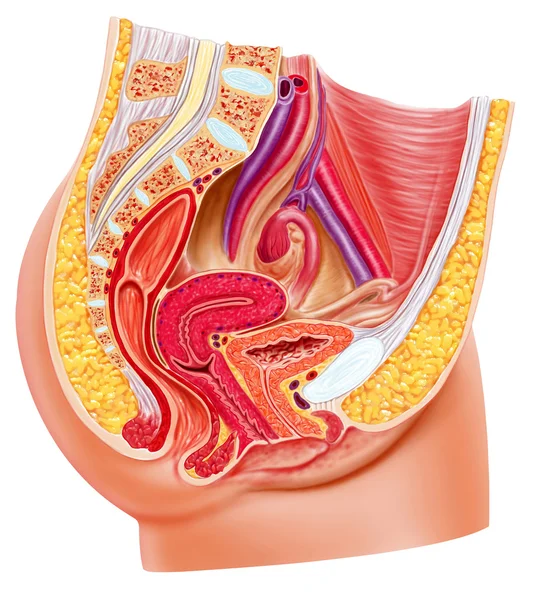
Anatomy female reproductive system, cutaway.
Anatomy female reproductive system, cutaway. — Stock Photo, Image
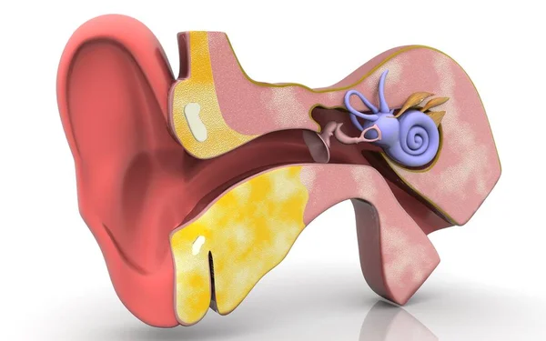
3D illustration of inner ear .Cross section of inner ear
Illustration Inner Ear Cross Section Inner Ear — Stock Photo, Image
Back bone with skull
Back bone with skull — Stock Photo, Image
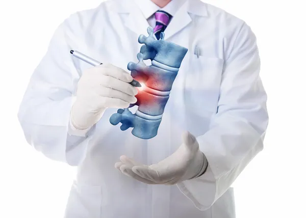
Doctor showing her patient a spine model
Doctor showing her patient a spine model — Stock Photo, Image
Man with visible spine
Man with visible spine — Stock Photo, Image
Brain anatomy MEDULLA and SPINAL CORD - cross section
Brain anatomy MEDULLA and SPINAL CORD - cross section — Stock Photo, Image
3d rendered medically accurate illustration of the brain
Rendered Medically Accurate Illustration Brain — Stock Photo, Image
Bulging Disc Degeneration - Spine problem
Bulging Disc Degeneration - Spine problem — Stock Photo, Image
Intervertebral disc with pressure on spinal cord top view
Intervertebral disc with pressure on spinal cord top view — Stock Photo, Image
Central organ of Human Nervous System Brain Anatomy. 3D
Central Organ Human Nervous System Brain Anatomy — Stock Photo, Image
Spinal cord, cross-section. 3D illustration which shows the white and the grey matter with dorsal and ventral horns
Spinal Cord Cross Section Illustration Which Shows White Grey Matter — Stock Photo, Image
Section of the spinal cord, vintage engraving.
Section of the spinal cord, vintage engraving. — Stock Photo, Image
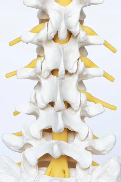
Posterior view of lumbar spine model on white background
Posterior View Lumbar Spine Model White Background — Stock Photo, Image
Spinal cord under pressure of bulging disc
Spinal cord under pressure of bulging disc — Stock Photo, Image
Spinal cord under pressure of bulging disc
Spinal cord under pressure of bulging disc — Stock Photo, Image
Female body with SPINE bone scan
Female body with SPINE bone scan — Stock Photo, Image
Spinal column with nerves and discs.
Spinal column with nerves and discs. — Stock Photo, Image
Spinal segment with a disk
Spinal segment with a disk — Stock Photo, Image
Lower Back Pain
Lower Back Pain — Stock Photo, Image
3d rendered medically accurate illustration of a male nervous system
Rendered Medically Accurate Illustration Male Nervous System — Stock Photo, Image
3D illustration, skeletal, spine, cord, spinal, painful, posture, isolated, human, cervical, medical, render,skeleton, diagram, osteoporosis, lumbar, backache, Scoliosis, kyphosis, injury, vertebra, medicine, tan, illustration, bone, healthy, segment
Illustration Skeletal Spine Cord Spinal Painful Posture Isolated Human Cervical — Stock Photo, Image
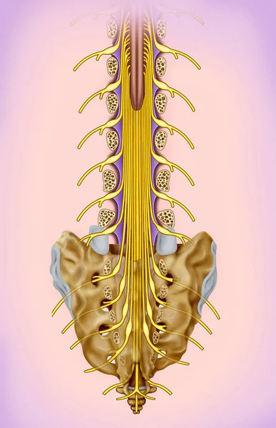
Lumbar and sacral nerve branch illustration. The spinal nerves are named according to the point of emergence of the spinal cord.
Lumbar Sacral Nerve Branch Illustration Spinal Nerves Named According Point — Stock Photo, Image
Spinal segment with a disk
Spinal segment with a disk — Stock Photo, Image
Spine Vertebrae - Lateral view Side view
Spine Vertebrae - Lateral view Side view — Stock Photo, Image
Back pain
Back pain — Stock Photo, Image
Illustration of human back pain with highlighted spinal cord
Illustration of human back pain with highlighted spinal cord — Stock Photo, Image
Nervous system
Nervous system — Stock Photo, Image
Doctor showing a female patient a part of a spine
Doctor showing a female patient a part of a spine — Stock Photo, Image
Male neurologist doctor examines cervical vertebrae of female patient spinal column in medical clinic. Neurological physical examination. Osteopathy, chiropractic, physiotherapy. Selective focus.
Male neurologist doctor examines cervical vertebrae of female patient spinal column in medical clinic. Neurological physical examination. Osteopathy, chiropractic, physiotherapy. Selective focus. — Stock Photo, Image
Modern digital illustration of human brain
Modern Digital Illustration Human Brain — Stock Photo, Image
Male Doctor explaining something to a woman
Male Doctor explaining something to a woman — Stock Photo, Image
Brain and spinal cord anatomy - cross section
Brain and spinal cord anatomy - cross section — Stock Photo, Image
Woman with kidneys disease on the blue background
Woman Kidneys Disease Blue Background — Stock Photo, Image
Neck brace
Neck brace — Stock Photo, Image
Spinal cord nerve energy impulses into brain
Spinal cord nerve energy impulses into brain — Stock Photo, Image
Woman has neck and back pain
Woman has neck and back pain — Stock Photo, Image
Spine
Spine — Stock Photo, Image
Spinal cord under pressure of bulging disc
Spinal cord under pressure of bulging disc — Stock Photo, Image
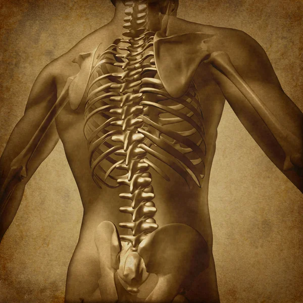
Human Back Grunge Texture
Human Back Grunge Texture — Stock Photo, Image
Male Hurt Backbone - Vertebrae Pain
Male Hurt Backbone - Vertebrae Pain — Stock Photo, Image
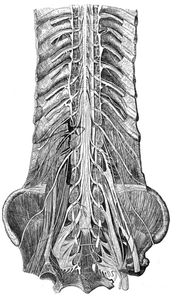
Nerves of the lower trunk, the spinal canal is open, front view
Nerves of the lower trunk, the spinal canal is open, front view — Stock Photo, Image
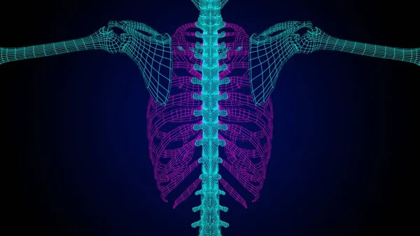
Rib cage anatomy 3D Illustration with wire frame skeleton for medical concept
Rib Cage Anatomy Illustration Wire Frame Skeleton Medical Concept — Stock Photo, Image
Human Central Nervous System brain Anatomy. 3D
Human Central Nervous System Brain Anatomy — Stock Photo, Image
Human Central Nervous System brain Anatomy. 3D
Human Central Nervous System Brain Anatomy — Stock Photo, Image
Human Internal Digestive Organ Liver Anatomy. 3D
Human Internal Digestive Organ Liver Anatomy — Stock Photo, Image
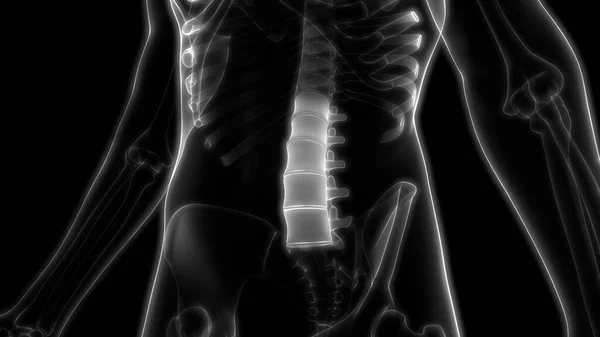
Vertebral Column of Lumbar Vertebrae Human Skeleton System Anatomy 3d rendering
Vertebral Column Lumbar Vertebrae Human Skeleton System Anatomy Rendering — Stock Photo, Image
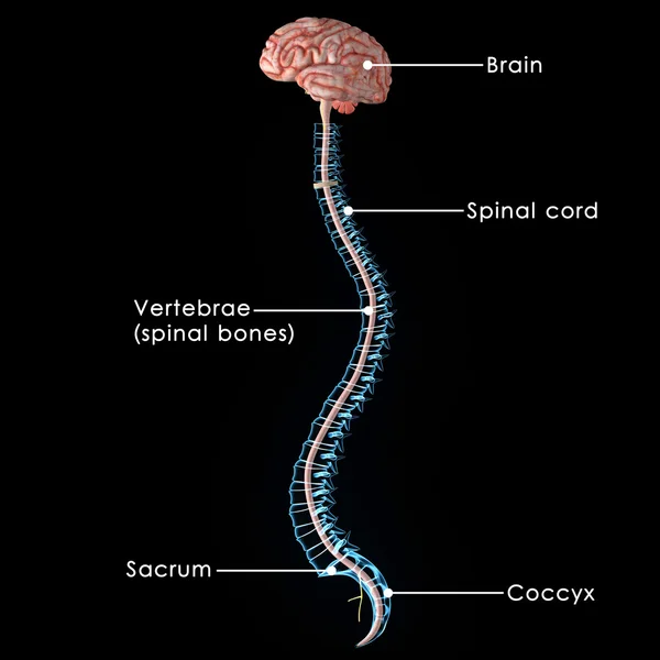
Spinal Cord
Spinal Cord — Stock Photo, Image
3d rendered medically accurate illustration of a male brain
Rendered Medically Accurate Illustration Male Brain — Stock Photo, Image
Vertebra on x ray human body, cg medicine 3d illustration
Vertebra Ray Human Body Medicine Illustration — Stock Photo, Image
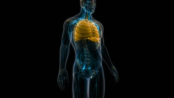
Human Respiratory System Lungs Anatomy. 3D
Human Respiratory System Lungs Anatomy — Stock Photo, Image

Related image searches

**Spinal Cord Images: Enhance Your Projects with Stunning Visuals**

**Introduction**

Welcome to our collection of stock images featuring stunning visuals of the spinal cord. Whether you're a medical professional, educator, or designer, these images are an invaluable resource for enhancing your projects. In this article, we will provide an overview of the available images, their file types, and offer practical advice on how to use them effectively. So, let's dive in and explore the incredible world of spinal cord imagery!

**Explore the Diverse Range of Spinal Cord Images**

Our collection offers an extensive range of high-quality spinal cord images in JPG, AI, and EPS formats. From detailed anatomical illustrations to artistic representations, we have something to suit every need. Our stock images depict various aspects of the spinal cord, such as its structure, functions, and common pathologies. Whether you require images for medical textbooks, educational presentations, or website designs, we have you covered.

**The Power of Visuals in Medical Projects**

When it comes to medical projects, visuals play a crucial role in conveying information effectively. Utilizing visually appealing and accurate spinal cord images can greatly enhance the understanding of complex medical concepts. For medical professionals, these images serve as valuable resources for patient education, presentations, and research publications. Moreover, educators can make use of these visuals to engage students and create interactive learning experiences.

**Practical Tips for Using Spinal Cord Images**

To make the most of our spinal cord images, here are a few practical tips:

1. Select the Most Relevant Images: Carefully choose images that align with the specific theme or topic of your project. For example, if you are creating a presentation about spinal cord injuries, focus on images that illustrate different types of injuries and their impact on the body.

2. Ensure Accuracy and Scientific Validity: When using spinal cord images in medical or educational contexts, accuracy is paramount. Verify that the images represent the anatomical structures and functions of the spinal cord correctly. This will ensure credibility and avoid any misinterpretations.

3. Customize Images to Fit Your Design: Stock images can be edited and customized to match your project's aesthetic requirements. Consider cropping, resizing, or adding text overlays to seamlessly integrate the images into your designs while maintaining a professional appearance.

4. Acknowledge Copyright and Usage Rights: It is essential to respect copyright and usage rights when utilizing stock images. Ensure that you have the necessary permissions and licenses to use the images in your specific project. Give proper attribution when required to avoid any legal complications.

**Conclusion**

Stock images of the spinal cord are invaluable resources for medical professionals, educators, and designers. By incorporating visually stunning and scientifically accurate visuals into your projects, you can effectively communicate complex concepts and engage your audience. Remember to choose the most relevant images, ensure accuracy, customize them to fit your design, and respect copyright and usage rights. Explore our collection of spinal cord images today and take your projects to new heights!

*Discover the Fascinating World of Spinal Cord Imagery* *Enhance Your Visual Presentations with Spinal Cord Images* *Unlock the Power of Visual Communication with Our Spinal Cord Images* *Transform Your Projects with High-Quality Spinal Cord Visuals*