Cercine Stock Vector Images

26 Cercine vector art & graphics are available under a royalty-free license

Bankart lesion as anterior part of the glenoid labrum trauma outline diagram
Anatomy of the Hip and Acetabular Labrum. Ligamentum teres and Articular cartilage. Lateral view with femur removed of right hip.
Anatomy Hip Acetabular Labrum Ligamentum Teres Articular Cartilage Lateral View Femur Removed Right Hip
Detailed diagram of shoulder labrum tears
Detailed Diagram Shoulder Labrum Tears
This is use for explain and compare when the head of the humerus
Hip labrum tear, Hip joint medical illustration flashcard
Hip Labrum Tear Hip Joint Medical Illustration Flashcard
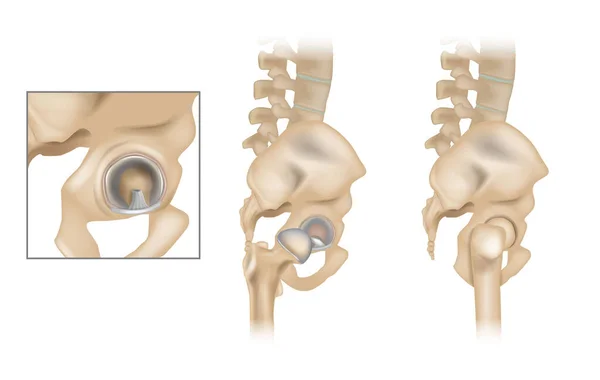
Total Hip Arthroplasty THA with Prosthetic Components and Normal Hip Joint Anatomy. Total Hip Replacement Surgery with Implant Components and Normal Hip Joint Anatomy
Total Hip Arthroplasty Tha Prosthetic Components Normal Hip Joint Anatomy Total Hip Replacement Surgery Implant Components Normal Hip Joint Anatomy
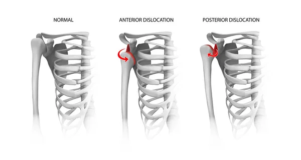
Shoulder dislocation options. Vector illustration.
Shoulder Dislocation Options Vector Illustration
Anatomy of the Hip and Acetabular Labrum. Ligamentum teres and Articular cartilage. Lateral view with femur removed of right hip.
Anatomy Hip Acetabular Labrum Ligamentum Teres Articular Cartilage Lateral View Femur Removed Right Hip
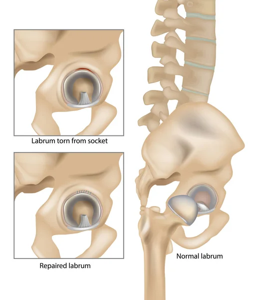
Hip Labral Tears. Labrum torn from socket and Repaired labrum. Surgery for Repairing a Torn Hip Labral. Vector
Hip Labral Tears Labrum Torn Socket Repaired Labrum Surgery Repairing Torn Hip Labral Vector
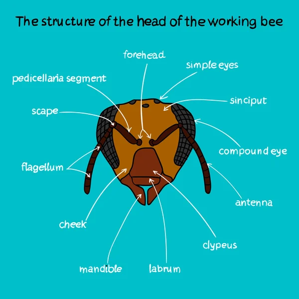
The structure of the head of the working bee color
Detailed diagram of SLAP tear in shoulder joint
Detailed Diagram Slap Tear Shoulder Joint
Hip Joint Anatomy with Acetabular Labrum and Ligamentum Teres, Anterior and Lateral Pelvis Medical Illustration. Human Pelvis and Hip Joint Anatomical Structure
Hip Joint Anatomy Acetabular Labrum Ligamentum Teres Anterior Lateral Pelvis Medical Illustration Human Pelvis Hip Joint Anatomical Structure
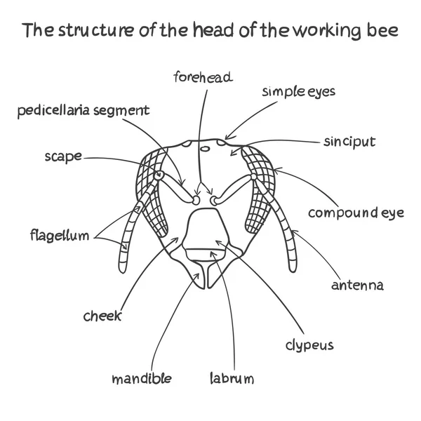
The structure of the head of the working bee
Hip labral tear diagram
Hip Labral Tear Diagram
Inflammation of shoulder bursae causing pain
Inflammation Shoulder Bursae Causing Pain
Glenohumeral joint coronal anatomy highlights humeral head, glenoid cavity, and supraspinatus tendon, conveying shoulder articulation and stabilizing soft tissues. Outline diagram
Glenohumeral Joint Coronal Anatomy Highlights Humeral Head Glenoid Cavity Supraspinatus Tendon Conveying Shoulder Articulation Stabilizing Soft Tissues Outline Diagram
Abstract Gradient Circles with white Labrum Glyph
Abstract Gradient Circles White Labrum Glyph
Grasshopper
Grasshopper
External anatomy of a grasshopper
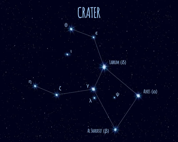
Crater constellation, vector illustration with the names of basic stars against the starry sky
Crater Constellation Vector Illustration Names Basic Stars Starry Sky
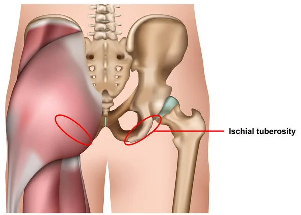
Ischial tuberosity 3d medical vector illustration isolated on white background infographic
Ischial Tuberosity Medical Vector Illustration Isolated White Background Infographic
Detailed anatomy of shoulder bursitis inflammation
Detailed Anatomy Shoulder Bursitis Inflammation
Antagonistic Muscles (Insects) concept vector illustration
Antagonistic Muscles Insects Concept Vector Illustration
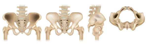
The pelvic girdle Lateral, anterior, bottom and posterior view. Human pelvis and sacrum bones isolated on white background. Vector of pelvic area in four different positions
Pelvic Girdle Lateral Anterior Bottom Posterior View Human Pelvis Sacrum Bones Isolated White Background Vector Pelvic Area Four Different Positions
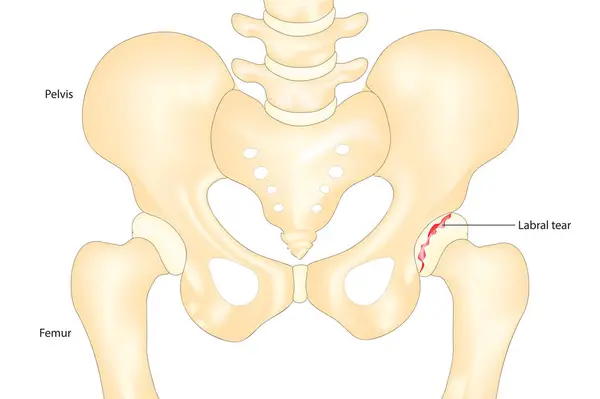
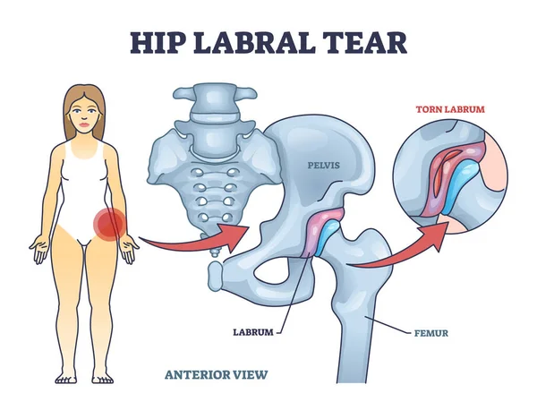
Hip labral tear anatomical explanation with medical labrum bone damage outline diagram. Labeled educational skeletal anatomy with pelvis, femur and torn labrum condition example vector illustration.
Hip Labral Tear Anatomical Explanation Medical Labrum Bone Damage Outline Diagram Labeled Educational Skeletal Anatomy Pelvis Femur Torn Labrum Condition Example Vector Illustration
Glenohumeral joint diagram shows labeled shoulder anatomy, highlighting humeral head, glenoid cavity, and rotator cuff tendons, communicates structure and stability. Outline diagram
Glenohumeral Joint Diagram Shows Labeled Shoulder Anatomy Highlighting Humeral Head Glenoid Cavity Rotator Cuff Tendons Communicates Structure Stability Outline Diagram