Ciliado Stock Vector Images

32 Ciliado vector art & graphics are available under a royalty-free license
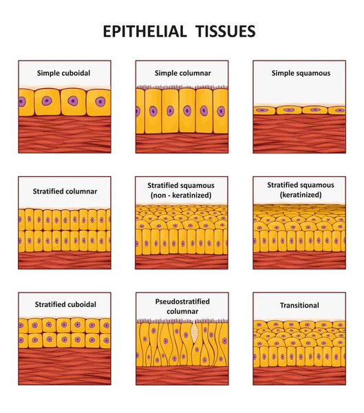
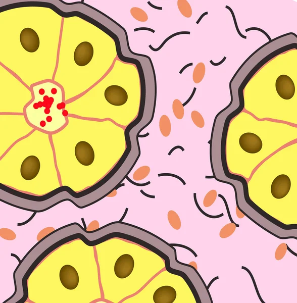
Different types of epithelial tissue collection.
Olfactory Bulb Anatomy. Epithelium receptor cells. Nasal mucosa
Olfactory Bulb Anatomy Epithelium Receptor Cells Nasal Mucosa
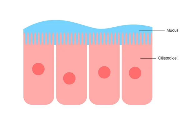
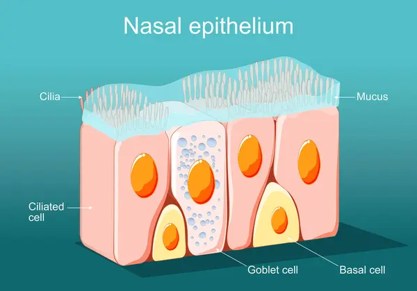
Nasal epithelium concept
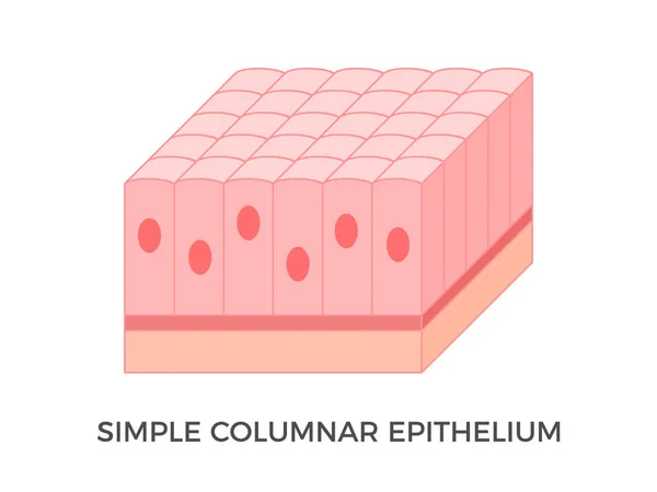
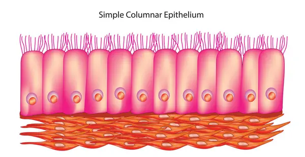
Simple columnar epithelium. Epithelial tissue types. Tall and slender cells with oval-shaped nuclei. Lines most organs of the digestive tract like stomach, intestines. Medical illustration. Vector.
Simple Columnar Epithelium Epithelial Tissue Types Tall Slender Cells Oval Shaped Nuclei Lines Most Organs Digestive Tract Stomach Intestines Medical Illustration Vector
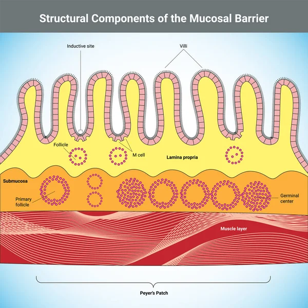
Structural Components of the Mucosal Barrier medical illustration
3D Isometric Flat Vector Conceptual Illustration of Nasal Mucosa Cells, Medical Educational Diagram
Isometric Flat Vector Conceptual Illustration Nasal Mucosa Cells Medical Educational Diagram
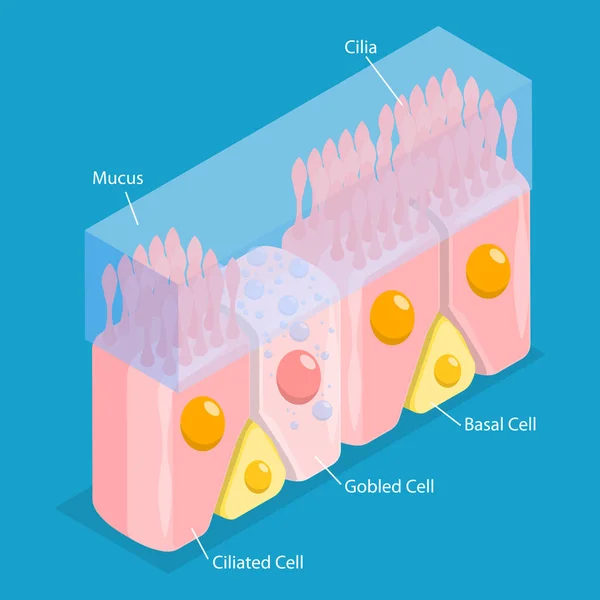
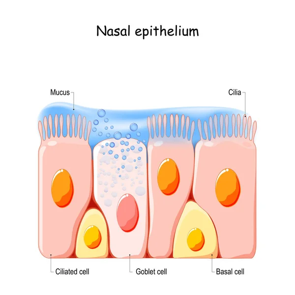
Nasal Epithelium structure anatomy, labeled with cilia, mucus, ciliated cell, goblet cell, basal cell. Human tissue nasal cavity, immune response to nasal diseases, diagram biology education.
Nasal Epithelium Structure Anatomy Labeled Cilia Mucus Ciliated Cell Goblet Cell Basal Cell Human Tissue Nasal Cavity Immune Response Nasal Diseases Diagram Biology Education
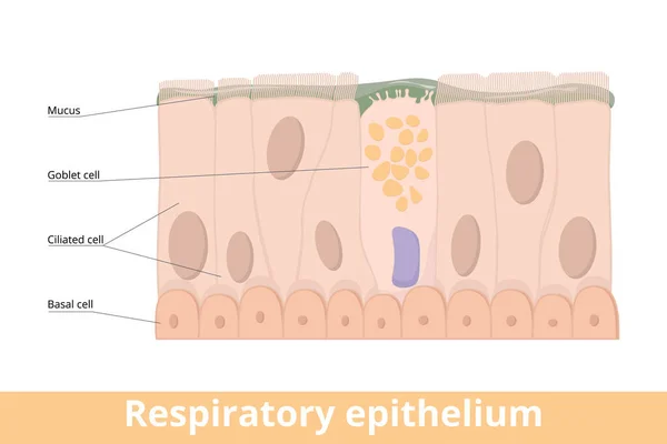
Respiratory epithelium. A type of ciliated columnar epithelium found lining most of the respiratory tract as respiratory mucosa including goblet cell, basal and ciliated cells, mucus.
Respiratory Epithelium Type Ciliated Columnar Epithelium Found Lining Most Respiratory Tract Respiratory Mucosa Including Goblet Cell Basal Ciliated Cells Mucus
Epithelium of nasal mucosa
Nasal epithelium concept
Epithelium of nasal mucosa
Respiratory Epithelium Cell Structure Illustration. Ciliated, Basal, and Goblet Cells Diagram. Mucociliary Epithelium Cross Section Illustration
Respiratory Epithelium Cell Structure Illustration Ciliated Basal Goblet Cells Diagram Mucociliary Epithelium Cross Section Illustration
Nasal epithelium. Ciliated columnar epithelium. epithelial cells forms the lining of the stomach and intestines, duodenum, fallopian tubes, uterus, central canal of the spinal cord, nose, ears and the taste buds. Ciliated cells. Respiratory defense
Nasal Epithelium Ciliated Columnar Epithelium Epithelial Cells Forms Lining Stomach Intestines Duodenum Fallopian Tubes Uterus Central Canal Spinal Cord Nose Ears Taste Buds Ciliated Cells Respiratory Defense
Ciliated epithelium medical background
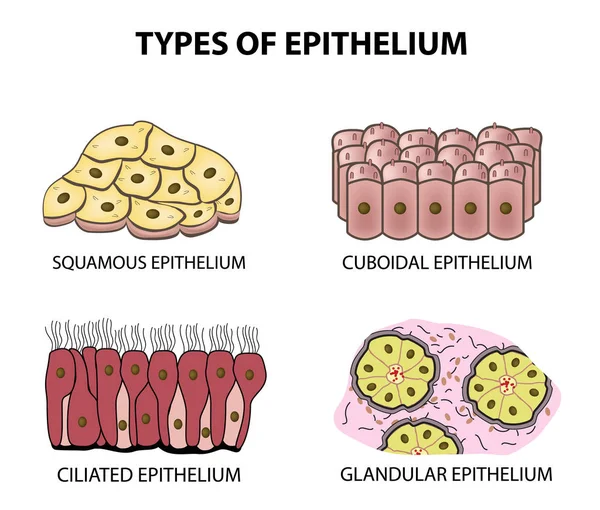
Types of epithelium. squamous, cubic, ciliated, glandular. Set. Infographics. Vector illustration on isolated background
Epithelium. squamous, cubic, ciliated, glandular. Set. Infographics. Vector illustration.
Ciliated epithelium. Epithelial tissue types. It performs the function of moving particles or fluid over the epithelial surface. Found in trachea, bronchial tubes, nasal cavities. Vector illustration
Ciliated Epithelium Epithelial Tissue Types Performs Function Moving Particles Fluid Epithelial Surface Found Trachea Bronchial Tubes Nasal Cavities Vector Illustration
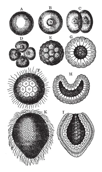
Coral Stages where Free swimming ciliiated gastrulais another labels, vintage line drawing or engraving illustration.
Coral Stages Free Swimming Ciliiated Gastrulais Another Labels Vintage Line Drawing Engraving Illustration
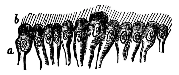
Ciliated epithelial tissue which is covered with long waving hair like projections, vintage line drawing or engraving illustration.
Ciliated Epithelial Tissue Which Covered Long Waving Hair Projections Vintage Line Drawing Engraving Illustration
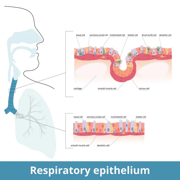
Respiratory epithelium. Human respiratory tract tissue with specific cells: ciliated, goblet, brush and basal cells. Smooth muscle cells innervated by dendritic cells and cartilage. Lungs histology.
Respiratory Epithelium Human Respiratory Tract Tissue Specific Cells Ciliated Goblet Brush Basal Cells Smooth Muscle Cells Innervated Dendritic Cells Cartilage Lungs Histology
Nasal mucosa cells. Nasal secretions. Ciliated, basal and goblet cells. Olfactory epithelium. Cells act as a low resistance filter. Vector illustration
Nasal Mucosa Cells Nasal Secretions Ciliated Basal Goblet Cells Olfactory Epithelium Cells Act Low Resistance Filter Vector Illustration
Simple columnar epithelium tissue
Simple Columnar Epithelium Tissue
Bronchus. Medical illustration depicting Respiratory cilia and mucus. Pseudostratified columnar epithelium. Structure Ciliated Epithelium Infographics
Bronchus Medical Illustration Depicting Respiratory Cilia Mucus Pseudostratified Columnar Epithelium Structure Ciliated Epithelium Infographics
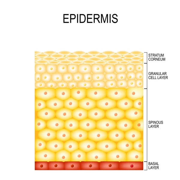
Skin Cells and Structure Layers of epidermis
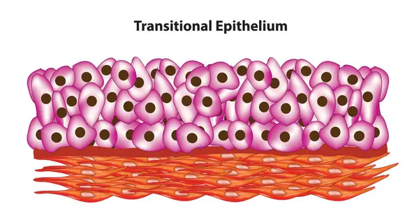
Cell arrangement of transitional epithelium
Cell Arrangement Transitional Epithelium
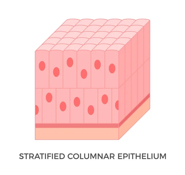
Stratified columnar epithelium. Epithelial tissue types. Tall and slender cells with oval-shaped nuclei. It is found in the conjunctiva, pharynx, anus, and male urethra. Medical illustration. Vector.
Stratified Columnar Epithelium Epithelial Tissue Types Tall Slender Cells Oval Shaped Nuclei Found Conjunctiva Pharynx Anus Male Urethra Medical Illustration Vector
Ciliated columnar epithelium. epithelial cells forms the lining of the stomach and intestines, duodenum, fallopian tubes, uterus, central canal of the spinal cord, nose, ears and the taste buds.
Ciliated Columnar Epithelium Epithelial Cells Forms Lining Stomach Intestines Duodenum Fallopian Tubes Uterus Central Canal Spinal Cord Nose Ears Taste Buds
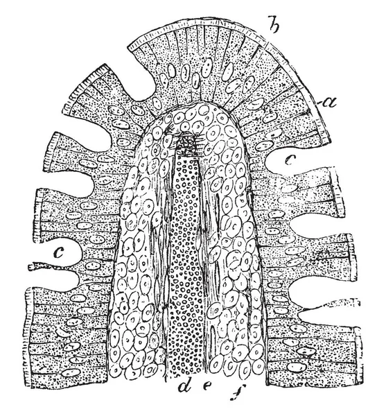
A vertical section of an intestinal villus of a cat have a striated basilar border of the epithelium, vintage line drawing or engraving illustration.
Vertical Section Intestinal Villus Cat Have Striated Basilar Border Epithelium Vintage Line Drawing Engraving Illustration
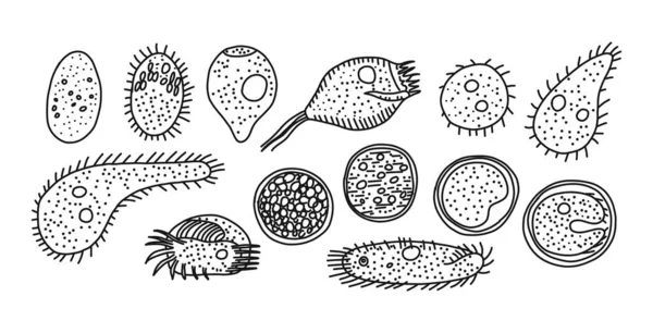
Ciliated infusoria origin and development modes, vector illustration.
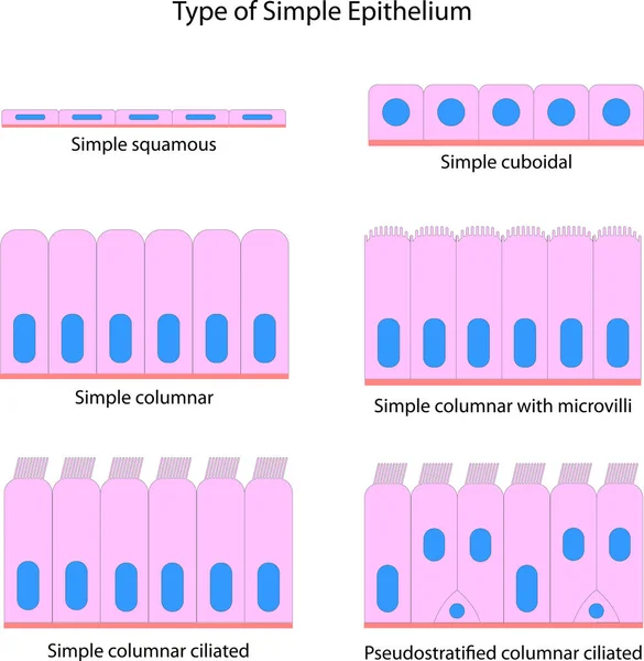
Type of Simple Epithelium
Type Simple Epithelium
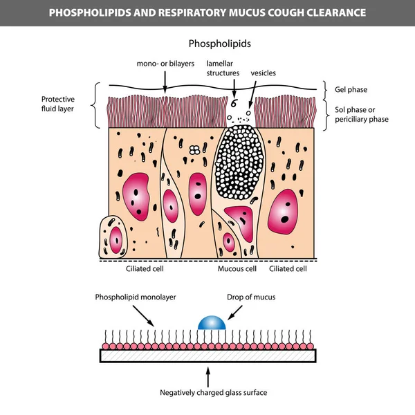
Medical vector illustration of phospholipids and respiratory mucus cough clearance. Ciliated and mucous cell structure.
Medical Vector Illustration Phospholipids Respiratory Mucus Cough Clearance Ciliated Mucous Cell Structure