Citocinas Stock Vector Images

90 Citocinas vector art & graphics are available under a royalty-free license
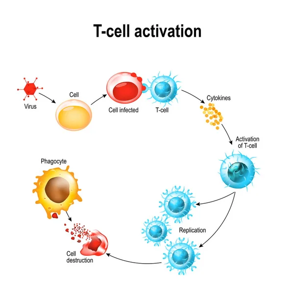
Activation of T-cell leukocytes
Activation of  T-cell leukocytes
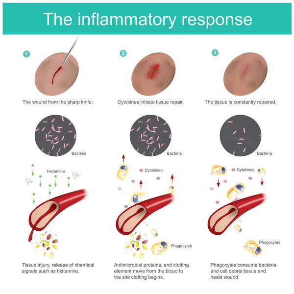
The inflammatory response. When the skin tissue is damaged, the body will produce immunity, eliminate bacteria, together with the new tissue regeneration process. all the response caused by blood vessel and cells fluid in the arteries.
Inflammatory Response Skin Tissue Damaged Body Produce Immunity Eliminate Bacteria Together New Tissue Regeneration Process All Response Caused Blood Vessel Cells Fluid Arteries
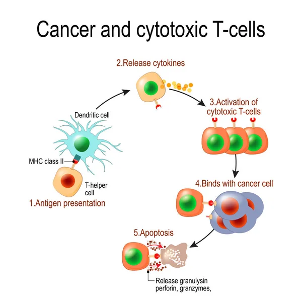
Cancer and cytotoxic T-cells. T lymphocyte kills cancer cells. T-cell (immune responses), release the perforin and granzymes, and attack cancerous cells. Through the action of perforin, granzymes enter the cytoplasm of the target cell, and lead to ap
Cancer Cytotoxic Cells Lymphocyte Kills Cancer Cells Cell Immune Responses Release Perforin Granzymes Attack Cancerous Cells Action Perforin Granzymes Enter Cytoplasm Target Cell Lead
Ovarian cyst, follicle and Corpus luteum.
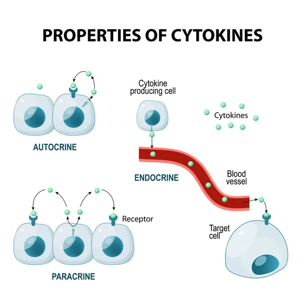
Properties of cytokines
Cytokine. Vector concept
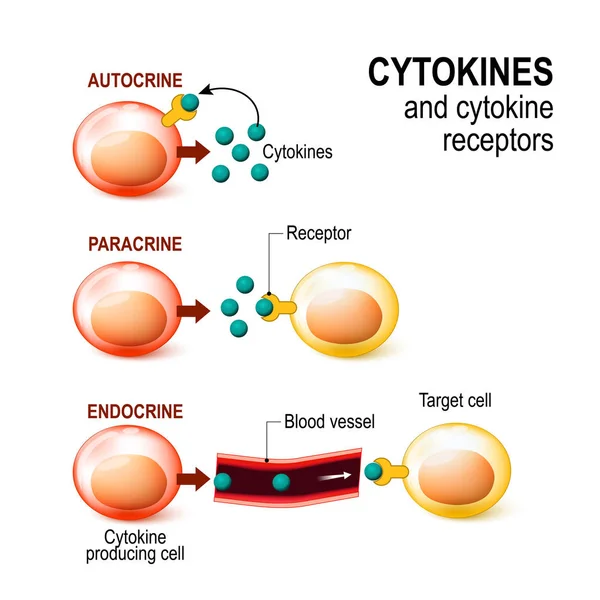
Cytokine receptor. signal transduction between cells.
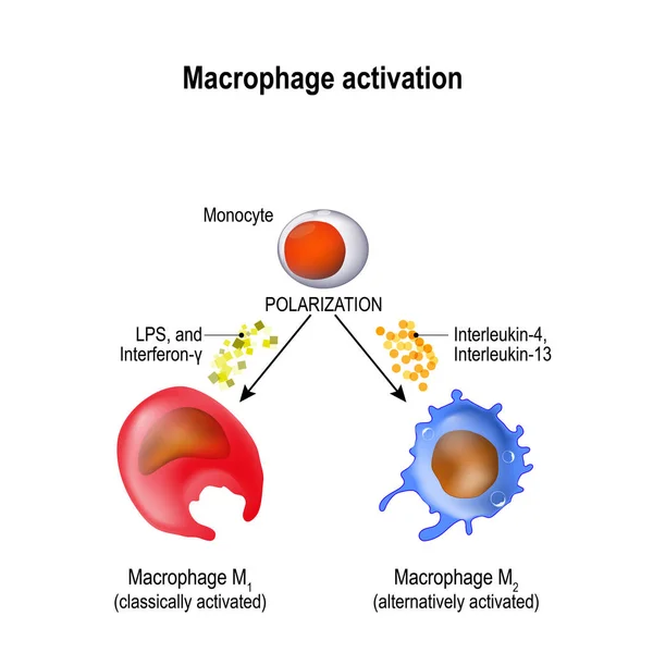
Macrophages are produced by the differentiation of monocytes in tissues. Macrophages M1 encourage inflammation. M2 decrease inflammation. Activation and polarization of the macrophage. Human immune system
Macrophages Produced Differentiation Monocytes Tissues Macrophages Encourage Inflammation Decrease Inflammation Activation Polarization Macrophage Human Immune System
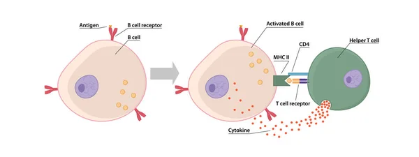
B-cell and T helper cells function.
IL - interleukin acronym, concept
Interleukin Acronym Concept
Epithelium
Epithelium
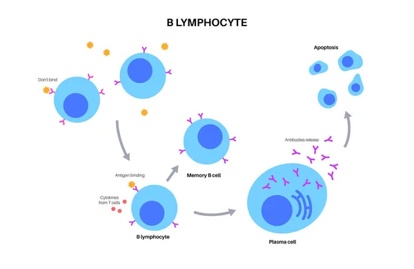
Detailed scheme of B cell activation. Mature B cell encounters antigen that binds to its B cell receptor and it becomes activated
Detailed Scheme Cell Activation Mature Cell Encounters Antigen Binds Its Cell Receptor Becomes Activated
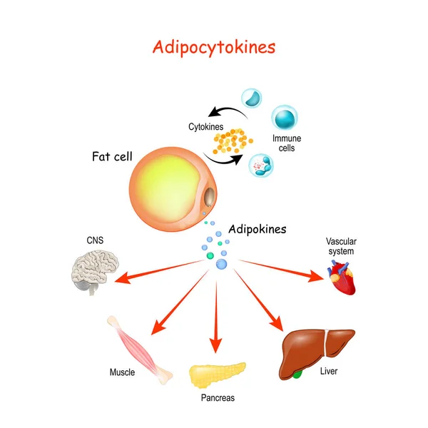
Adipocytokines, immune cells and metabolism. Vector illustration for medical, education and science use.
Adipocytokines Immune Cells Metabolism Vector Illustration Medical Education Science Use
B cell activation
B cell activation
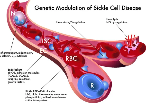
Medical illustration of the genetic modulation of sickle cell disease
Medical Illustration Genetic Modulation Sickle Cell Disease
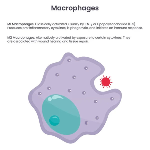
Macrophage monocyte white blood cell vector illustration graphic
Macrophage Monocyte White Blood Cell Vector Illustration Graphic
Activation of B cell leukocytes. transparent realistic cells of Adaptive and Innate immune system. vector poster
Activation Cell Leukocytes Transparent Realistic Cells Adaptive Innate Immune System Vector Poster
B cell activation process. Humoral immunity. T cell-dependent activation. Adaptive immunity. Vector illustration. Medical poster. Schematic diagram
Cell Activation Process Humoral Immunity Cell Dependent Activation Adaptive Immunity Vector Illustration Medical Poster Schematic Diagram
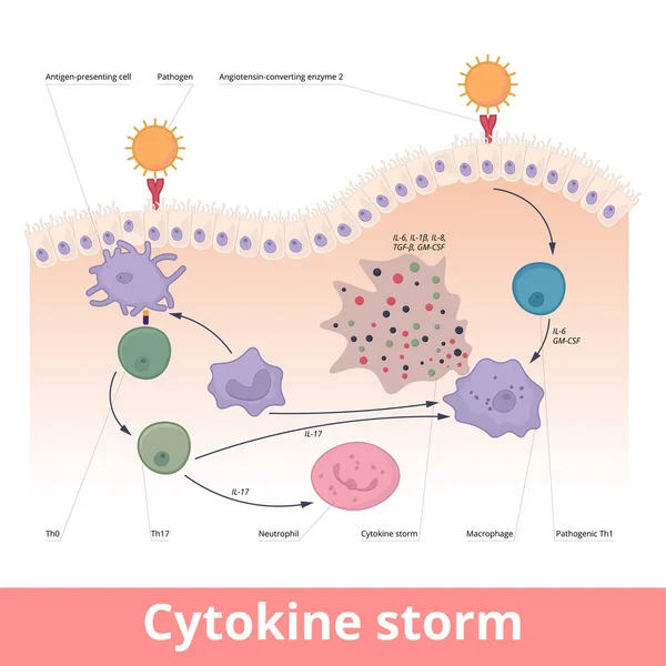
Cytokine storm. Hypercytokinemia during which the immune system causes a release of cytokines with help of macrophages, t helper cells, neutrophils.
Cytokine Storm Hypercytokinemia Which Immune System Causes Release Cytokines Help Macrophages Helper Cells Neutrophils
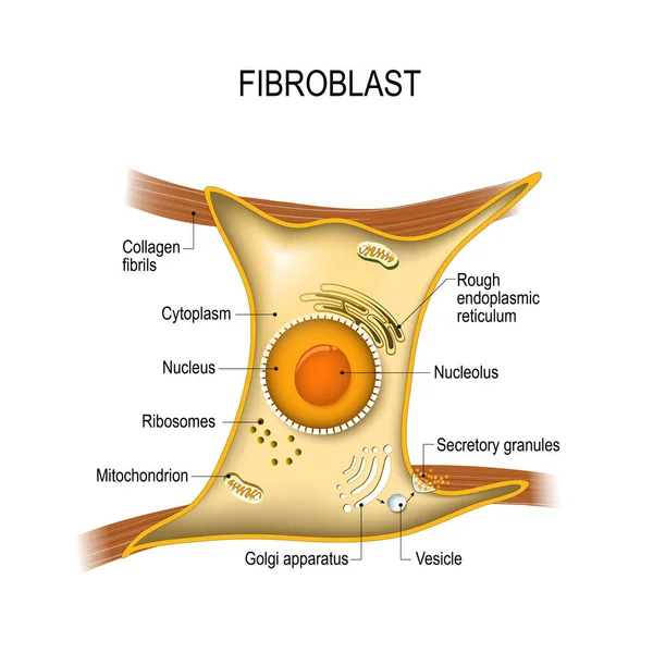
Fibroblast is vital to the skin's strength and elasticity. Struc
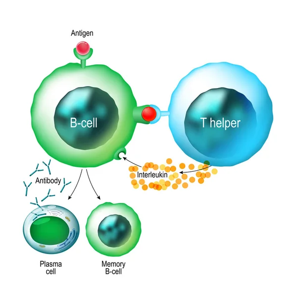
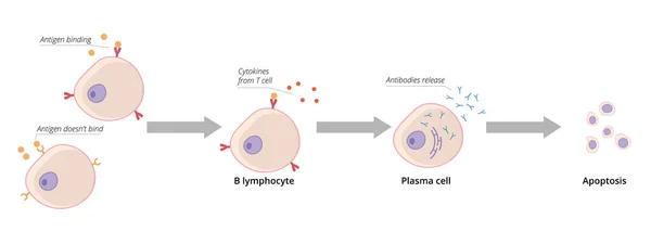
B-cell leukocyte activation by Antigen. From antigen binding to B cell receptor, and Chemical Signal of T-cell helper to Becomes plasma cell and Antibodies Releases. White blood cell. Vector illustration
Cell Leukocyte Activation Antigen Antigen Binding Cell Receptor Chemical Signal Cell Helper Becomes Plasma Cell Antibodies Releases White Blood Cell Vector Illustration
Granulocytes and Agranulocytes. White blood cells. Two types of leukocytes. Close-up of Cells of Immune system Lymphocyte, Monocyte, Neutrophil, Eosinophil, Basophil and Mast cell. Vector illustration
Granulocytes Agranulocytes White Blood Cells Two Types Leukocytes Close Cells Immune System Lymphocyte Monocyte Neutrophil Eosinophil Basophil Mast Cell Vector Illustration
Macrophage polarization process. Functions of M1 and M2 macrophages. immune response. Vector illustration. Medical poster. Schematic diagram
Macrophage Polarization Process Functions Macrophages Immune Response Vector Illustration Medical Poster Schematic Diagram
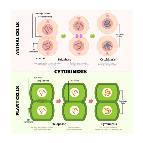
Cytokines in animal cell and plant cell diagram process, produce protein for immune system, labeled telophase, cytokinesis, cleavage furrow, parent and daughter cell division. Vector illustration
Cytokines Animal Cell Plant Cell Diagram Process Produce Protein Immune System Labeled Telophase Cytokinesis Cleavage Furrow Parent Daughter Cell Division Vector Illustration
Lymphocyte B and T-cell. Cells of immune system (immune response). Vector illustration on white background. Didatic illustration.
Lymphocyte Cell Cells Immune System Immune Response Vector Illustration White Background Didatic Illustration
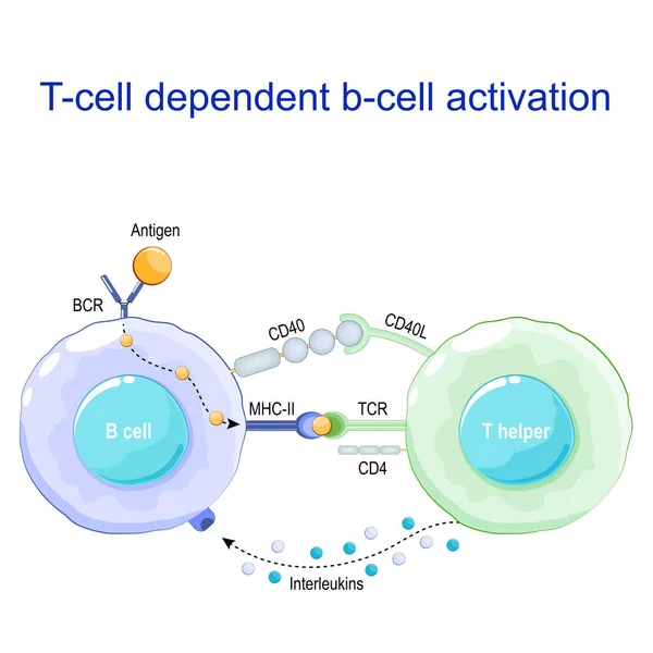
T-cell dependent b-cell activation. B lymphocyte binds an antigen, receive help from a T helper, and differentiate into a plasma cell that secretes of antibodies. Receptors on surface of white blood cells. Human immune system. Vector poster
Cell Dependent Cell Activation Lymphocyte Binds Antigen Receive Help Helper Differentiate Plasma Cell Secretes Antibodies Receptors Surface White Blood Cells Human Immune System Vector Poster
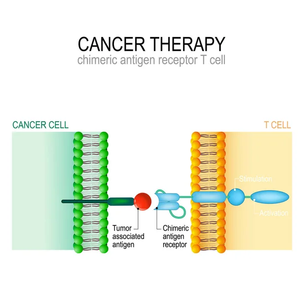
Cancer therapy. CAR T immunotherapy.
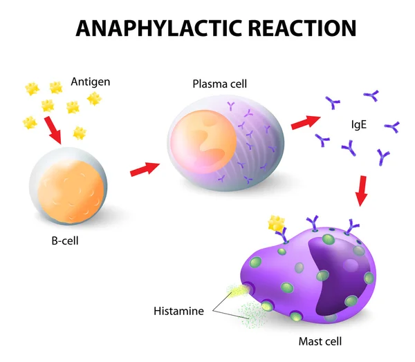
Allergy and anaphylaxis
B cell activation
B cell activation
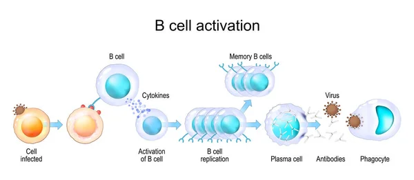
B-cell activation process diagram poster, Activation of B cell leukocytes, Adaptive and Innate immune system, labeled cytokines, memory b cell, antibodies, antigen, phagocyte, virus,b cell replication
Cell Activation Process Diagram Poster Activation Cell Leukocytes Adaptive Innate Immune System Labeled Cytokines Memory Cell Antibodies Antigen Phagocyte Virus Cell Replication
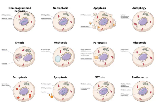
Cell death types: general differences between cell death processes, including common necrosis, autophagy, apoptosis and specific entosis, paraptosis and ferroptosis.
Cell Death Types General Differences Cell Death Processes Including Common Necrosis Autophagy Apoptosis Specific Entosis Paraptosis Ferroptosis
Cellular senescence. From Normal to Senescent cell. Telomere and DNA damaged, mitochondrial dysfunction are primary drivers of damage in aging. Anti-aging therapy. Senescence-associated secretory phenotype SASP. Anti aging medicine. vector poster
Cellular Senescence Normal Senescent Cell Telomere Dna Damaged Mitochondrial Dysfunction Primary Drivers Damage Aging Aging Therapy Senescence Associated Secretory Phenotype Sasp Aging Medicine Vector Poster
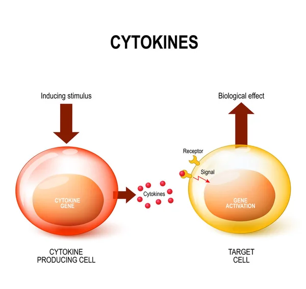
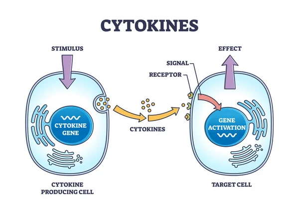
Cytokines process as micro proteins for cell signaling outline diagram. Labeled educational scheme with immune response and producing or target cells vector illustration. Stimulus and antibody effect.
Cytokines Process Micro Proteins Cell Signaling Outline Diagram Labeled Educational Scheme Immune Response Producing Target Cells Vector Illustration Stimulus Antibody Effect
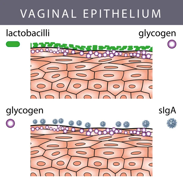
Vaginal Epithelium with Glycogen
Aging process. Frailty Biomarkers. Cellular senescence. Epigenetic clocks. Vector diagram
Aging Process Frailty Biomarkers Cellular Senescence Epigenetic Clocks Vector Diagram
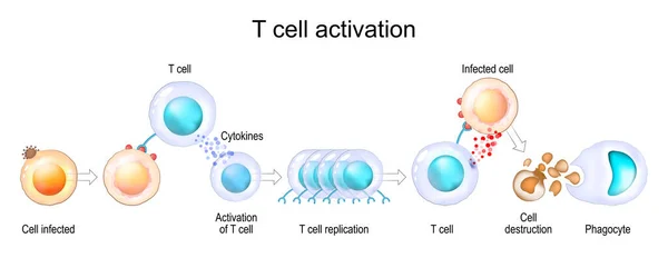
Activation of leukocytes. T-cell encounters its cognate antigen on the surface of an infected cell. T-cells direct and regulate immune responses and attack infected or cancerous cells. Cell-mediated immunity. The Adaptive and Innate immune system. ve
Activation Leukocytes Cell Encounters Its Cognate Antigen Surface Infected Cell Cells Direct Regulate Immune Responses Attack Infected Cancerous Cells Cell Mediated Immunity Adaptive Innate Immune System
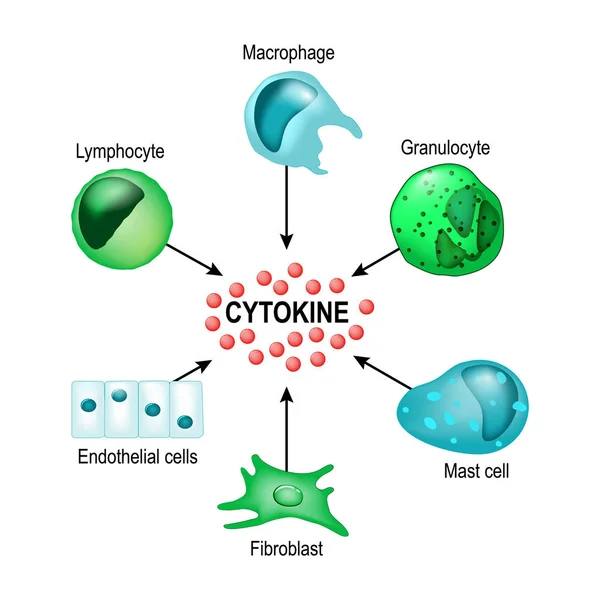
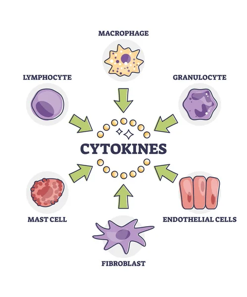
Cytokines releasing cells list for immune system response outline diagram. Labeled educational scheme with macrophage, granulocyte, endothelial, fibroblast, mast and lymphocyte vector illustration.
Cytokines Releasing Cells List Immune System Response Outline Diagram Labeled Educational Scheme Macrophage Granulocyte Endothelial Fibroblast Mast Lymphocyte Vector Illustration
T and B Cell binding to elicit a response to a T cell-dependent antigen, the B and T cells must come close together. B cell must receive two signals from the native antigen and the T cells cytokines
Cell Binding Elicit Response Cell Dependent Antigen Cells Must Come Close Together Cell Must Receive Two Signals Native Antigen Cells Cytokines
Regulatory T cell function. Suppressor T-cells. Prevent autoimmune disease. Immune homeostasis. Adaptive immune response. Vector illustration
Regulatory Cell Function Suppressor Cells Prevent Autoimmune Disease Immune Homeostasis Adaptive Immune Response Vector Illustration
Acute inflammation overview, skin injury with splinter, neutrophils in blood vessels, and mast cell signals explain redness, swelling, pain, loss of function. Outline diagram
Acute Inflammation Overview Skin Injury Splinter Neutrophils Blood Vessels Mast Cell Signals Explain Redness Swelling Pain Loss Function Outline Diagram
Sickle cell disease
Sickle cell disease
Bone remodeling from resorption to new bone formation. Close-up of Trabecular bone matrix, Osteoblasts, Osteoclasts and Osteocytes. Vector illustration
Bone Remodeling Resorption New Bone Formation Close Trabecular Bone Matrix Osteoblasts Osteoclasts Osteocytes Vector Illustration
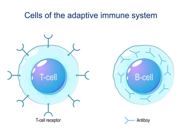
Cells of the adaptive immune system, (immune response). B lymphocyte and T-cell. Labeled T-cell receptor, antigen presenting, secreting cytokines. Infographic Diagram Biology Education Poster Design.
Cells Adaptive Immune System Immune Response Lymphocyte Cell Labeled Cell Receptor Antigen Presenting Secreting Cytokines Infographic Diagram Biology Education Poster Design
Activation of B-cell leukocytes
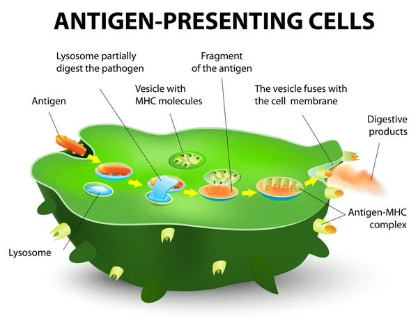
Antigen-presenting cell
Dendritic cells process stage diagram, labeled with immature and mature, pathogen cytokines, antigen-presenting cells. immune system, medical poster, biology education. antigens, immune system cells
Dendritic Cells Process Stage Diagram Labeled Immature Mature Pathogen Cytokines Antigen Presenting Cells Immune System Medical Poster Biology Education Antigens Immune System Cells
Immune response brief flow shows innate to adaptive stages with T cells, B cells, and antibodies defeating pathogens, cells interact over time from exposure to memory. Outline diagram
Immune Response Brief Flow Shows Innate Adaptive Stages Cells Cells Antibodies Defeating Pathogens Cells Interact Time Exposure Memory Outline Diagram
Resistin is a hormone from adipose tissue, regulator of inflammation, autoimmune processes, obesity and insulin resistance. Atherosclerosis and its related complications. Target cells for resistin: macrophage, PBMC, osteoclast, osteoblast, plasma and
Resistin Hormone Adipose Tissue Regulator Inflammation Autoimmune Processes Obesity Insulin Resistance Atherosclerosis Its Related Complications Target Cells Resistin Macrophage Pbmc Osteoclast Osteoblast Plasma
Hormones produced by adipocytes. Brown adipose tissue (BAT) and White adipose tissue (WAT). Cytokines, Resistin, Leptin, and Estrogen. Human endocrine system. Vector illustration for medical, education and science use.
Hormones Produced Adipocytes Brown Adipose Tissue Bat White Adipose Tissue Wat Cytokines Resistin Leptin Estrogen Human Endocrine System Vector Illustration Medical Education Science Use
Physiological Aging. Cellular senescence and Adipocyte changes with age. Aging process, Insulin resistance and diabetes. Vector illustration
Physiological Aging Cellular Senescence Adipocyte Changes Age Aging Process Insulin Resistance Diabetes Vector Illustration
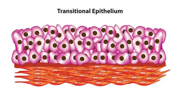
Cell arrangement of transitional epithelium
Cell Arrangement Transitional Epithelium
Adaptive immune system. Structure and Anatomy of T-cell and B-cell. Immunological memory. Lymphocytes of Cell-mediated immunity. Vector illustration. Medical poster. Schematic diagram
Adaptive Immune System Structure Anatomy Cell Cell Immunological Memory Lymphocytes Cell Mediated Immunity Vector Illustration Medical Poster Schematic Diagram
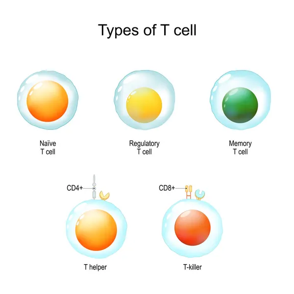
Types of T cell. Helper and Regulatory white blood cell, Natural killer, Memory T-cell, Naive Lymphocyte. Vector illustration. Medical poster. Schematic diagram
Types Cell Helper Regulatory White Blood Cell Natural Killer Memory Cell Naive Lymphocyte Vector Illustration Medical Poster Schematic Diagram
Cell mediated immunity diagram process, Immune response. activation of phagocytes, labeled macrophage, pathogen, t-lymphocyte, release cytokines in response to antigen, cytotoxic. Biology education,
Cell Mediated Immunity Diagram Process Immune Response Activation Phagocytes Labeled Macrophage Pathogen Lymphocyte Release Cytokines Response Antigen Cytotoxic Biology Education
Types of Lymphocytes. T and B cell. Human adaptive immune system. immune response and component of humoral immunity. Antibody, Plasma cell, T helper, and T-killer. Vector illustration
Types Lymphocytes Cell Human Adaptive Immune System Immune Response Component Humoral Immunity Antibody Plasma Cell Helper Killer Vector Illustration
Different types of T Cells types. Labeled Naive, Regulatory, Memory, T Helper, T Killer. Immunology infographic diagram poster. Healthcare poster biology education diagram.
Different Types Cells Types Labeled Naive Regulatory Memory Helper Killer Immunology Infographic Diagram Poster Healthcare Poster Biology Education Diagram
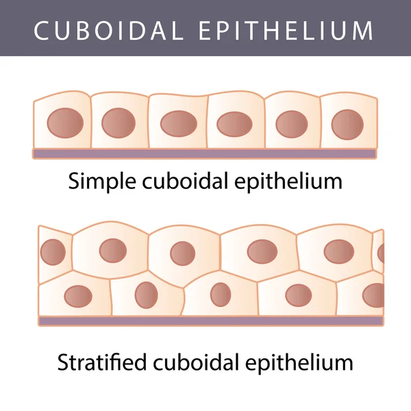
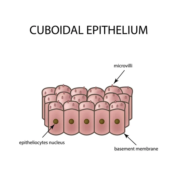
The structure of cubic epithelium. Infographics. Vector illustration on isolated background
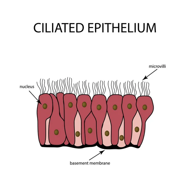
The structure of the ciliated epithelium. Infographics. Vector illustration on isolated background
Neuroinflammation. Development of Neurodegenerative Diseases. From injury of brain, damage, infection or stress to Microglia activation, release of cytokines, chronic inflammation and Neuronal damage
Neuroinflammation Development Neurodegenerative Diseases Injury Brain Damage Infection Stress Microglia Activation Release Cytokines Chronic Inflammation Neuronal Damage
Mechanism of allergy. Mast cells and allergic reaction. B-cell is exposed to allergen, plasma cells will initiate an overproduction of IgE antibodies. The IgE molecules attach themselves to mast cells. When allergen enters the body for the second tim
Mechanism Allergy Mast Cells Allergic Reaction Cell Exposed Allergen Plasma Cells Initiate Overproduction Ige Antibodies Ige Molecules Attach Themselves Mast Cells Allergen Enters Body Second Tim
Types of T cell. From Naive and memory cells to T helper and T-killer. Immunology infographic. vector Illustration
Types Cell Naive Memory Cells Helper Killer Immunology Infographic Vector Illustration
T-cell and B-cell. Cells of Adaptive immune system. immune response and lymphocytes. Vector illustration on white background.
Cell Cell Cells Adaptive Immune System Immune Response Lymphocytes Vector Illustration White Background
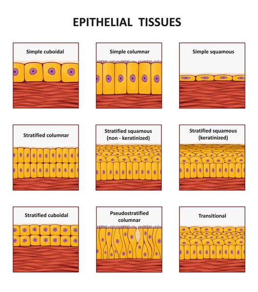
Different types of epithelial tissue collection.
The gut-brain axis. Connection between the brain and intestines via Immune signals, Gut Microbiota, Hormones, and Vagus nerve. Mental health. Schematic illustration. medical diagram. Detailed Vector poster
Gut Brain Axis Connection Brain Intestines Immune Signals Gut Microbiota Hormones Vagus Nerve Mental Health Schematic Illustration Medical Diagram Detailed Vector Poster
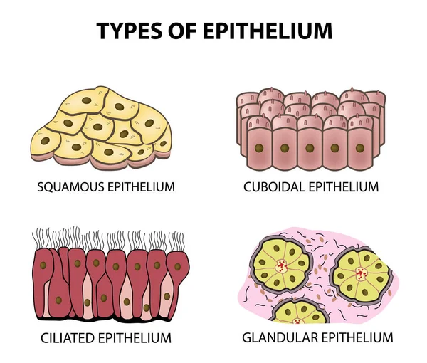
Types of epithelium. squamous, cubic, ciliated, glandular. Set. Infographics. Vector illustration on isolated background
Epithelium
Epithelium
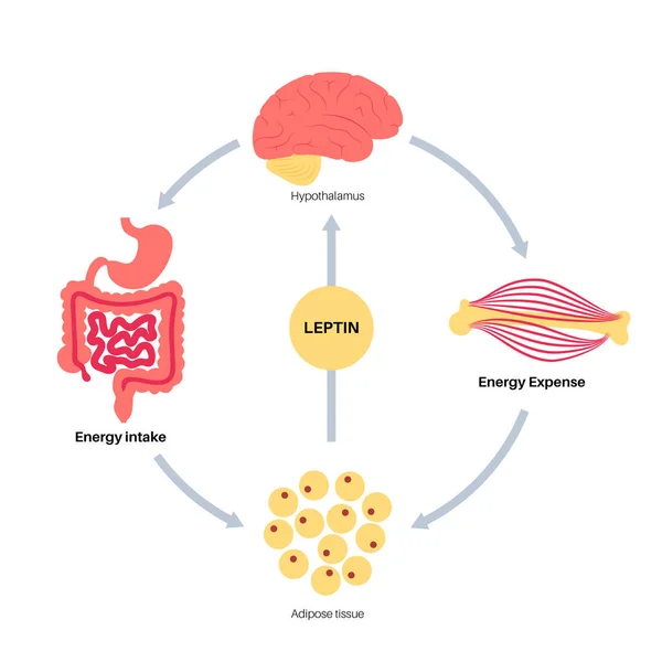
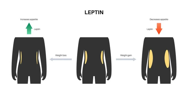
Hunger appetite hormones
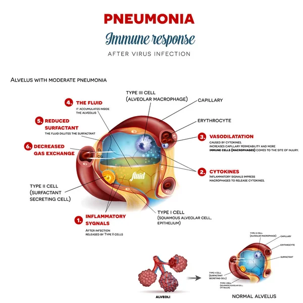
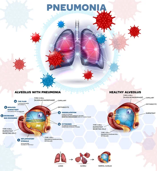
Pneumonia immune response step by step after virus infection, detailed alveolus anatomy illustration
Pneumonia Immune Response Step Step Virus Infection Detailed Alveolus Anatomy Illustration
Epithelium
Epithelium
Pneumonia explained info poster, body immune response, human lungs and detailed alveolus anatomy illustration
Pneumonia Explained Info Poster Body Immune Response Human Lungs Detailed Alveolus Anatomy Illustration
Hunger appetite hormones
Visceral fat and Chronic inflammation. Cross section of female abdomen with Abdominal muscle, Subcutaneous and Visceral fat. Close-up of Adipose tissue with T cells, macrophages, and Enlarged fat cells. Vector illustration. Metabolic syndrome
Visceral Fat Chronic Inflammation Cross Section Female Abdomen Abdominal Muscle Subcutaneous Visceral Fat Close Adipose Tissue Cells Macrophages Enlarged Fat Cells Vector Illustration Metabolic Syndrome
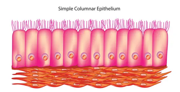
Simple columnar epithelium tissue
Simple Columnar Epithelium Tissue
Pneumonia info poster beautiful colorful design, body immune response, human lungs abstract protection lines around and detailed alveolus anatomy illustration.
Pneumonia Info Poster Beautiful Colorful Design Body Immune Response Human Lungs Abstract Protection Lines Detailed Alveolus Anatomy Illustration
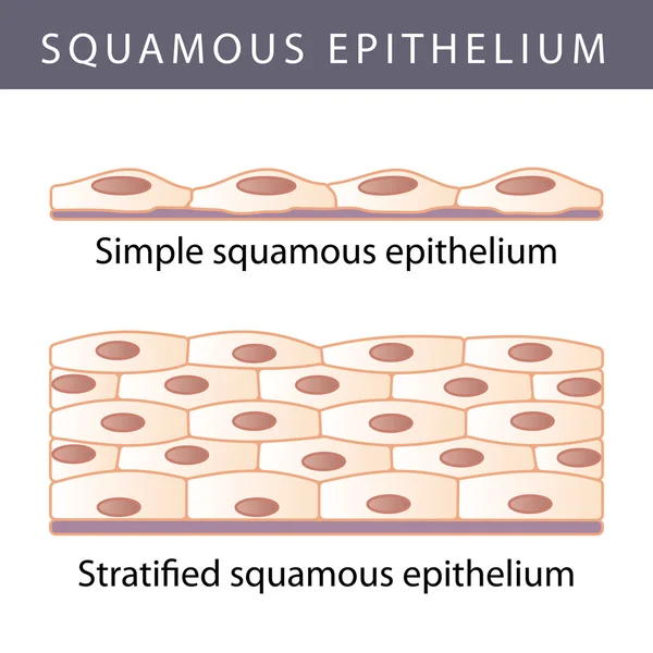
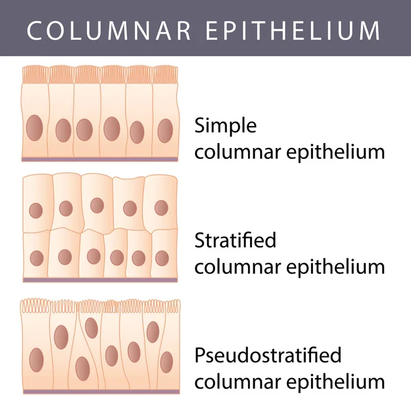
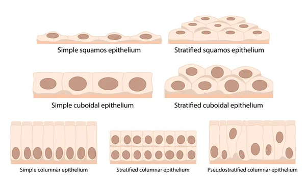
Cells of epithelial tissue: squamous (flattened and thin), cuboidal (boxy, as wide as it is tall), columnar (rectangular, taller than it is wide), pseudostratified.
Cells Epithelial Tissue Squamous Flattened Thin Cuboidal Boxy Wide Tall Columnar Rectangular Taller Wide Pseudostratified
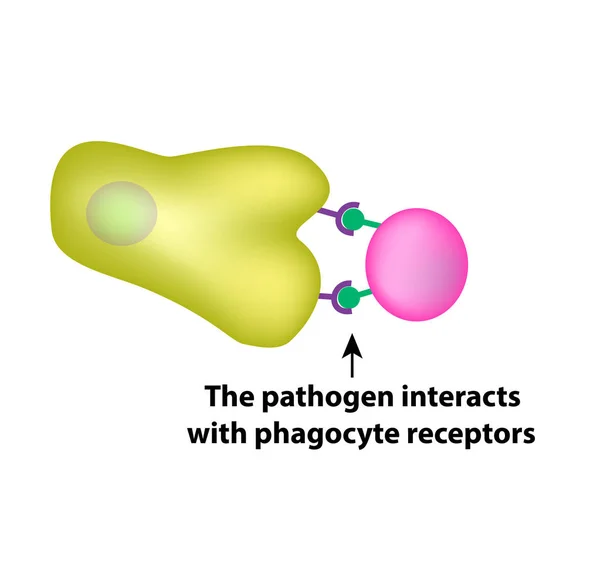
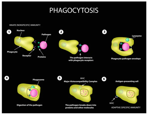
Innate immunity. Adaptive specific . Phagocytosis. Infographics. vector illustration
Innate immunity. Adaptive specific . Phagocytosis. Infographics. vector
Lymphocyte cell Cartoon character style with a crazy face
B cell activation
B cell activation
B cell activation
B cell activation
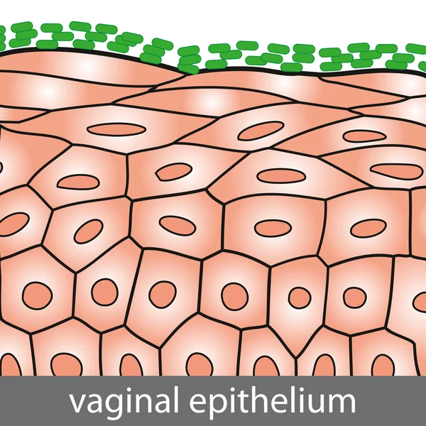
Vaginal Epithelium
Vaginal Epithelium
Tumor microenvironment TME. Cellular diagram cancer cells, CAFs, TAMs, angiogenesis, and immunosuppressive stroma. Immune and vascular components. medical illustration. science explainer
Tumor Microenvironment Tme Cellular Diagram Cancer Cells Cafs Tams Angiogenesis Immunosuppressive Stroma Immune Vascular Components Medical Illustration Science Explainer
Innate immunity. Adaptive specific . Phagocytosis. Infographics. vector illustration
B-cell activation diagram shows naive B cell, helper T cell, and antibodies progressing from recognition to clonal expansion and response for humoral immunity. Outline diagram
Cell Activation Diagram Shows Naive Cell Helper Cell Antibodies Progressing Recognition Clonal Expansion Response Humoral Immunity Outline Diagram
One Finger lymphocyte cell in mascot cartoon character style
The structure of the glandular epithelium. Infographics. Vector illustration on isolated background
Epithelium. squamous, cubic, ciliated, glandular. Set. Infographics. Vector illustration.
T-cell dependent b-cell activation. B lymphocyte cell and T-helper. Close-up of White blood cells, leucocytes. Immune response. Adaptive immunity. Humoral immunity. vector illustration
Cell Dependent Cell Activation Lymphocyte Cell Helper Close White Blood Cells Leucocytes Immune Response Adaptive Immunity Humoral Immunity Vector Illustration
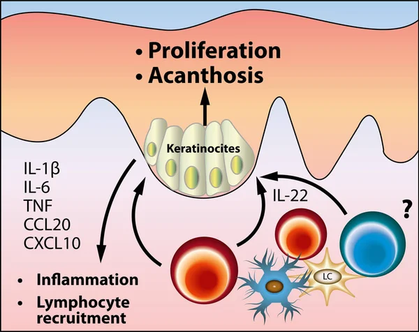
The pathogenesis of acanthosis nigricans scheme, vector medical illustration
Pathogenesis Acanthosis Nigricans Scheme Vector Medical Illustration
A funny cartoon character of lymphocyte cell with a menu