Dendritico Stock Vector Images

90 Dendritico vector art & graphics are available under a royalty-free license
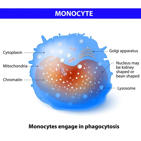
Monocyte - type of White Blood Cell
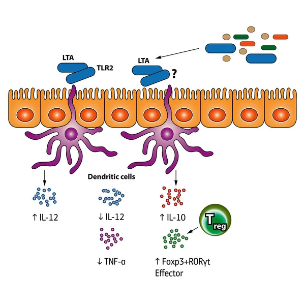
Stimulation of the Regulatory T cells vector medical illustration
Stimulation Regulatory Cells Vector Medical Illustration
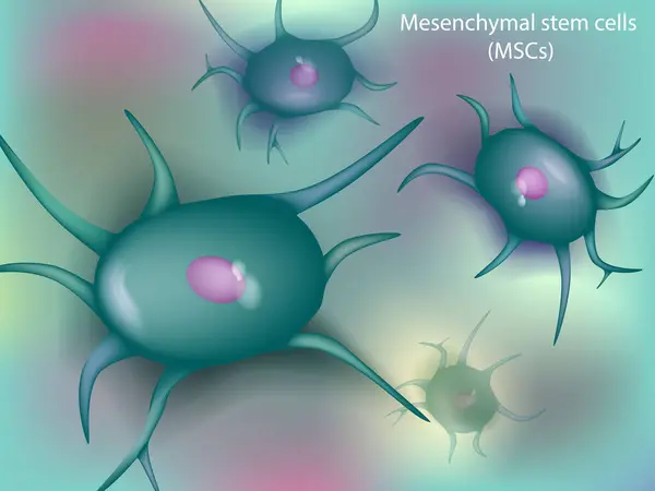
Mesenchymal stem cells or MSCs are stromal cells. Bone Marrow
Mesenchymal Stem Cells Mscs Stromal Cells Bone Marrow
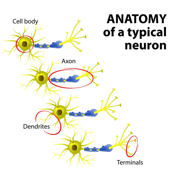
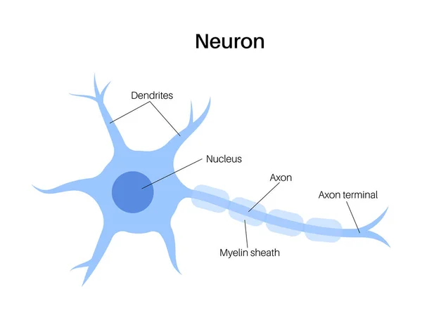
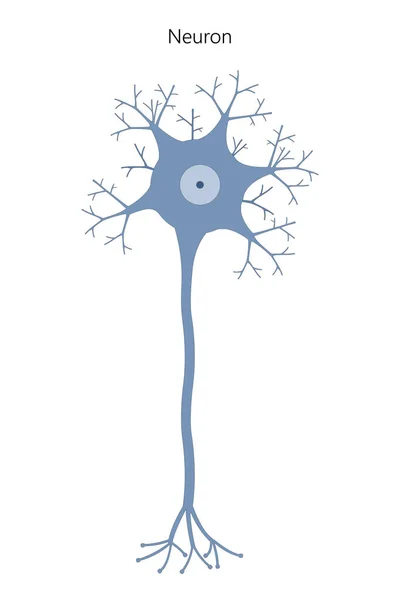
Anatomy of a typical neuron
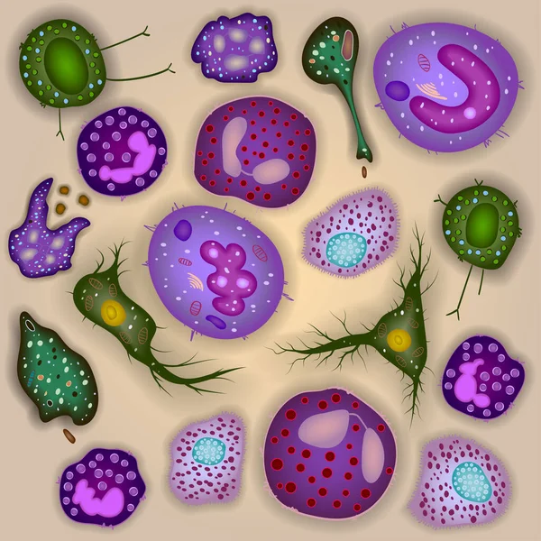
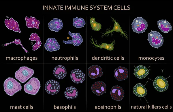
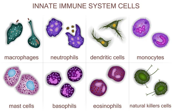
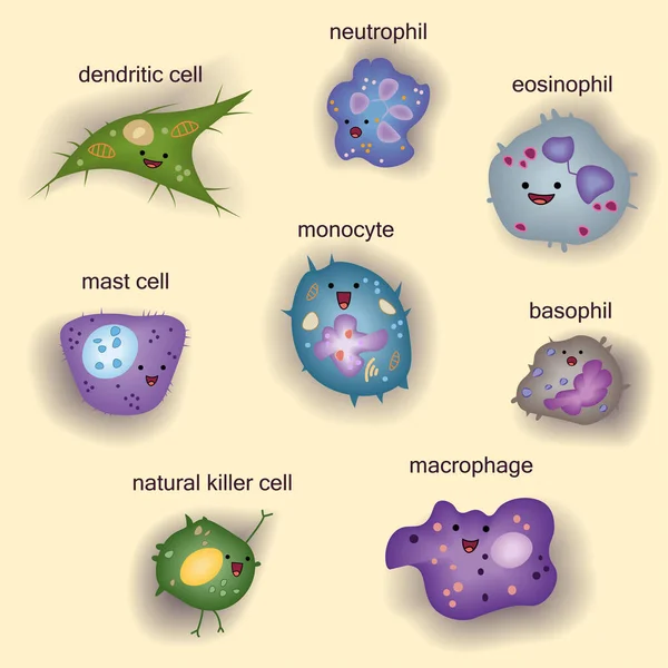
Set of innate immune system cells, vector illustration
Set Innate Immune System Cells Vector Illustration
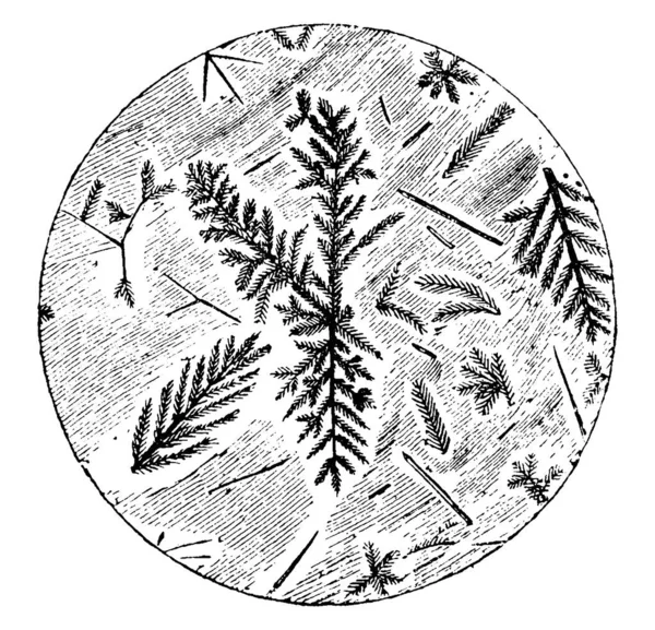
Microscopic section of Pitchstone, showing dendritic crystals, a crystal that develops with a typical multi-branching tree-like form, vintage line drawing or engraving illustration.
Microscopic Section Pitchstone Showing Dendritic Crystals Crystal Develops Typical Multi Branching Tree Form Vintage Line Drawing Engraving Illustration
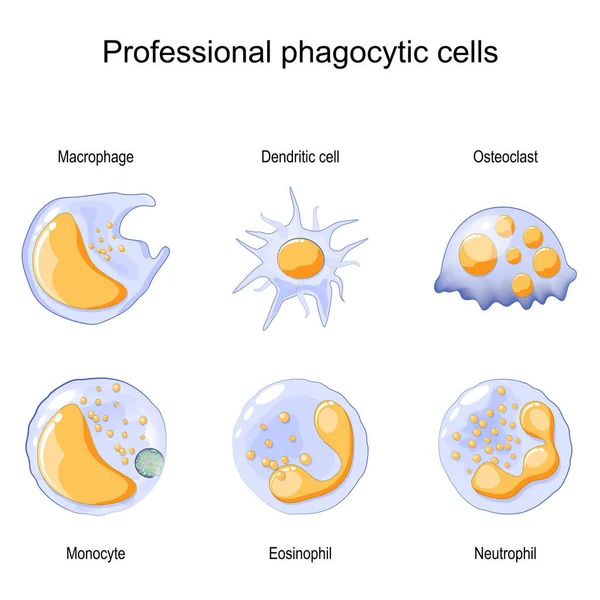
Phagocytosis. Professional phagocytic cells. Neutrophils, macrophages, monocytes, dendritic cells, osteoclasts and eosinophils are immune response to most infections. Vector illustration. Medical poster.
Phagocytosis Professional Phagocytic Cells Neutrophils Macrophages Monocytes Dendritic Cells Osteoclasts Eosinophils Immune Response Most Infections Vector Illustration Medical Poster
Herpes simplex keratitis HSV1 infection detailed infographic poster showing human eyeball cross section anatomy with highlighted focus zone over corneal lesion vector illustration.
Herpes Simplex Keratitis Hsv1 Infection Detailed Infographic Poster Showing Human Eyeball Cross Section Anatomy Highlighted Focus Zone Corneal Lesion Vector Illustration
Set of innate immune system cells, vector illustration
Set Innate Immune System Cells Vector Illustration
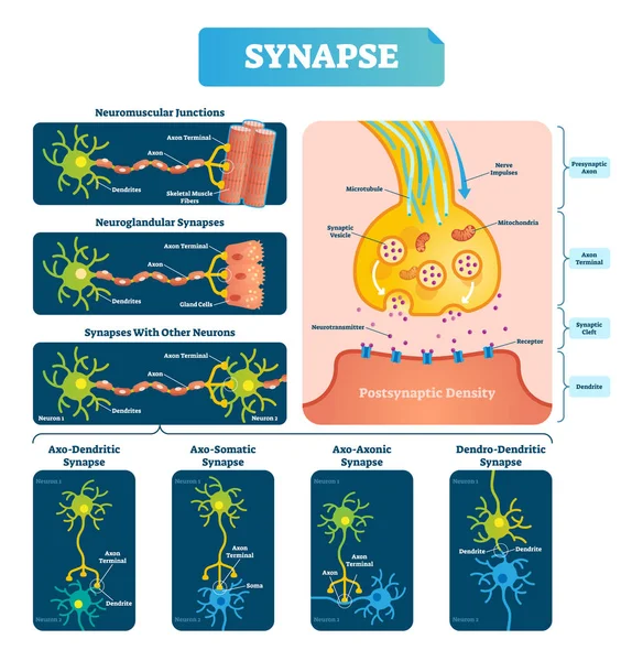
Synapse vector illustration. Labeled diagram with neuromuscular example.
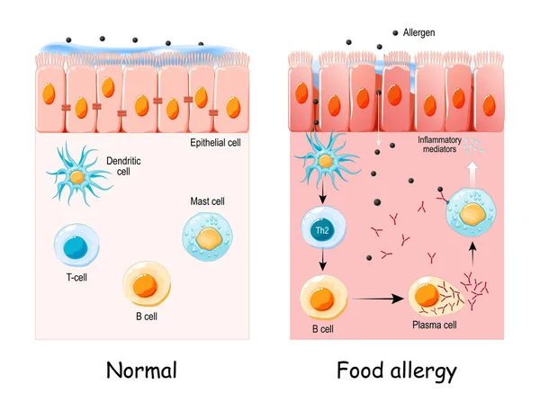
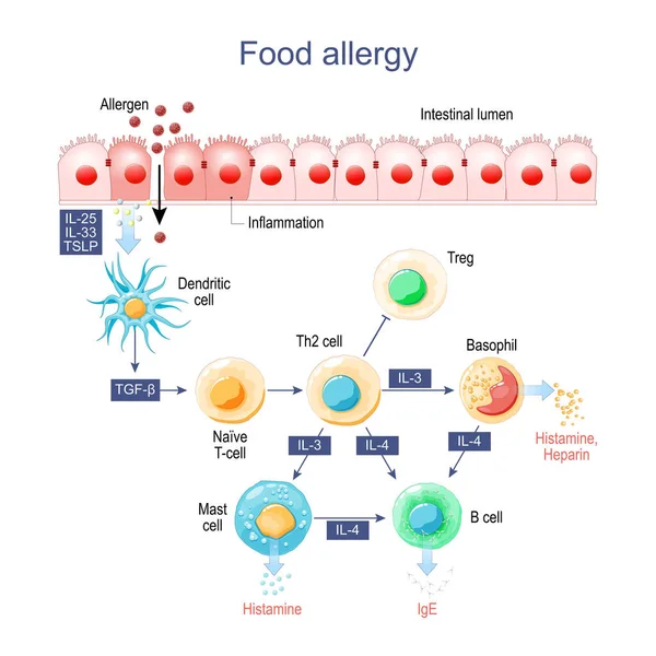
Food allergy is an abnormal immune response to food. Cell of normal Epithelium and Food allergy. development of an allergic reaction from allergens penetration to Inflammatory mediators synthesis. vector
Food Allergy Abnormal Immune Response Food Cell Normal Epithelium Food Allergy Development Allergic Reaction Allergens Penetration Inflammatory Mediators Synthesis Vector
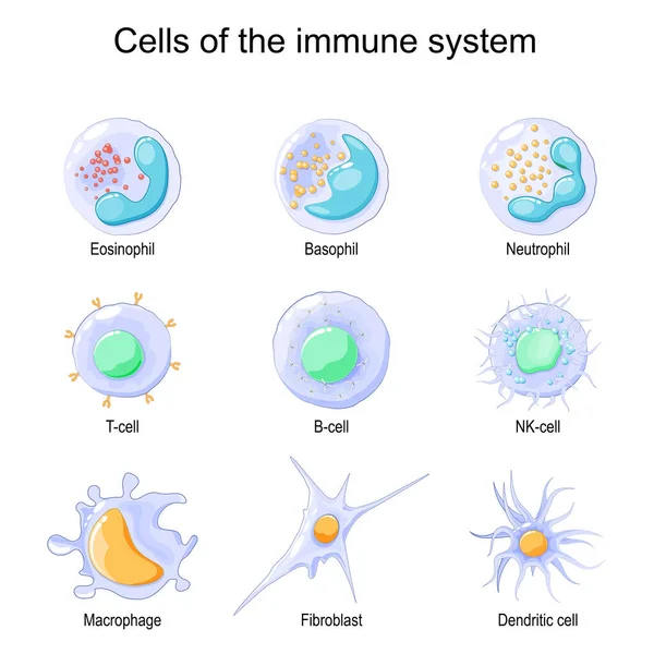
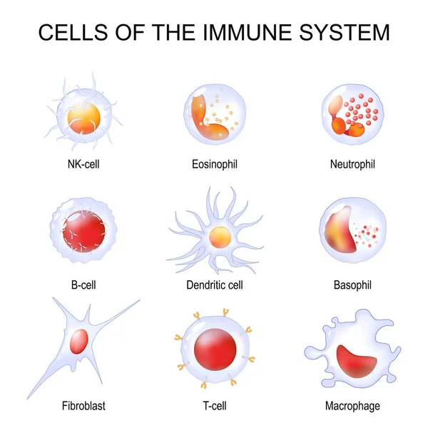
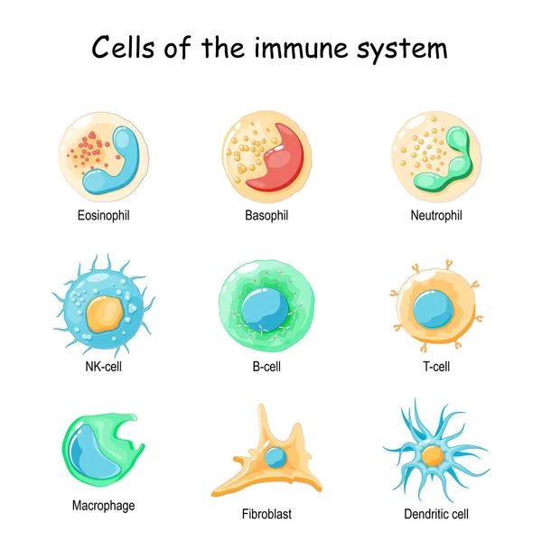
Cells of the immune system. White blood cells or leukocytes Eosinophil, Neutrophil, Basophil, Macrophage, Fibroblast, and Dendritic cell. Vector illustration
Cells Immune System White Blood Cells Leukocytes Eosinophil Neutrophil Basophil Macrophage Fibroblast Dendritic Cell Vector Illustration
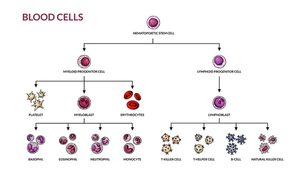
Human hematopoiesis blood cell formation from bone marrow, hematopoietic stem cells differentiation structure diagram hand drawn schematic vector illustration. Medical science educational illustration
Human Hematopoiesis Blood Cell Formation Bone Marrow Hematopoietic Stem Cells Differentiation Structure Diagram Hand Drawn Schematic Vector Illustration Medical Science Educational Illustration
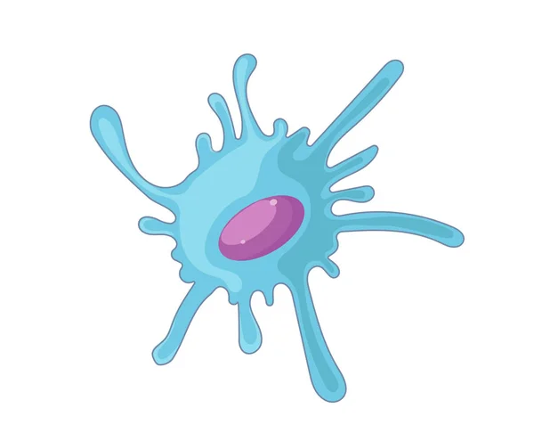
Dendritic cell vector illustration
Immune system cells diagram showing innate and adaptive branches, highlighting hematopoietic stem cell, T lymphocyte, and B lymphocyte to explain defense pathways. Outline diagram
Immune System Cells Diagram Showing Innate Adaptive Branches Highlighting Hematopoietic Stem Cell Lymphocyte Lymphocyte Explain Defense Pathways Outline Diagram
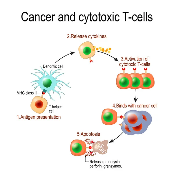
Cancer and cytotoxic T-cells. T lymphocyte kills cancer cells. T-cell (immune responses), release the perforin and granzymes, and attack cancerous cells. Through the action of perforin, granzymes enter the cytoplasm of the target cell, and lead to ap
Cancer Cytotoxic Cells Lymphocyte Kills Cancer Cells Cell Immune Responses Release Perforin Granzymes Attack Cancerous Cells Action Perforin Granzymes Enter Cytoplasm Target Cell Lead
Cells of the immune system. White blood cells or leukocytes Plasma cell, Eosinophil, Neutrophil, Basophil, Macrophage, T-cell, NK-cell, B-cell. Isometric Flat vector illustration
Cells Immune System White Blood Cells Leukocytes Plasma Cell Eosinophil Neutrophil Basophil Macrophage Cell Cell Cell Isometric Flat Vector Illustration
Herpes simplex keratitis HSV1 corneal infection infographic showing human eyeball front view anatomy with green dendritic lesion on blue iris over black background vector illustration.
Herpes Simplex Keratitis Hsv1 Corneal Infection Infographic Showing Human Eyeball Front View Anatomy Green Dendritic Lesion Blue Iris Black Background Vector Illustration
Minimalist Black Glyph Style Neuron Cell Structure Science Biology Icon
Moose River Flat Mountains Sunburst Circle Mono Line
Gray-orange Immunoglobulin Round Icon
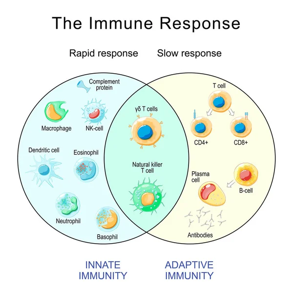
Adaptive and Innate immunity. Cells of The Immune System. Immune response. Immunology infographic. Rapid and slow response. Isometric flat vector Illustration
Adaptive Innate Immunity Cells Immune System Immune Response Immunology Infographic Rapid Slow Response Isometric Flat Vector Illustration
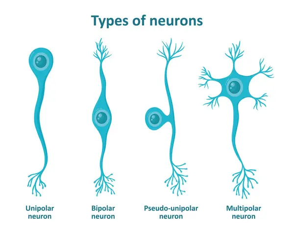
Different types of neurons, nerve cells. Transmission of information in the human body. Poster with educational information.
Different Types Neurons Nerve Cells Transmission Information Human Body Poster Educational Information
Set of innate immune system cells, vector illustration
Set Innate Immune System Cells Vector Illustration
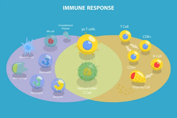
3D Isometric Flat Vector Conceptual Illustration of Immune Response, Cells of The Immune System
Isometric Flat Vector Conceptual Illustration Immune Response Cells Immune System
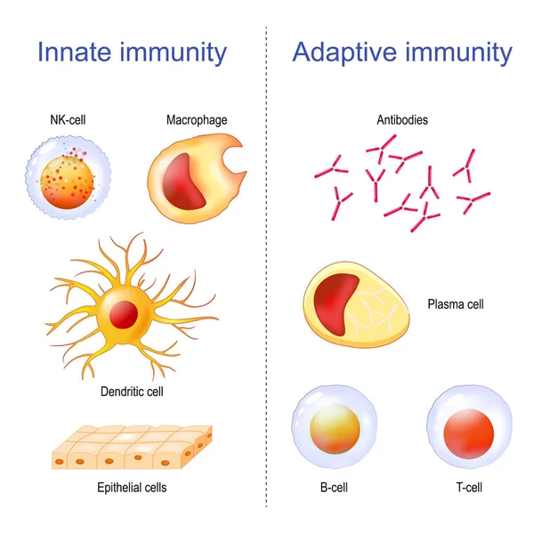
Adaptive immunity: T-cell, Antibodies, Plasma cell and B-cell. Innate immunity: Macrophage, Dendritic, Epithelial, and NK cells. Immunology infographic. vector Illustration
Adaptive Immunity Cell Antibodies Plasma Cell Cell Innate Immunity Macrophage Dendritic Epithelial Cells Immunology Infographic Vector Illustration
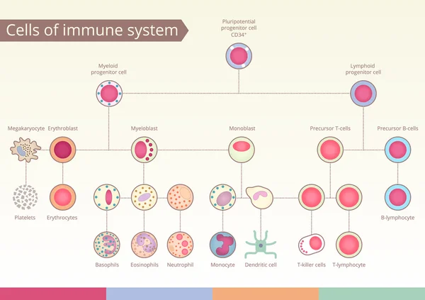
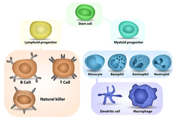
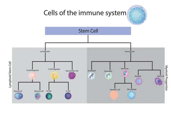
Origin of Cells of immune system.
Minimalist Linear Neuron Nerve Cell Anatomy Biology Science Symbol Icon
Herpes simplex keratitis HSV1 infection pathway infographic showing human head profile silhouette with trigeminal nerve branches and green dendritic lesion diagram vector illustration.
Herpes Simplex Keratitis Hsv1 Infection Pathway Infographic Showing Human Head Profile Silhouette Trigeminal Nerve Branches Green Dendritic Lesion Diagram Vector Illustration
Herpes simplex keratitis HSV1 infection pathway infographic showing human head profile silhouette with trigeminal nerve pathway and macro eye layout with corneal lesion vector illustration.
Herpes Simplex Keratitis Hsv1 Infection Pathway Infographic Showing Human Head Profile Silhouette Trigeminal Nerve Pathway Macro Eye Layout Corneal Lesion Vector Illustration
Herpes simplex keratitis HSV1 infection infographic showing human eye macro layout and detailed cornea cross section anatomy with dendritic lesion on corneal surface vector illustration.
Herpes Simplex Keratitis Hsv1 Infection Infographic Showing Human Eye Macro Layout Detailed Cornea Cross Section Anatomy Dendritic Lesion Corneal Surface Vector Illustration
Tumor microenvironment TME. Cellular diagram cancer cells, CAFs, TAMs, angiogenesis, and immunosuppressive stroma. Immune and vascular components. medical illustration. science explainer
Tumor Microenvironment Tme Cellular Diagram Cancer Cells Cafs Tams Angiogenesis Immunosuppressive Stroma Immune Vascular Components Medical Illustration Science Explainer
Neuron. Neuronal structure. Vector illustration
Neuron Neuronal Structure Vector Illustration
Neuron anatomy poster
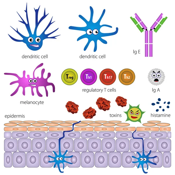
The cells of the protective layer of the skin. Protection against inflammation
Immune cell lineage brief diagram maps stem-to-progenitor branching, showing hematopoietic stem cell splitting into myeloid and lymphoid paths, key T cell, B cell, macrophage. Outline diagram
Immune Cell Lineage Brief Diagram Maps Stem Progenitor Branching Showing Hematopoietic Stem Cell Splitting Myeloid Lymphoid Paths Key Cell Cell Macrophage Outline Diagram
IgA immunity. Peyers patch. Lymphoid follicles of the small intestine generate IgA immune response with B cells, T cells, dendritic cells, and plasma cells secreting IgA.
Iga Immunity Peyers Patch Lymphoid Follicles Small Intestine Generate Iga Immune Response Cells Cells Dendritic Cells Plasma Cells Secreting Iga
Herpes simplex keratitis HSV1 infection detailed infographic showing human eyeball cross section anatomy with lines pointing to internal structures and focus zone vector illustration.
Herpes Simplex Keratitis Hsv1 Infection Detailed Infographic Showing Human Eyeball Cross Section Anatomy Lines Pointing Internal Structures Focus Zone Vector Illustration
Herpes zoster ophthalmicus corneal infection minimal black outline icon showing human eye cross section anatomy with red highlighted target focus zone over lesion vector illustration.
Herpes Zoster Ophthalmicus Corneal Infection Minimal Black Outline Icon Showing Human Eye Cross Section Anatomy Red Highlighted Target Focus Zone Lesion Vector Illustration
Monocyte to Macrophage. Circulating Monocyte Cell. In Vitro Differentiation of Macrophages and Dendritic Cells
Monocyte Macrophage Circulating Monocyte Cell Vitro Differentiation Macrophages Dendritic Cells
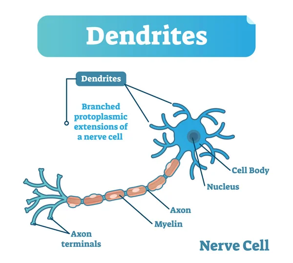
Dendrite biological anatomy vector illustration diagram with nerve cell structure.
Immune Response. Rapid and slow response of Adaptive and Innate Immunity and antibody activation. Cells of The Immune System. Immunology infographic. vector illustration
Immune Response Rapid Slow Response Adaptive Innate Immunity Antibody Activation Cells Immune System Immunology Infographic Vector Illustration
Herpes simplex keratitis HSV1 corneal infection infographic showing human eyeball side view anatomy with green dendritic lesion over blue iris on black background vector illustration.
Herpes Simplex Keratitis Hsv1 Corneal Infection Infographic Showing Human Eyeball Side View Anatomy Green Dendritic Lesion Blue Iris Black Background Vector Illustration
Seamless multicolored texture with stripes. Polygonal background. Colored mosaic pattern. Abstract geometric wallpaper of the surface. Stripe tiled backdrop
Seamless Multicolored Texture Stripes Polygonal Background Colored Mosaic Pattern Abstract Geometric Wallpaper Surface Stripe Tiled Backdrop
Antitumor Immunity. Immune Response. Immunotherapy. Rapid and slow response of Adaptive and Innate Immunity and antibody activation. Cells of The Immune System. Immunology infographic. vector illustration
Antitumor Immunity Immune Response Immunotherapy Rapid Slow Response Adaptive Innate Immunity Antibody Activation Cells Immune System Immunology Infographic Vector Illustration
Cell-Mediated Immunity from antigen presentation to cytotoxic response and Memory T Cell formation. Adaptive immunity. Vector illustration. Medical poster. Schematic diagram
Cell Mediated Immunity Antigen Presentation Cytotoxic Response Memory Cell Formation Adaptive Immunity Vector Illustration Medical Poster Schematic Diagram
Mesenchymal stem cells or mesenchymal stromal cells or Medicinal signaling cells are stromal cells. Cells bone Marrow
Mesenchymal Stem Cells Mesenchymal Stromal Cells Medicinal Signaling Cells Stromal Cells Cells Bone Marrow
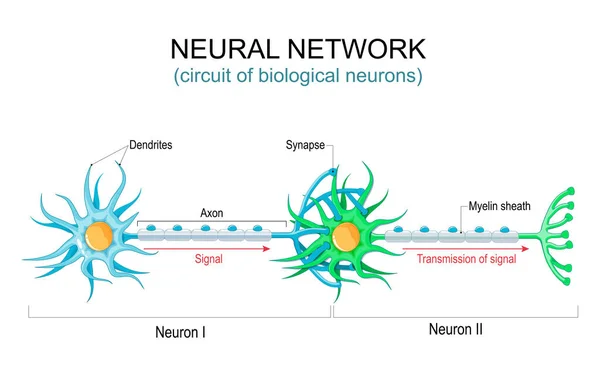
Neural network. circuit or connections of the biological neurons. nervous system. Communication structure of neurons. Transmission of signal in the human brain. Vector illustration
Neural Network Circuit Connections Biological Neurons Nervous System Communication Structure Neurons Transmission Signal Human Brain Vector Illustration
Food allergy. Inflammation of Intestine. Allergens, Histamine and immune cells, that release allergic reaction.
Food Allergy Inflammation Intestine Allergens Histamine Immune Cells Release Allergic Reaction
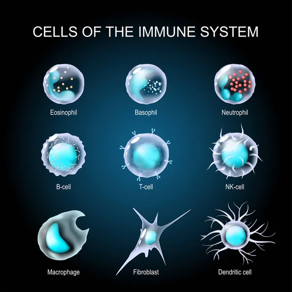
Cells of the immune system. White blood cells or leukocytes Eosinophil, Neutrophil, Basophil, Macrophage, Fibroblast, and Dendritic cell. Set of transparent realistic cells on a dark background. Vector illustration
Cells Immune System White Blood Cells Leukocytes Eosinophil Neutrophil Basophil Macrophage Fibroblast Dendritic Cell Set Transparent Realistic Cells Dark Background Vector Illustration
Herpes simplex keratitis HSV1 corneal infection detailed diagram showing eye cross section anatomy with highlighted red target focus over dendritic lesion vector illustration.
Herpes Simplex Keratitis Hsv1 Corneal Infection Detailed Diagram Showing Eye Cross Section Anatomy Highlighted Red Target Focus Dendritic Lesion Vector Illustration
Herpes zoster ophthalmicus corneal infection development scheme showing eyeball cross section anatomy and transition to magnified detailed dendritic lesion focus zone vector illustration.
Herpes Zoster Ophthalmicus Corneal Infection Development Scheme Showing Eyeball Cross Section Anatomy Transition Magnified Detailed Dendritic Lesion Focus Zone Vector Illustration
Herpes simplex keratitis HSV1 infection pathway infographic showing human head profile silhouette with trigeminal nerve diagram and eyeball cross section with lesion vector illustration.
Herpes Simplex Keratitis Hsv1 Infection Pathway Infographic Showing Human Head Profile Silhouette Trigeminal Nerve Diagram Eyeball Cross Section Lesion Vector Illustration
Huntington's Disease HD. Huntingtons chorea from genetic mutation Chromosome and DNA location, mutant huntingtin protein to neuronal degeneration. Fatal neurodegenerative inherited disease. Neurology. Medical illustration of pathogenesis. Scientific
Huntington Disease Huntingtons Chorea Genetic Mutation Chromosome Dna Location Mutant Huntingtin Protein Neuronal Degeneration Fatal Neurodegenerative Inherited Disease Neurology Medical Illustration Pathogenesis Scientific
Set of innate immune system cells, cartoon cute funny vector illustration
Set Innate Immune System Cells Cartoon Cute Funny Vector Illustration
Moose River Mountains Sun Circle Mono line
White Blood cell formation
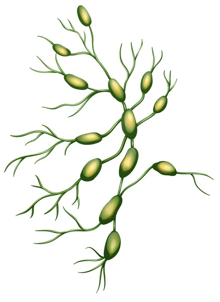
Lymph nodes
Lymph nodes
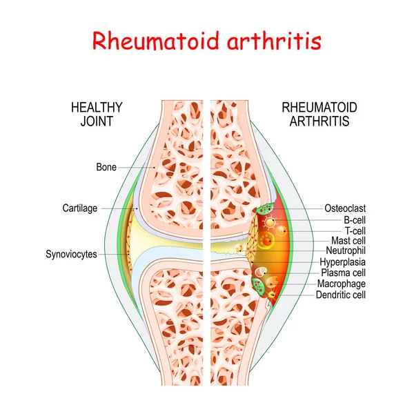
Rheumatoid arthritis. Healthy and damage joint. Close-up of bone, cartilage, and Cells in a joint capsule: synoviocytes, osteoclast, neutrophil, T lymphocyte, B-cell, macrophage, mast, plasma, and dendritic cell,
Rheumatoid Arthritis Healthy Damage Joint Close Bone Cartilage Cells Joint Capsule Synoviocytes Osteoclast Neutrophil Lymphocyte Cell Macrophage Mast Plasma Dendritic Cell
Regulatory T cell function. Suppressor T-cells. Prevent autoimmune disease. Immune homeostasis. Adaptive immune response. Vector illustration
Regulatory Cell Function Suppressor Cells Prevent Autoimmune Disease Immune Homeostasis Adaptive Immune Response Vector Illustration
Herpes simplex keratitis HSV1 infection infographic showing detailed human eyeball cross section anatomy with red highlighted target focus zone over corneal lesion vector illustration.
Herpes Simplex Keratitis Hsv1 Infection Infographic Showing Detailed Human Eyeball Cross Section Anatomy Red Highlighted Target Focus Zone Corneal Lesion Vector Illustration
Herpes simplex keratitis HSV1 corneal infection infographic. Human eyeball cross section anatomy, lines pointing to structures dendritic lesion looked through magnifying glass vector illustration
Herpes Simplex Keratitis Hsv1 Corneal Infection Infographic Human Eyeball Cross Section Anatomy Lines Pointing Structures Dendritic Lesion Looked Magnifying Glass Vector Illustration
Healthy vs damaged motor neuron comparison. Neuronal degeneration. Abnormal neuron anatomy. Medical illustration. infographic diagram for science explainer. differential diagnosis
Healthy Damaged Motor Neuron Comparison Neuronal Degeneration Abnormal Neuron Anatomy Medical Illustration Infographic Diagram Science Explainer Differential Diagnosis
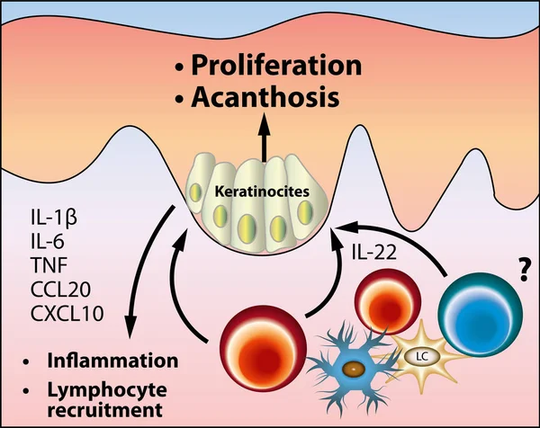
The pathogenesis of acanthosis nigricans scheme, vector medical illustration
Pathogenesis Acanthosis Nigricans Scheme Vector Medical Illustration
Cells of the immune system. Vector diagram
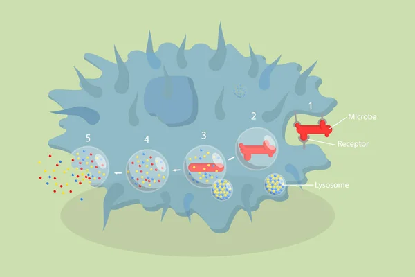
3D Isometric Flat Vector Conceptual Illustration of Phagocytosis, Labeled endocytosis Educational Scheme
Isometric Flat Vector Conceptual Illustration Phagocytosis Labeled Endocytosis Educational Scheme
Also known as dendritic cell or accessory cells of immune system phagocytosis and antigen presentation. Anatomy, biology, science concept vector illustration.
Also Known Dendritic Cell Accessory Cells Immune System Phagocytosis Antigen Presentation Anatomy Biology Science Concept Vector Illustration
Immune system cell types. Cartoon macrophage, dendritic, monocyte, mast, b and t cells. Adaptive and innate immunity, lymphocyte vector set
Neuron. Neuronal structure. Vector illustration
Neuron Neuronal Structure Vector Illustration
Hematopoiesis diagram. Human blood cells types with names. Scientific microbiology vector illustration in sketch style. blood cellular components formation
Hematopoiesis Diagram Human Blood Cells Types Names Scientific Microbiology Vector Illustration Sketch Style Blood Cellular Components Formation
Dendritic cells process stage diagram, labeled with immature and mature, pathogen cytokines, antigen-presenting cells. immune system, medical poster, biology education. antigens, immune system cells
Dendritic Cells Process Stage Diagram Labeled Immature Mature Pathogen Cytokines Antigen Presenting Cells Immune System Medical Poster Biology Education Antigens Immune System Cells
Immune response brief flow shows innate to adaptive stages with T cells, B cells, and antibodies defeating pathogens, cells interact over time from exposure to memory. Outline diagram
Immune Response Brief Flow Shows Innate Adaptive Stages Cells Cells Antibodies Defeating Pathogens Cells Interact Time Exposure Memory Outline Diagram
Dendritic spine. Neuron anatomy. Learning and memory. Neuronal plasticity. Neural communication. Transmission of the nerve signal between two neurons with axon and synapse. Vector illustration
Dendritic Spine Neuron Anatomy Learning Memory Neuronal Plasticity Neural Communication Transmission Nerve Signal Two Neurons Axon Synapse Vector Illustration
Blood cells macrophage cell drawing. Anatomy, biology, science concept. Scientific Designing of Macrophage. The White Blood Cell Structure.
Blood Cells Macrophage Cell Drawing Anatomy Biology Science Concept Scientific Designing Macrophage White Blood Cell Structure
Cells of the immune system. White blood cells or leukocytes Eosinophil, Neutrophil, Basophil, Macrophage, Fibroblast, and Dendritic cell. Set of transparent realistic cells on a white background. Vector illustration
Cells Immune System White Blood Cells Leukocytes Eosinophil Neutrophil Basophil Macrophage Fibroblast Dendritic Cell Set Transparent Realistic Cells White Background Vector Illustration
Innate immunity from Fever and Complement system (protein for holes in the plasma membrane), to Macrophage, NK and Dendritic cells. Adaptive immunity from Antibodies and Plasma cell to B-cell, T helper, T-killer. comparison and difference
Innate Immunity Fever Complement System Protein Holes Plasma Membrane Macrophage Dendritic Cells Adaptive Immunity Antibodies Plasma Cell Cell Helper Killer Comparison Difference
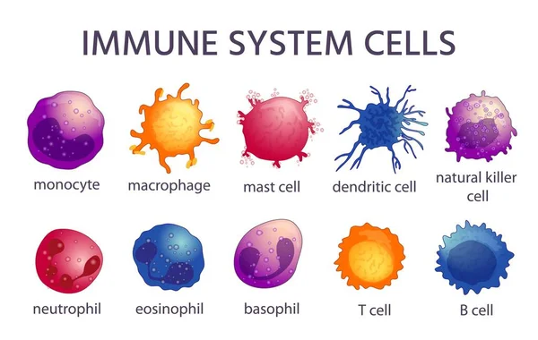
Illustration of immune system cells
Illustration Immune System Cells
Herpes simplex keratitis HSV1 infection pathway infographic showing human head profile silhouette with trigeminal nerve diagram and eyeball cross section vector illustration.
Herpes Simplex Keratitis Hsv1 Infection Pathway Infographic Showing Human Head Profile Silhouette Trigeminal Nerve Diagram Eyeball Cross Section Vector Illustration
Herpes simplex keratitis HSV1 infection infographic showing eyeball cross section transition to detailed cornea anatomy diagram with highlighted red dendritic lesion focus vector illustration.
Herpes Simplex Keratitis Hsv1 Infection Infographic Showing Eyeball Cross Section Transition Detailed Cornea Anatomy Diagram Highlighted Red Dendritic Lesion Focus Vector Illustration
Herpes simplex keratitis HSV1 infection pathway infographic showing human head profile silhouette with trigeminal nerve branches and circle icon zoom of corneal lesion vector illustration.
Herpes Simplex Keratitis Hsv1 Infection Pathway Infographic Showing Human Head Profile Silhouette Trigeminal Nerve Branches Circle Icon Zoom Corneal Lesion Vector Illustration
Neuromuscular junction. A synaptic connection between the terminal end of a motor nerve and a muscle. Presynaptic (nerve terminal), postsynaptic part, synaptic cleft.
Neuromuscular Junction Synaptic Connection Terminal End Motor Nerve Muscle Presynaptic Nerve Terminal Postsynaptic Part Synaptic Cleft
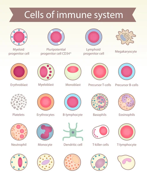
Cells of immune system.
Biological structure of Neuron cell
Biological Structure Neuron Cell
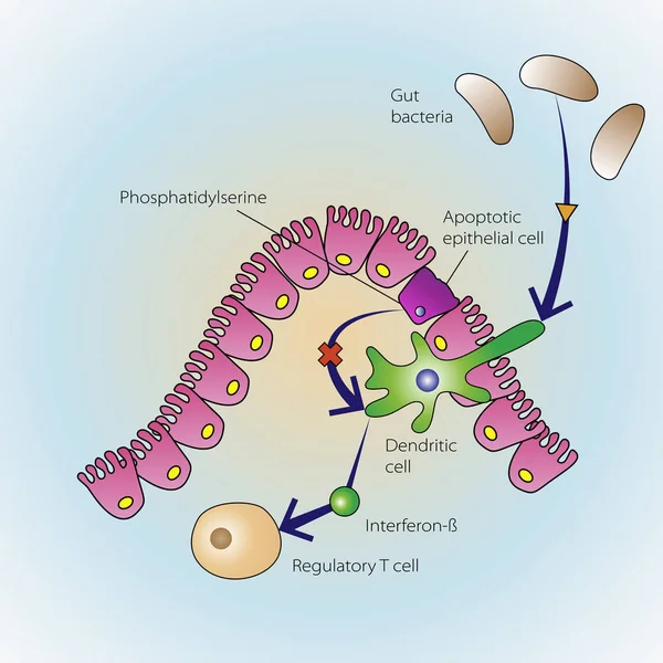
Dying epithelial cells
Cells of the immune system. White blood cells or leukocytes: Eosinophil, Neutrophil, Basophil, Macrophage, Fibroblast, and Dendritic cell. Vector diagram
Cells Immune System White Blood Cells Leukocytes Eosinophil Neutrophil Basophil Macrophage Fibroblast Dendritic Cell Vector Diagram
Herpes simplex keratitis HSV1 corneal infection flat minimal poster showing human eyeball cross section anatomy with green dendritic lesion on purple gradient background vector illustration.
Herpes Simplex Keratitis Hsv1 Corneal Infection Flat Minimal Poster Showing Human Eyeball Cross Section Anatomy Green Dendritic Lesion Purple Gradient Background Vector Illustration
Herpes simplex keratitis HSV1 development scheme showing human head profile silhouette with trigeminal nerve transition to eyeball anatomy and corneal dendritic lesion focus vector illustration
Herpes Simplex Keratitis Hsv1 Development Scheme Showing Human Head Profile Silhouette Trigeminal Nerve Transition Eyeball Anatomy Corneal Dendritic Lesion Focus Vector Illustration
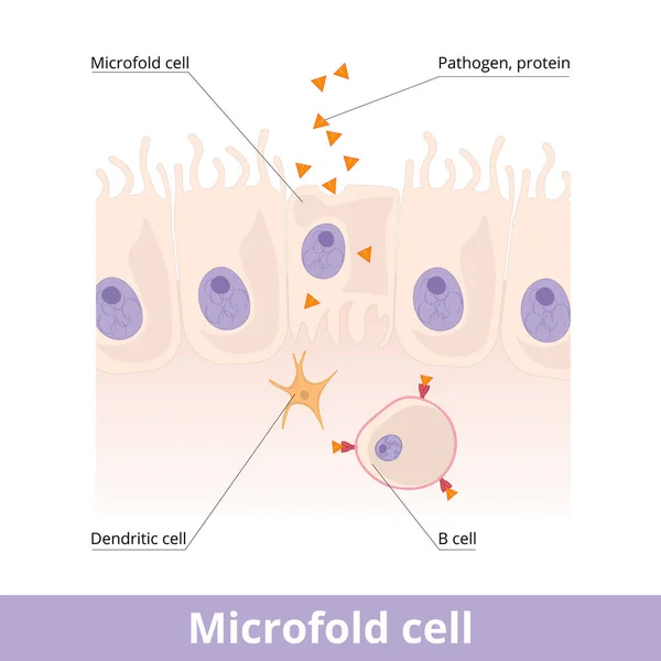
Microfold cell. Found in the lymphoid tissue (GALT) of the Peyer's patches in the small intestine, and in the mucosa-associated lymphoid tissue (MALT) of the gastrointestinal tract.
Microfold Cell Found Lymphoid Tissue Galt Peyer Patches Small Intestine Mucosa Associated Lymphoid Tissue Malt Gastrointestinal Tract
Ocular herpes simplex virus keratitis infection symptoms infographic showing human eye anatomy with eyelid blisters shingles and dendritic lesion on cornea medical vector illustration.
Ocular Herpes Simplex Virus Keratitis Infection Symptoms Infographic Showing Human Eye Anatomy Eyelid Blisters Shingles Dendritic Lesion Cornea Medical Vector Illustration
Herpes simplex keratitis HSV1 corneal infection infographic showing human eye closeup view with green dendritic lesion on pupil and highlighted red target focus zone vector illustration.
Herpes Simplex Keratitis Hsv1 Corneal Infection Infographic Showing Human Eye Closeup View Green Dendritic Lesion Pupil Highlighted Red Target Focus Zone Vector Illustration