Fibroblasti Stock Vector Images

54 Fibroblasti vector art & graphics are available under a royalty-free license
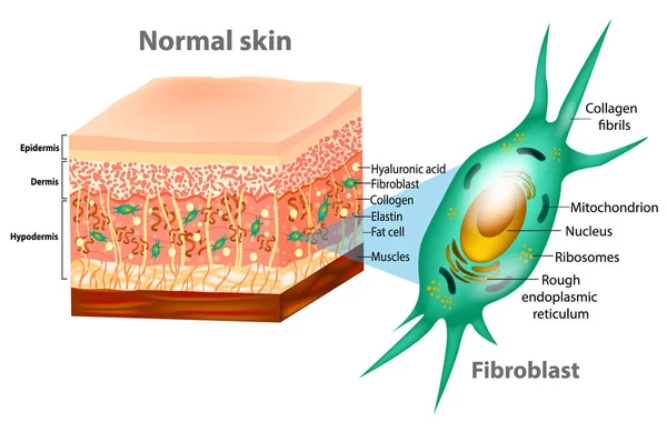
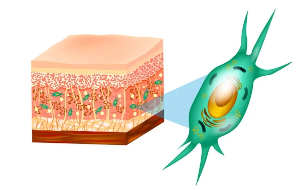
Fibroblast and Human skin structure (Muscles, Fat cell, Hyaluronic acid, Elastin, Collagen, Fibroblast).
Fibroblast Human Skin Structure Muscles Fat Cell Hyaluronic Acid Elastin Collagen Fibroblast
Fibroblast is vital to the skin's strength and elasticity. Struc
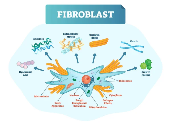
Fibroblast vector illustration. Scheme with extracellular, collagen fibrils, elastin, hyaluronic acid, microtubule, golgi apparatus, nucleus and ribosomes.
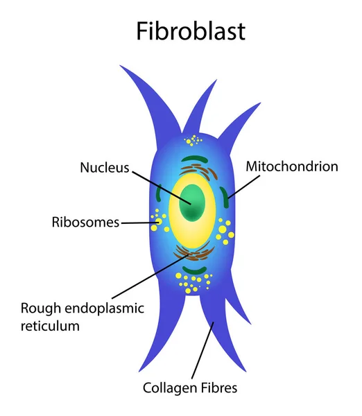
Fibroblast anatomy. structure of cell. Diagram with golgi apparatus, nucleus, mitochondrion and ribosomes. vector illustration. Poster
Fibroblast Anatomy Structure Cell Diagram Golgi Apparatus Nucleus Mitochondrion Ribosomes Vector Illustration Poster
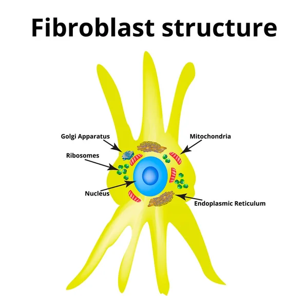
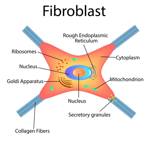
Fibroblast structure. Fibroblast cell. Vector illustration on isolated background
Skin icon in flat style isolated on white background. Organs symbol stock vector illustration.
Illustration showing the structure of the dermis.Not notation.
Illustration Showing Structure Dermis Notation
Aging process comparison. The difference between younger and older skin on woman face vector illustration. Cross section of high and low density of collagen, elastin fibers and fibroblasts in dermis.
Aging Process Comparison Difference Younger Older Skin Woman Face Vector Illustration Cross Section High Low Density Collagen Elastin Fibers Fibroblasts Dermis
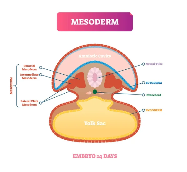
Mesoderm vector illustration. Labeled medical diagram with embryo structure
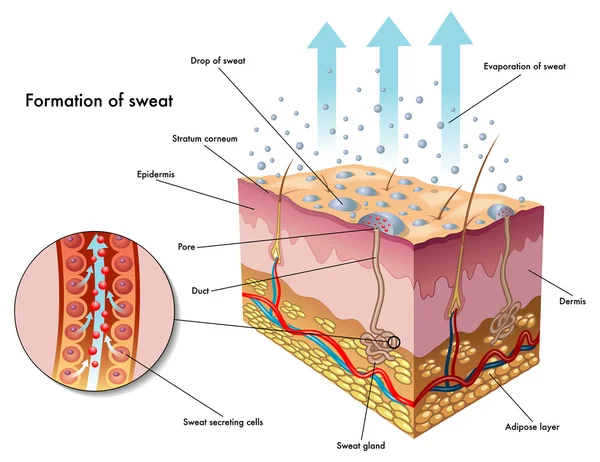
Sweating
Sweating
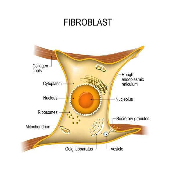
Fibroblast is a dermis cell. Structure of Fibroblast cell.
Cross section of skin layer with intact collagen, elastin fibers and fibroblasts in the dermis vector illustration on white background. Synthesis of collagen and elastin are produced by fibroblasts.
Cross Section Skin Layer Intact Collagen Elastin Fibers Fibroblasts Dermis Vector Illustration White Background Synthesis Collagen Elastin Produced Fibroblasts
The old woman with aging and wrinkled skin vector on white background. Cross section of depletion of collagen, elastin fibers and fibroblasts in the dermis layer. Skin care and beauty concept.
Old Woman Aging Wrinkled Skin Vector White Background Cross Section Depletion Collagen Elastin Fibers Fibroblasts Dermis Layer Skin Care Beauty Concept
Cross section of high density of collagen, elastin fibers and fibroblasts in the dermis layer vector illustration on white background. Formation of collagen and elastin are produced by fibroblasts.
Cross Section High Density Collagen Elastin Fibers Fibroblasts Dermis Layer Vector Illustration White Background Formation Collagen Elastin Produced Fibroblasts
Hair in skin icon, cartoon style
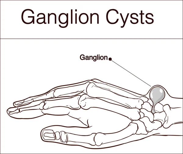
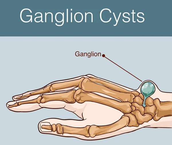
Vector illustration of a Ganglion cyst
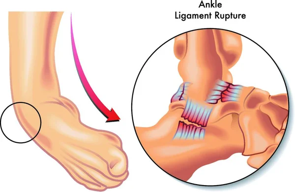
Medical Illustration of ankle ligament rupture
Medical Illustration Ankle Ligament Rupture
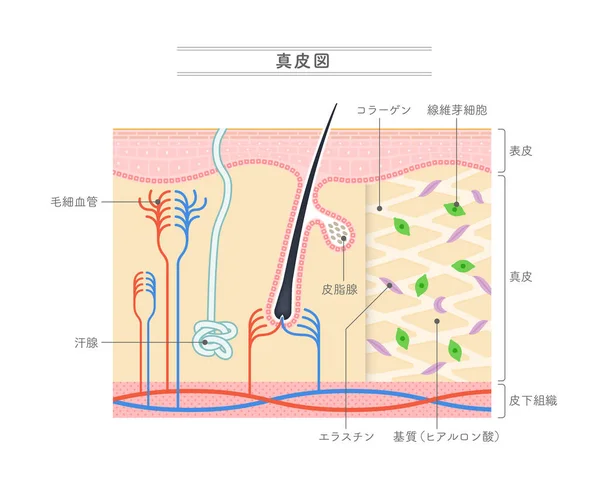
Illustration showing the structure of the dermis.Japanese notation.
Illustration Showing Structure Dermis Japanese Notation
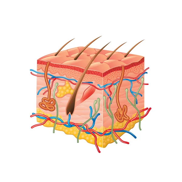
Human skin anatomy isolated on white vector
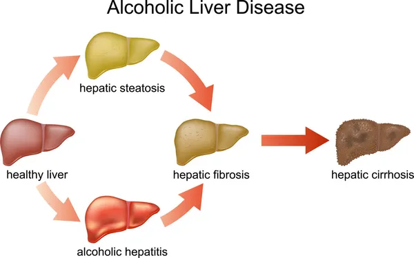
Alcoholic Liver Disease.
Fibroblast structure. Fibroblast cell. Vector illustration isolated
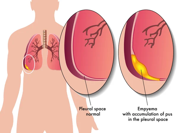
Pleural empyema
Pleural empyema
Asbestosis pathophysiology, inhaled asbestos fibers trigger macrophage injury and collagen scarring in alveoli, impairing gas exchange, key objects, lungs, alveoli, fibers. Outline diagram
Asbestosis Pathophysiology Inhaled Asbestos Fibers Trigger Macrophage Injury Collagen Scarring Alveoli Impairing Gas Exchange Key Objects Lungs Alveoli Fibers Outline Diagram
Tissue repair structure diagram hand drawn schematic vector illustration. Medical science educational illustration
Tissue Repair Structure Diagram Hand Drawn Schematic Vector Illustration Medical Science Educational Illustration
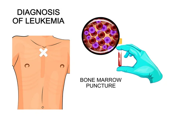
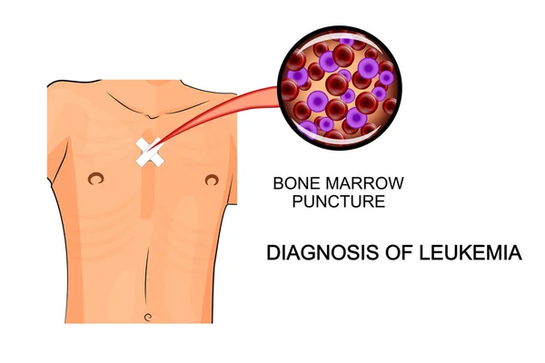
Diagnosis of leukemia. Bone marrow puncture
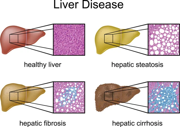
Liver Disease with micrograph
Skin icon in monochrome style isolated on white background. Organs symbol stock vector illustration.
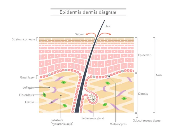
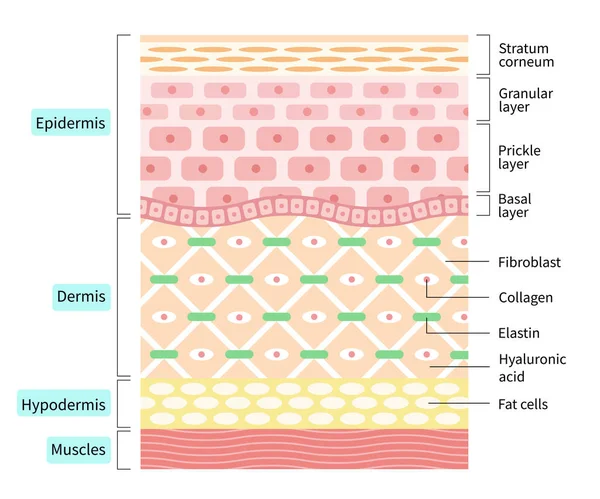
Illustration showing the structure of the epidermis and dermis.English notation.
Illustration Showing Structure Epidermis Dermis English Notation
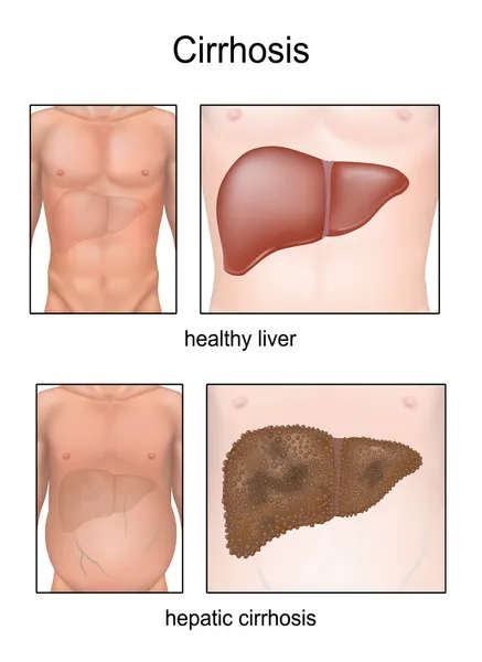
Cirrhosis.
Cirrhosis.
Fibroblast and Human skin structure (Muscles, Fat cell, Hyaluronic acid, Elastin, Collagen, Fibroblast).
Fibroblast Human Skin Structure Muscles Fat Cell Hyaluronic Acid Elastin Collagen Fibroblast
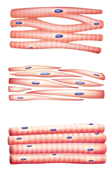
Types of muscles
Types of muscles
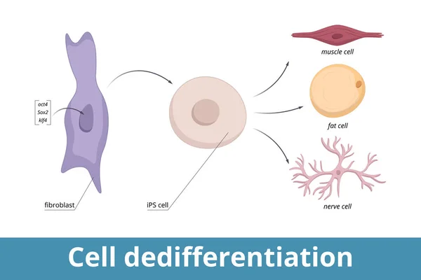
Cell dedifferentiation. Genes encoding three transcription regulators are artificially introduced into fibroblast nucleus, cell divides in culture (iPS cell), induced to de-differentiate.
Cell Dedifferentiation Genes Encoding Three Transcription Regulators Artificially Introduced Fibroblast Nucleus Cell Divides Culture Ips Cell Induced Differentiate
Skin icon in cartoon style isolated on white background. Organs symbol stock vector illustration.
Vector illustration. Fibroblast is a dermis cell. Structure of Fibroblast cell.
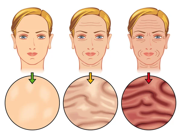
Aging process comparison with 3 generation women face vector illustration. Cross section of high density, depletion and low density of collagen, elastin fibers and fibroblasts in the dermis.
Aging Process Comparison Generation Women Face Vector Illustration Cross Section High Density Depletion Low Density Collagen Elastin Fibers Fibroblasts Dermis
Skin icon in black style isolated on white background. Organs symbol stock vector illustration.
Human dry skin
Human dry skin
Hygroma wrist joint
Hygroma wrist joint
Bone marrow puncture leukemia
Structure cells of dermis illustration isolated on white background. skin and health care concept
Structure Cells Dermis Illustration Isolated White Background Skin Health Care Concept
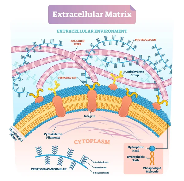
Extracellular matrix labeled infographic vector illustration scheme.
Illustration of collagen types and their locations
Illustration Collagen Types Locations
Vector illustration of a Ganglion cyst
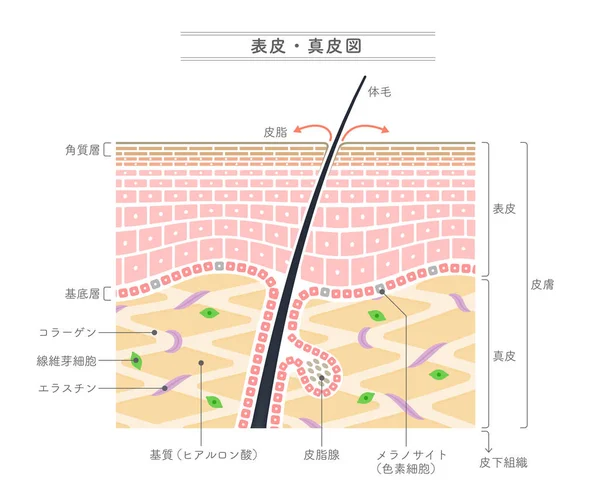
Illustration showing the structure of the epidermis and dermis.Japanese notation.
Illustration Showing Structure Epidermis Dermis Japanese Notation
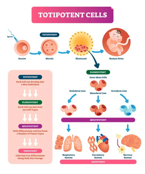
Totipotent cells vector illustration. Multi, uni and pluripotent diagram.
Human Skin aging
Human Skin aging
Layers of the human skin. skin and health care concept
Layers Human Skin Skin Health Care Concept
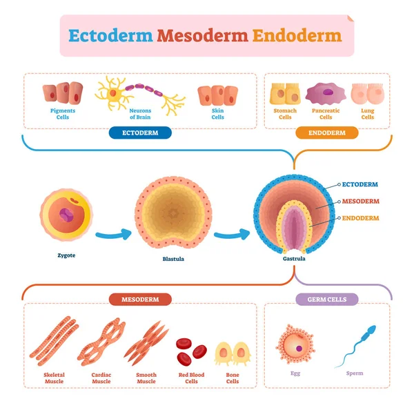
Endoderm, mesoderm and ectoderm vector illustration labeled infographic.
The old woman with lines and wrinkles skin vector on white background. Cross section of low density of collagen, elastin fibers and fibroblasts in the dermis layer. Skin care and beauty concept.
Old Woman Lines Wrinkles Skin Vector White Background Cross Section Low Density Collagen Elastin Fibers Fibroblasts Dermis Layer Skin Care Beauty Concept
Fibroblast cell structure. Connective tissue cell. Vector illustration
Cross section of skin layer with intact and damaged collagen, elastin fibers and fibroblasts in the dermis vector illustration on white background. Comparison of young healthy and aging wrinkled skin.
Cross Section Skin Layer Intact Damaged Collagen Elastin Fibers Fibroblasts Dermis Vector Illustration White Background Comparison Young Healthy Aging Wrinkled Skin
Cross section of aging process skin comparison vector illustration isolated on white background. High density, depletion and low density of collagen, elastin fibers and fibroblasts in the dermis.
Cross Section Aging Process Skin Comparison Vector Illustration Isolated White Background High Density Depletion Low Density Collagen Elastin Fibers Fibroblasts Dermis
Intact collagen, elastin fibers and fibroblasts with skin layer vector illustration on white background. Formation of young and healthy skin. Synthesis of collagen and elastin are made of fibroblasts.
Intact Collagen Elastin Fibers Fibroblasts Skin Layer Vector Illustration White Background Formation Young Healthy Skin Synthesis Collagen Elastin Made Fibroblasts
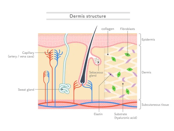
Illustration showing the structure of the dermis.English notation.
Illustration Showing Structure Dermis English Notation