Gram positive and Gram negative bacteria
Gram stain test. A glass Petri dish with pathogen bacterial culture before and after use Crystal violet. Bacteria stained with gentian violet. Gram-positive bacteria are violet-stained like cocci. Gram-negative bacteria are pink-stained like bacilli.
Gram Stain Test Glass Petri Dish Pathogen Bacterial Culture Use Crystal Violet Bacteria Stained Gentian Violet Gram Positive Bacteria Violet Stained Cocci Gram Negative Bacteria Pink Stained Bacilli
Diagram showing gram staining microbiology lab technique steps - microbiology laboratory using Crystal violet and Safranin
Diagram Showing Gram Staining Microbiology Lab Technique Steps Microbiology Laboratory Using Crystal Violet Safranin
Gram stain test. glass Petri dish with culture Gram-negative bacteria Escherichia coli after used of crystal violet stain. Close-up of Gram-positive bacteria Clostridium perfringens. Vector illustration
Gram Stain Test Glass Petri Dish Culture Gram Negative Bacteria Escherichia Coli Used Crystal Violet Stain Close Gram Positive Bacteria Clostridium Perfringens Vector Illustration
Diagram showing gram staining technique steps - microbiology laboratory using Crystal violet and Safranin
Diagram Showing Gram Staining Technique Steps Microbiology Laboratory Using Crystal Violet Safranin
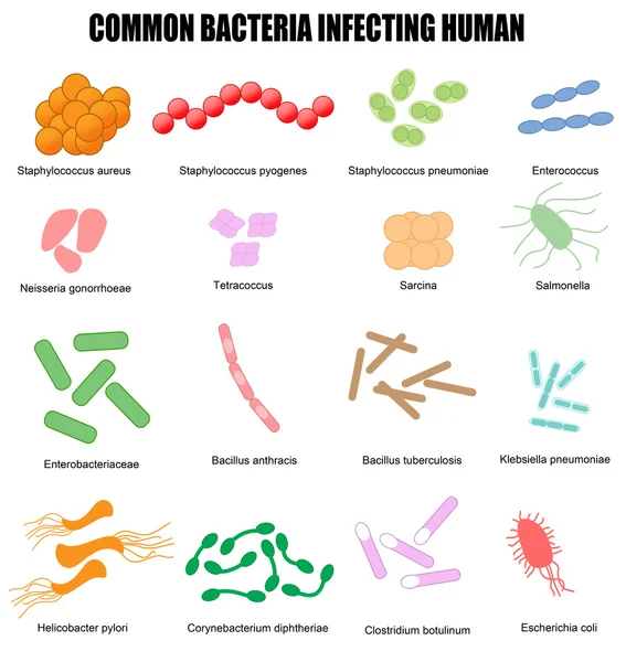
Common bacteria infecting human
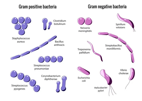
Gram positive bacteria or Gram negative bacteria. Gram stain test. Classification of bacteria by type of cell wall. Vector illustration. Medical diagram.
Gram Positive Bacteria Gram Negative Bacteria Gram Stain Test Classification Bacteria Type Cell Wall Vector Illustration Medical Diagram
Bacteria vector illustration
Common bacteria infecting human
This differential staining procedure separates most bacteria into two groups on the basis of cell wall composition, Bacteria gram posotive and gram negative
Differential Staining Procedure Separates Most Bacteria Two Groups Basis Cell Wall Composition Bacteria Gram Posotive Gram Negative
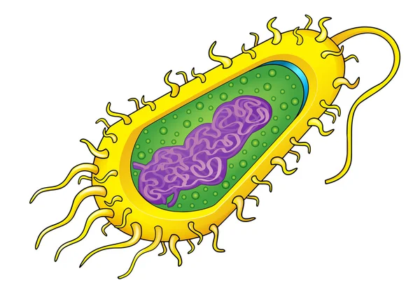
Pink bacterial cell
Pink bacterial cell
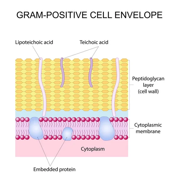
Gram-positive cell envelope
Bacteria cell
Bacteria cell
Gram negative bacteria do not retain the crystal violet stain. Gram positive bacteria give a positive result in the Gram stain test. comparison and difference type of bacterial cell wall. vector poster
Gram Negative Bacteria Retain Crystal Violet Stain Gram Positive Bacteria Give Positive Result Gram Stain Test Comparison Difference Type Bacterial Cell Wall Vector Poster
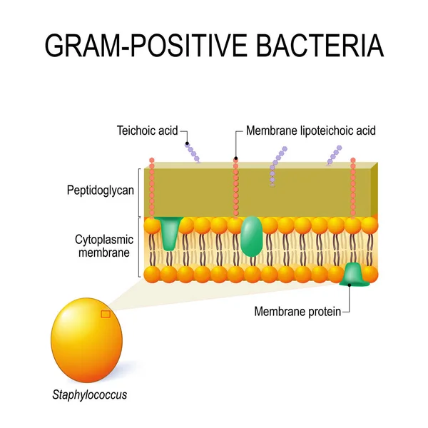
Cell wall structure of Gram-positive Bacteria for example Staphylococcus. Vector diagram for educational, medical, biological and science use
Cell Wall Structure Gram Positive Bacteria Example Staphylococcus Vector Diagram Educational Medical Biological Science Use
Bacteria cell structure
Gram Positive and Gram Negative Bacteria Microscopy Comparison Diagram for Microbiology Science Education
Gram Positive Gram Negative Bacteria Microscopy Comparison Diagram Microbiology Science Education
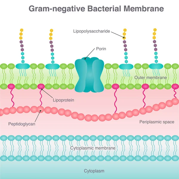
Gram-negative bacterial membrane diagram vector illustration
Gram Negative Bacterial Membrane Diagram Vector Illustration
Step-by-Step Gram Staining Procedure Diagram for Microbiology Laboratory Cell Wall Differentiation
Step Step Gram Staining Procedure Diagram Microbiology Laboratory Cell Wall Differentiation
Gram Positive vs Gram Negative Bacteria Comparison Infographic Diagram for Microbiology Science Education
Gram Positive Gram Negative Bacteria Comparison Infographic Diagram Microbiology Science Education
Bacteria cell structure
Bacteria cell
Bacteria cell
Types of bacterial cell wall. Gram-negative bacteria and Gram-negative bacteria. comparison, structure, and composition. Vector illustration
Types Bacterial Cell Wall Gram Negative Bacteria Gram Negative Bacteria Comparison Structure Composition Vector Illustration
Mixed Gram Staining Microscopic Field Diagram with Gram Positive Cocci and Gram Negative Bacilli
Mixed Gram Staining Microscopic Field Diagram Gram Positive Cocci Gram Negative Bacilli
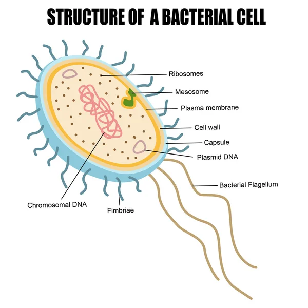
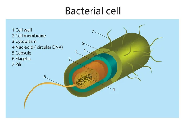
Structure of a bacterial cell
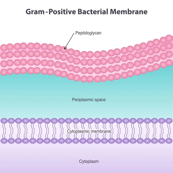
Gram-Positive Bacterial Membrane Diagram Illustration
Gram Positive Bacterial Membrane Diagram Illustration
Educational Microbiology Infographic Diagram Explaining the Four Steps of Gram Staining Procedure
Educational Microbiology Infographic Diagram Explaining Four Steps Gram Staining Procedure
Bacteria cell
Bacteria cell
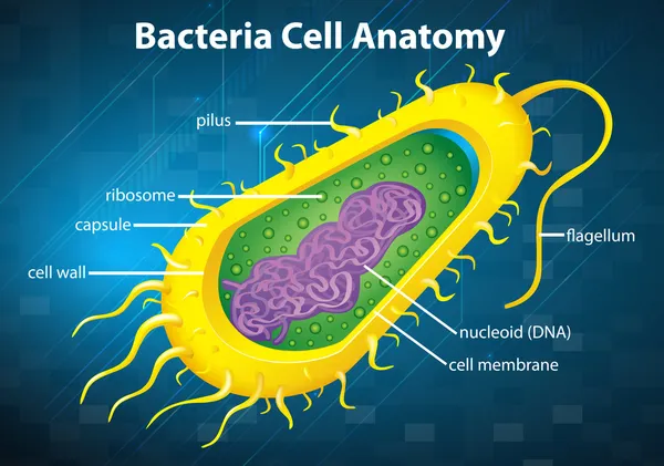
Illustration of the bacteria cell structure
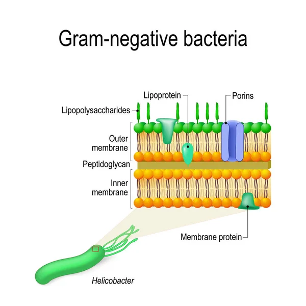
Cell wall structure of Gram-negative Bacteria for example Helicobacter. Vector diagram for educational, medical, biological and science use
Cell Wall Structure Gram Negative Bacteria Example Helicobacter Vector Diagram Educational Medical Biological Science Use
Gram-negative bacterial membrane diagram vector illustration
Gram Negative Bacterial Membrane Diagram Vector Illustration
Classification of bacteria. Grams method. Gram-positive and Gram-negative bacterium. Cross section of Cell wall, Plasma membrane and Peptidoglycan. Detailed Vector poster
Classification Bacteria Grams Method Gram Positive Gram Negative Bacterium Cross Section Cell Wall Plasma Membrane Peptidoglycan Detailed Vector Poster