Hyalin Stock Vector Images

26 Hyalin vector art & graphics are available under a royalty-free license
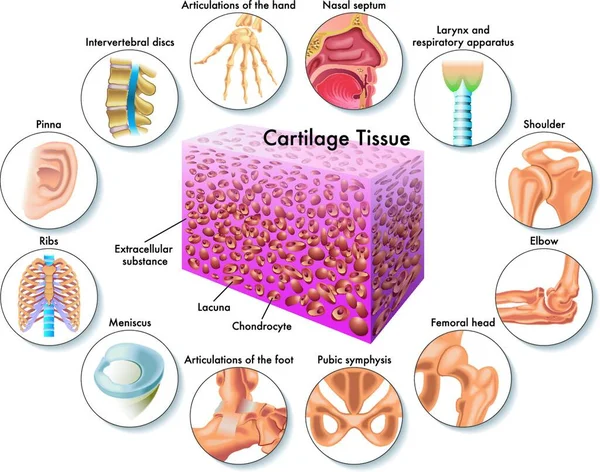
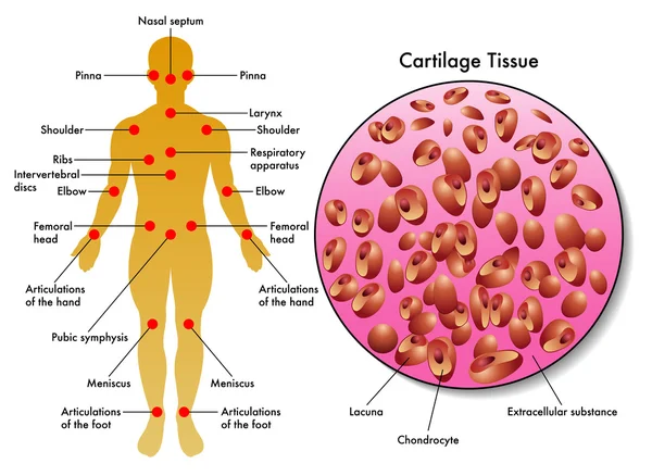
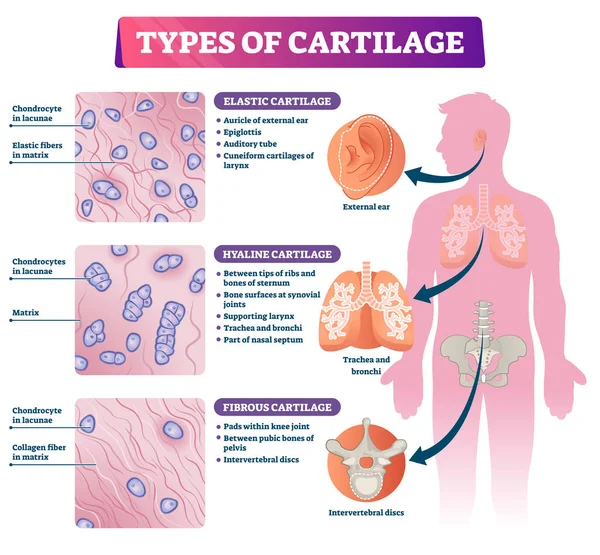
Medical illustration of cartilage tissue and its position in the human body
Medical Illustration Cartilage Tissue Its Position Human Body
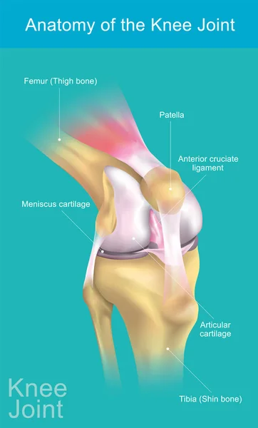
The knee joint joins the thigh with the leg and consists of two articulations: one between the femur and tibia (tibiofemoral joint),and one between the femur and patella. It is the largest joint in the human body. Illustration anatomy body.
Knee Joint Joins Thigh Leg Consists Two Articulations One Femur Tibia Tibiofemoral Joint One Femur Patella Largest Joint Human Body Illustration Anatomy Body
Human cartilage tissue
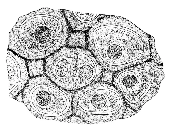
The cells of hyaline cartilage are irregular in shape, grouped together and are enclosed singly or in pairs in a capsule of hyaline substance, vintage line drawing or engraving illustration.
Cells Hyaline Cartilage Irregular Shape Grouped Together Enclosed Singly Pairs Capsule Hyaline Substance Vintage Line Drawing Engraving Illustration
Pearl heart
Pearl heart
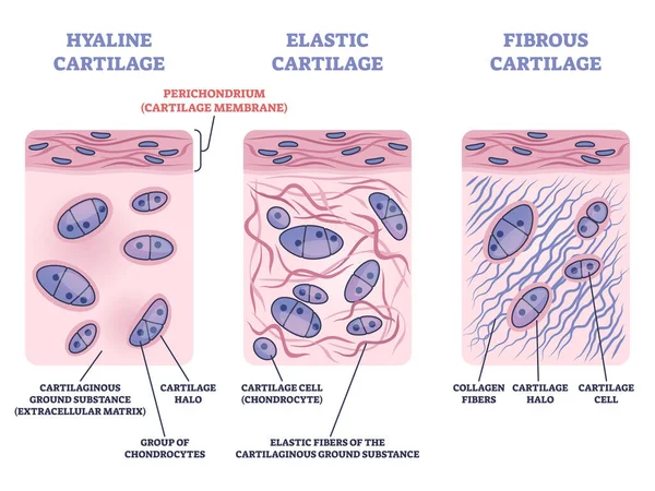
Perichondrium as hyaline and elastic cartilage membrane outline diagram
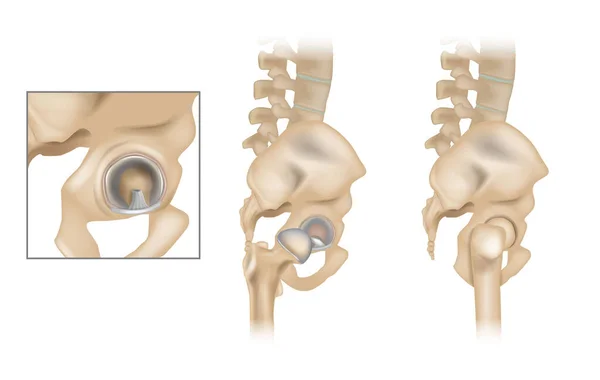
Anatomy of the Hip and Acetabular Labrum. Ligamentum teres and Articular cartilage. Lateral view with femur removed of right hip.
Anatomy Hip Acetabular Labrum Ligamentum Teres Articular Cartilage Lateral View Femur Removed Right Hip
Detailed medical illustration of the human esophagus and trachea showing their anatomical structure and cross sectional anatomy.
Detailed Medical Illustration Human Esophagus Trachea Showing Anatomical Structure Cross Sectional Anatomy
Transparent Vector Editable Text Effect Template
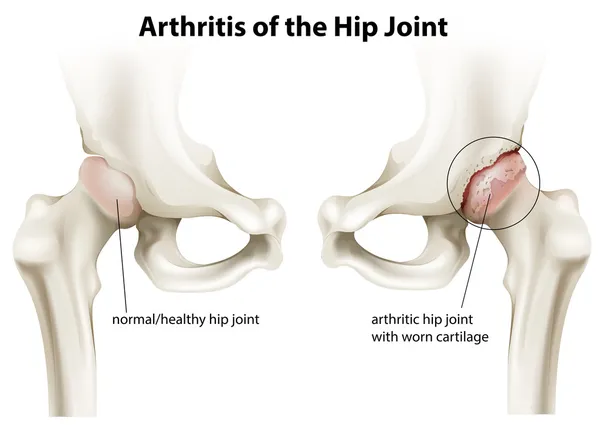
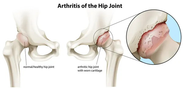
Arthritis of the hip joint
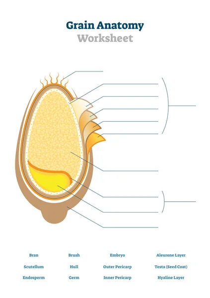
Grain anatomy worksheet vector illustration. Blank seed diagram template.
Arthritis of the hip joint
Types of cartilage vector illustration. Labeled educational tissue scheme.
Educational medical illustration showing the location and anatomy of the human esophagus and trachea in the neck and upper respiratory and digestive systems. The diagram includes the pharynx, thyroid gland, esophagus and trachea.
Educational Medical Illustration Showing Location Anatomy Human Esophagus Trachea Neck Upper Respiratory Digestive Systems Diagram Includes Pharynx Thyroid Gland Esophagus Trachea
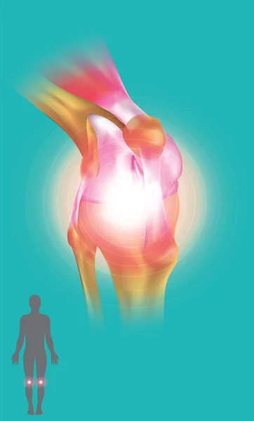
Knee Joint symptom
Knee Joint symptom
The matrix of hyaline cartilage has a dimly granular appearance like that of ground glass, and in man and higher mammals has no apparent structure, vintage line drawing or engraving illustration.
Matrix Hyaline Cartilage Has Dimly Granular Appearance Ground Glass Man Higher Mammals Has Apparent Structure Vintage Line Drawing Engraving Illustration
White blood cell mascot responsible for devesa of the body, diseases and other infections. Ideal for informative and institutional materials of medicine and health
White Blood Cell Mascot Responsible Devesa Body Diseases Other Infections Ideal Informative Institutional Materials Medicine Health
Anatomy of the Hip and Acetabular Labrum. Ligamentum teres and Articular cartilage. Lateral view with femur removed of right hip.
Anatomy Hip Acetabular Labrum Ligamentum Teres Articular Cartilage Lateral View Femur Removed Right Hip
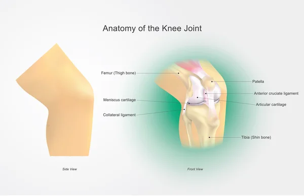
Anatomy of the Knee Joint.
Cartilaginous joints brief overview illustrating bone connection by cartilage, shows synchondrosis and symphysis with intervertebral disc and epiphyseal plate. Diagram
Cartilaginous Joints Brief Overview Illustrating Bone Connection Cartilage Shows Synchondrosis Symphysis Intervertebral Disc Epiphyseal Plate Diagram
Anatomy of the Knee Joint. Illustration, Vector Arts.
Types Collagen in cartilage tissues. Human skeleton with Costal cartilage. Parts of human body that have collagen: skull, knee, Auricle, Intervertebral disc and Meniscus. vector infographics.
Types Collagen Cartilage Tissues Human Skeleton Costal Cartilage Parts Human Body Have Collagen Skull Knee Auricle Intervertebral Disc Meniscus Vector Infographics
Lung surfactant function illustrated, comparison of open and collapsed alveolus, surfactant molecules and type II pneumocyte reduce surface tension to keep alveoli open. Outline diagram
Lung Surfactant Function Illustrated Comparison Open Collapsed Alveolus Surfactant Molecules Type Pneumocyte Reduce Surface Tension Keep Alveoli Open Outline Diagram
Heart
Heart
Types of cartilage: Fibrocartilage, Hyaline and Elastic. Human skeleton with joints. Close-up of cartilage cells. Magnification of Chondrocyte in a lacuna, Elastic fibers, Matrix, and Collagen fibers.
Types Cartilage Fibrocartilage Hyaline Elastic Human Skeleton Joints Close Cartilage Cells Magnification Chondrocyte Lacuna Elastic Fibers Matrix Collagen Fibers
Articular capsule outline shows a joint with bone ends, synovial membrane, and fluid, explaining cushioning and mobility. Outline diagram
Articular Capsule Outline Shows Joint Bone Ends Synovial Membrane Fluid Explaining Cushioning Mobility Outline Diagram