Lisosomas Stock Vector Images

29 Lisosomas vector art & graphics are available under a royalty-free license
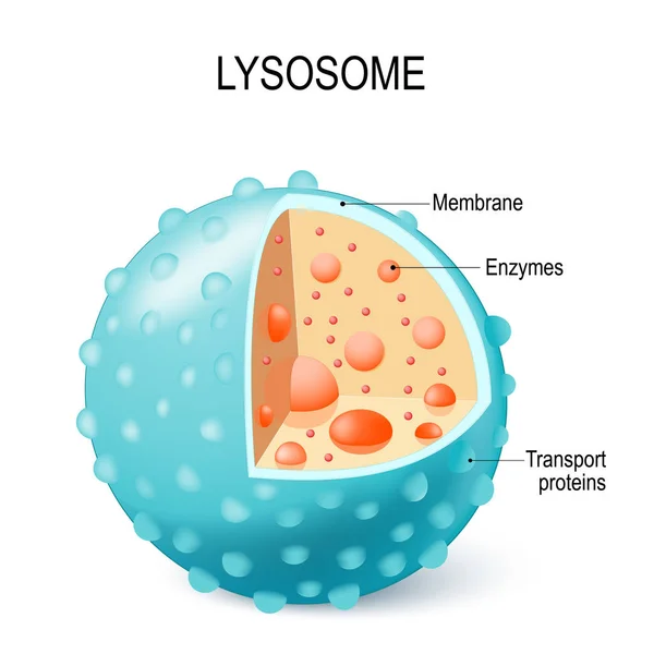
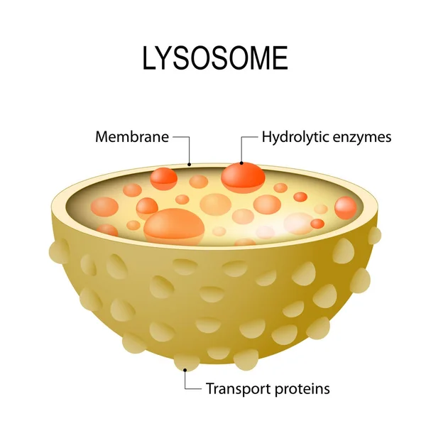
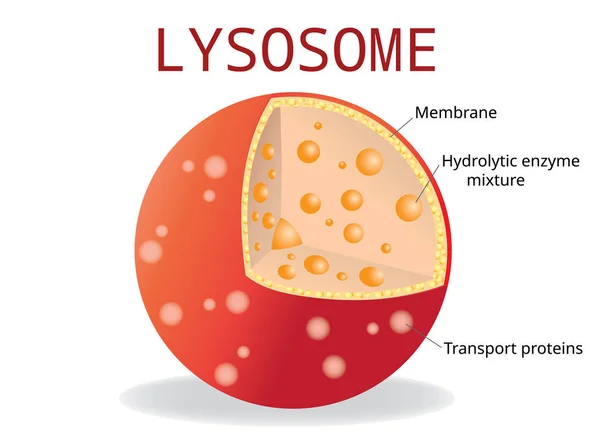
Anatomy of the Lysosome: Hydrolytic enzymes, Membrane and transport proteins. This organelle use the enzymes to break down and digest food particles, engulfed viruses or bacteria in the cell. Vector diagram for medical use
Anatomy Lysosome Hydrolytic Enzymes Membrane Transport Proteins Organelle Use Enzymes Break Digest Food Particles Engulfed Viruses Bacteria Cell Vector Diagram Medical Use
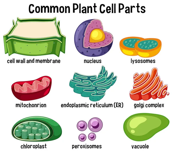
Common plant cell parts illustration
Common Plant Cell Parts Illustration
Illustration of biology and medical, Bone marrow is a spongy substance found in the center of the bones, Bone marrow produces white blood cells red blood cells and platelets, Anatomy of the bone
Illustration Biology Medical Bone Marrow Spongy Substance Found Center Bones Bone Marrow Produces White Blood Cells Red Blood Cells Platelets Anatomy Bone
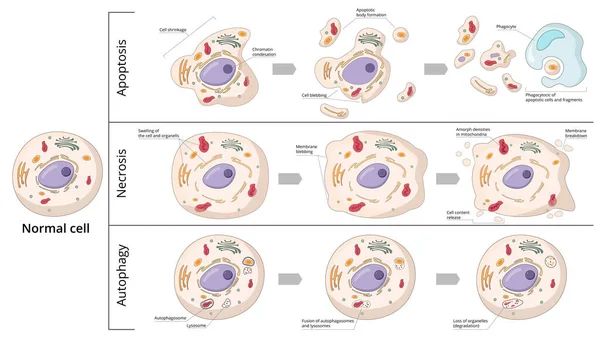
Three basic forms of cell death: apoptosis (chromosome condensation, nuclear fragmentation), autophagy (autophagosome formation), necrosis (membrane rupture, organelles swelling).
Three Basic Forms Cell Death Apoptosis Chromosome Condensation Nuclear Fragmentation Autophagy Autophagosome Formation Necrosis Membrane Rupture Organelles Swelling
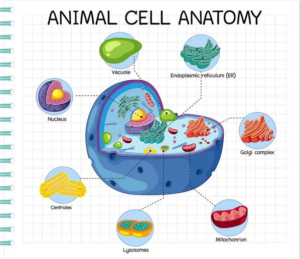
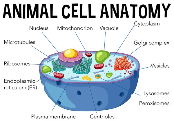
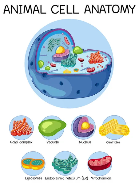
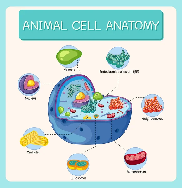
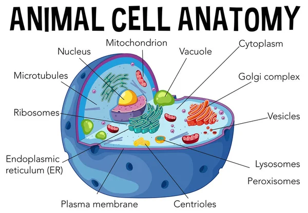
Anatomy of animal cell (Biology Diagram) illustration
Anatomy Animal Cell Biology Diagram Illustration
Autophagy of mitochondria. Diagram of the process of autophagy from Forming a membrane and autophagosome to fuse phagosome and lysosome when contents of the vesicles are degraded and recycled. Autophagy defects linked to various diseases and cancer d
Autophagy Mitochondria Diagram Process Autophagy Forming Membrane Autophagosome Fuse Phagosome Lysosome Contents Vesicles Degraded Recycled Autophagy Defects Linked Various Diseases Cancer
The chlamydial life cycle
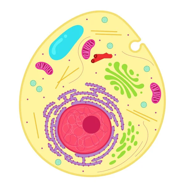
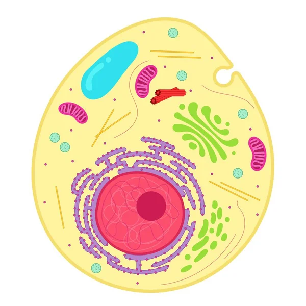
An animal cell is a type of eukaryotic cell.
Animal Cell Type Eukaryotic Cell
Plant cell anatomy. Vacuole structure. Vector illustration. poster. Schematic diagram
Plant Cell Anatomy Vacuole Structure Vector Illustration Poster Schematic Diagram
Anatomy of the Lysosome.
The Structure Of Human Cell
Structure Human Cell
An animal cell is a type of eukaryotic cell.
Animal Cell Type Eukaryotic Cell
Animal cell anatomy diagram illustration
Animal Cell Anatomy Diagram Illustration
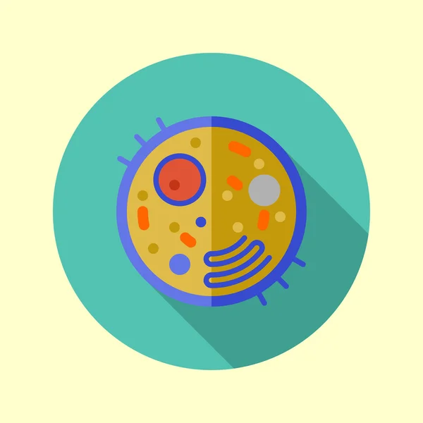
Cell icon
Cell icon
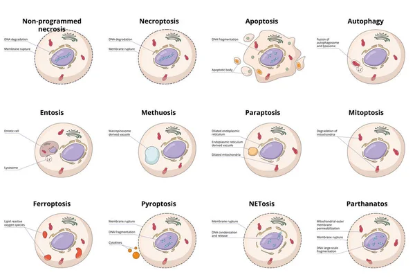
Cell death types: general differences between cell death processes, including common necrosis, autophagy, apoptosis and specific entosis, paraptosis and ferroptosis.
Cell Death Types General Differences Cell Death Processes Including Common Necrosis Autophagy Apoptosis Specific Entosis Paraptosis Ferroptosis
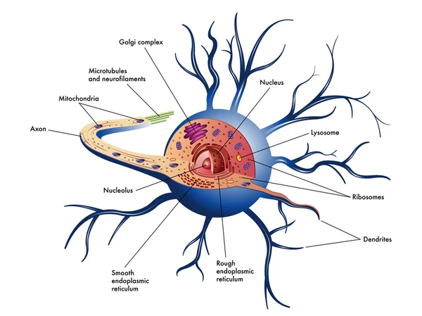
Nerve cell scheme
Nerve cell scheme
Detailed process of phagocytosis in four stages: entrapment of food particle, formation of food vacuole within cell, fusion of vacuole and lysosomes, digestion of food particle.
Detailed Process Phagocytosis Four Stages Entrapment Food Particle Formation Food Vacuole Cell Fusion Vacuole Lysosomes Digestion Food Particle
Lysosome Hydrolytic enzymes, Membrane and transport proteins, science and medical use, Vector Illustration
Lysosome Hydrolytic Enzymes Membrane Transport Proteins Science Medical Use Vector Illustration
The Structure Of Plant Cell
Structure Plant Cell
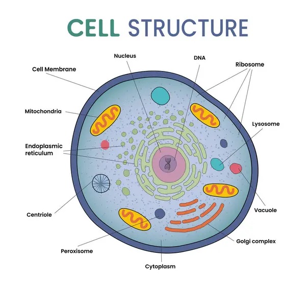
Illustration of cell structure infographic
Illustration Cell Structure Infographic
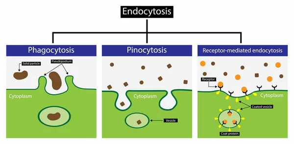
Illustration of biology,Endocytosis is a cellular process in which substances are brought into the cell, Endocytosis includes pinocytosis and phagocytosis, It is a form of active transport
Illustration Biology Endocytosis Cellular Process Which Substances Brought Cell Endocytosis Includes Pinocytosis Phagocytosis Form Active Transport
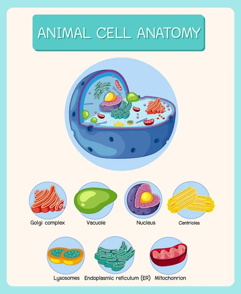
Anatomy of animal cell (Biology Diagram) illustration
Anatomy Animal Cell Biology Diagram Illustration
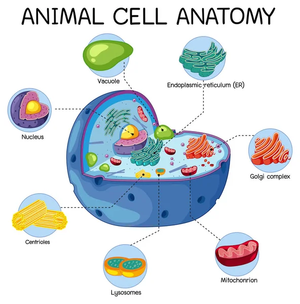
Anatomy of animal cell (Biology Diagram) illustration
Anatomy Animal Cell Biology Diagram Illustration
Neutrophil granulocytes. Characteristics and structure of lymphocytes
Plant cell anatomy. cross section. structure of a Eukaryotic cell. Vector diagram for your design, educational, medical, biological and science use
Plant Cell Anatomy Cross Section Structure Eukaryotic Cell Vector Diagram Your Design Educational Medical Biological Science Use
Anatomy of animal cell (Biology Diagram) illustration
Anatomy Animal Cell Biology Diagram Illustration
Anatomy of animal cell (Biology Diagram) illustration
Anatomy Animal Cell Biology Diagram Illustration
Plant cell. Structure and Anatomy. Close-up of layers of a cell wall. Vector illustration
Plant Cell Structure Anatomy Close Layers Cell Wall Vector Illustration
Diagram of animal cell anatomy illustration
Diagram Animal Cell Anatomy Illustration