Megalite Stock Vector Images

77 Megalite vector art & graphics are available under a royalty-free license
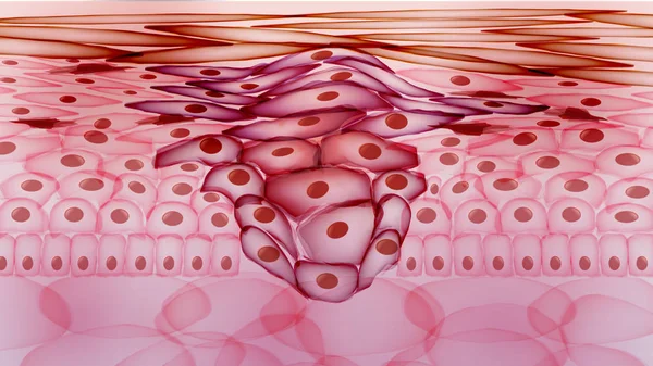
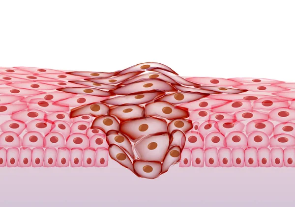
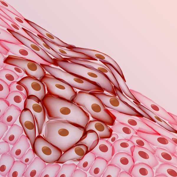
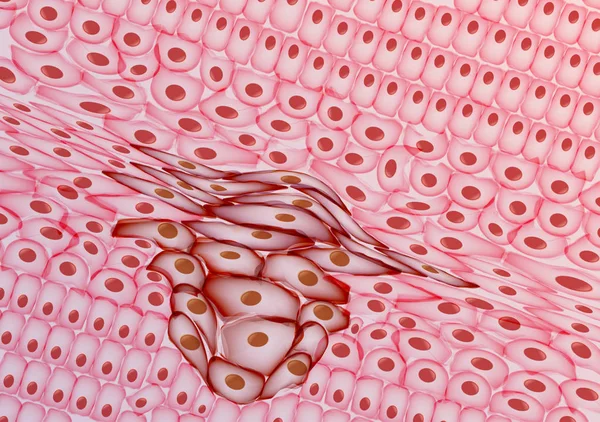
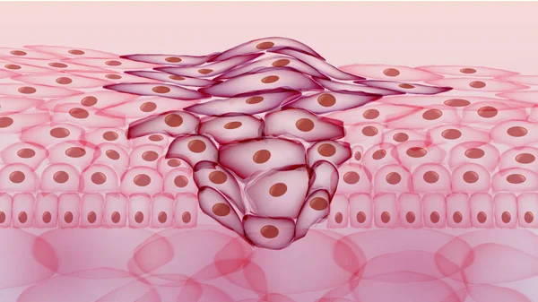
Growing Tumor, Tissue Section Inflamed Skin - Vector Illustratio
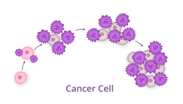
Cencer cell stage, Process, Development , vector
Cencer Cell Stage Process Development Vector
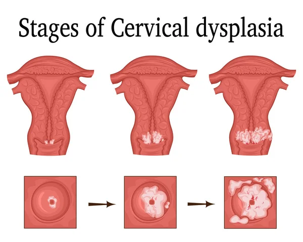
Illustration of Cervical dysplasia
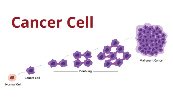
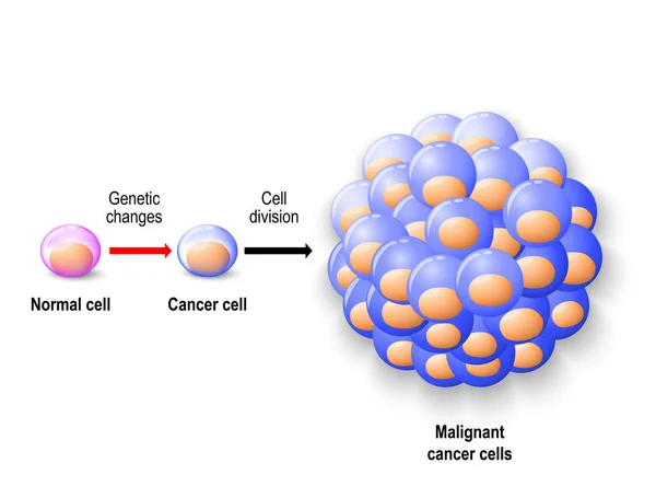
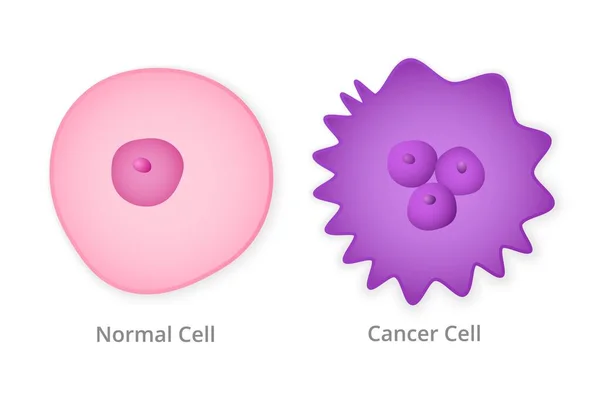
Normal human cell, cancer cell and malignant cancer
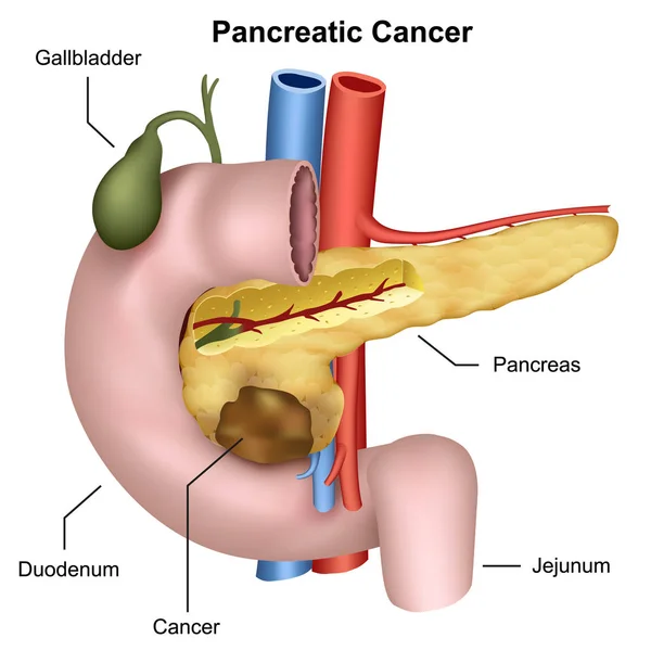
Pancreatic cancer medical vector illustration, pancreas anatomy
Pancreatic Cancer Medical Vector Illustration Pancreas Anatomy
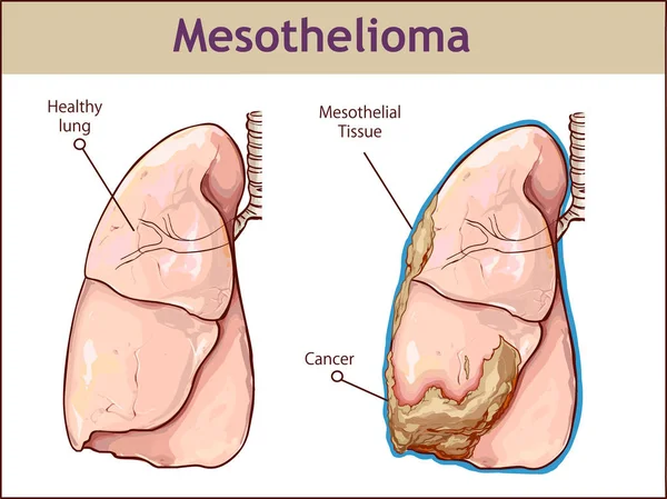
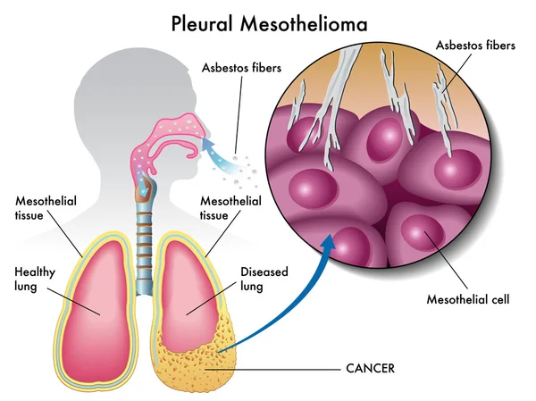
Vector illustration of a Pleural mesothelioma.
Vector Illustration Pleural Mesothelioma
Cervical cancer cells elongated Dysplasia stages infographic Female reproductive system squamous cell carcinoma in uterus pap smear test. Human anatomy internal organs flat style icon
Cervical Cancer Cells Elongated Dysplasia Stages Infographic Female Reproductive System Squamous Cell Carcinoma Uterus Pap Smear Test Human Anatomy Internal Organs Flat Style Icon
Human kidneys isolate on white background. Vector illustration.
Tumor angiogenesis. transition of tumors from a benign state to a malignant. tumor grow. Cancer cells and Spread of metastasis. Vector poster
Tumor Angiogenesis Transition Tumors Benign State Malignant Tumor Grow Cancer Cells Spread Metastasis Vector Poster
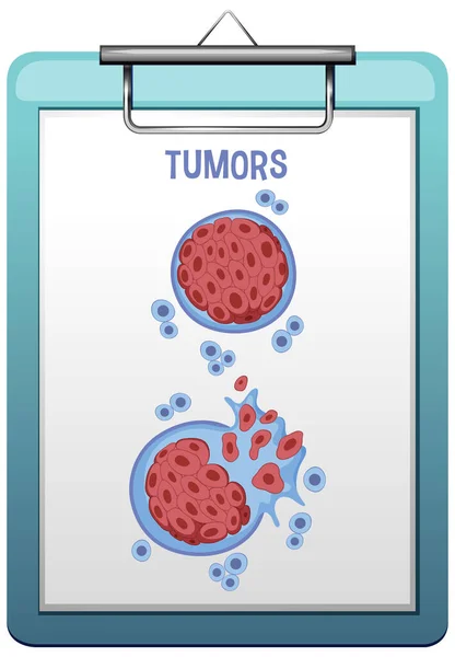
Tumor cells concept
Tumor cells concept
Process of development of cancer cells
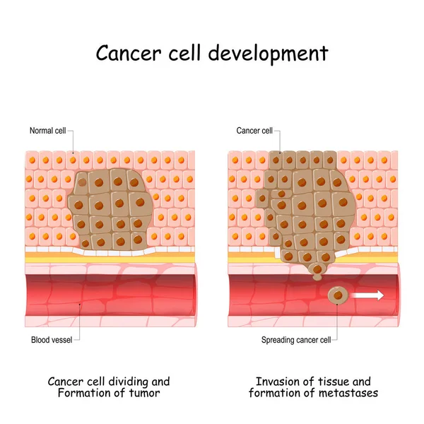
Cancer development from normal cell to Formation of tumor, Spreading cancer cells in blood flow, Invasion of other tissue, and formation of metastases. vector illustration
Cancer Development Normal Cell Formation Tumor Spreading Cancer Cells Blood Flow Invasion Other Tissue Formation Metastases Vector Illustration
ReedSternberg cells. Six variants: classic, mononuclear cell variant or Hodgkin cell, lacunar, mummified and anaplastic cell, predominant (LP cell).
Reedsternberg Cells Six Variants Classic Mononuclear Cell Variant Hodgkin Cell Lacunar Mummified Anaplastic Cell Predominant Cell
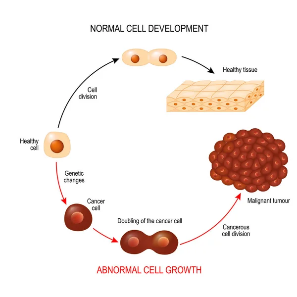
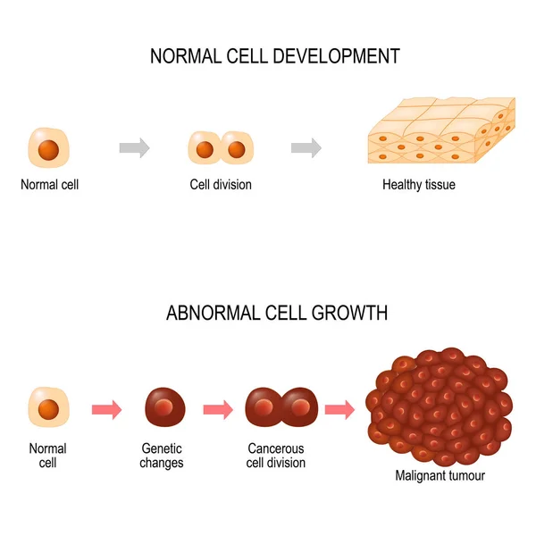
Cancer cell and normal cell. Healthy tissue and Malignant tumour. illustration showing cancer disease development. Vector diagram for your design, educational, biological, science and medical use
Cancer Cell Normal Cell Healthy Tissue Malignant Tumour Illustration Showing Cancer Disease Development Vector Diagram Your Design Educational Biological Science Medical Use
Tumor cells concept
Tumor cells concept
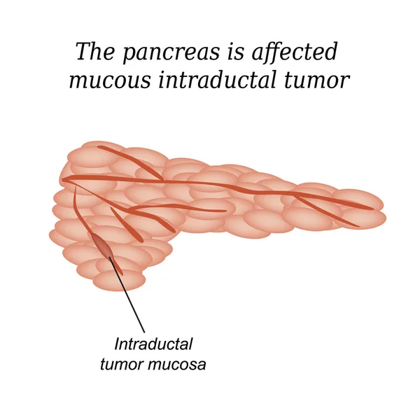
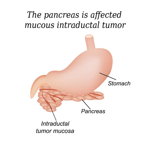
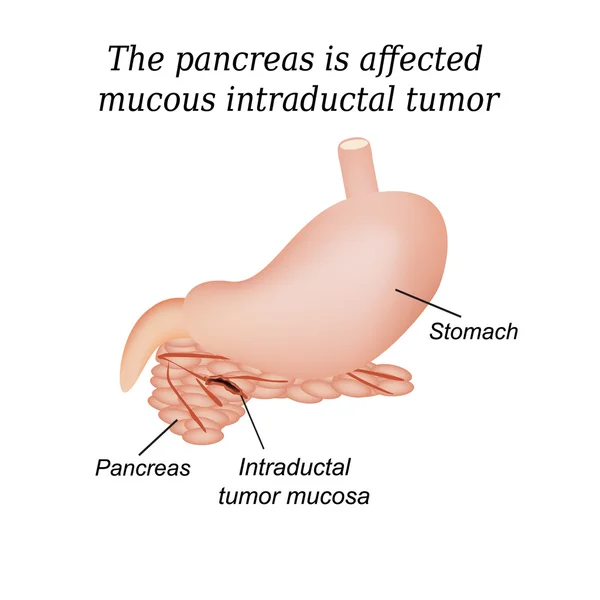
The pancreas is affected mucous intraductal tumor. Vector illustration on isolated background
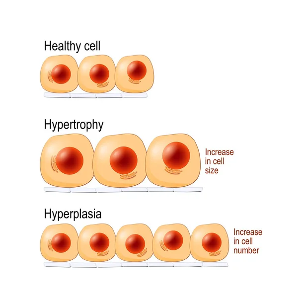
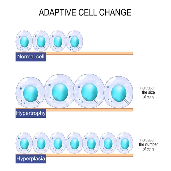
Normal cells, hypertrophy is an increase in cell size, hyperplasia results from an increase in cell number. different. Vector diagram for educational, medical, biological and science use
Normal Cells Hypertrophy Increase Cell Size Hyperplasia Results Increase Cell Number Different Vector Diagram Educational Medical Biological Science Use
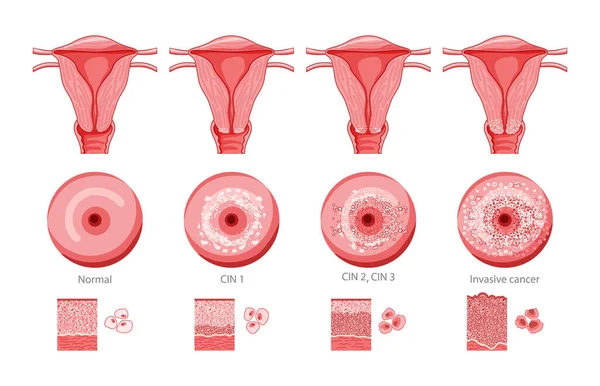
Set of Cervical invasive cancer intraepithelial neoplasia Dysplasia 1 2,3 and normal stage cell morphology on pap smear test infographic Female reproductive system Carcinoma view. medical flat style
Set Cervical Invasive Cancer Intraepithelial Neoplasia Dysplasia Normal Stage Cell Morphology Pap Smear Test Infographic Female Reproductive System Carcinoma View Medical Flat Style
Cervical dysplasia. Close-up of a cervix. Cervical cancer. Cervical intraepithelial neoplasia development from normal tissue and CIN I with mild dysplasia to moderate dysplasia and squamous cell carcinoma. vector illustration isolated on white backgr
Cervical Dysplasia Close Cervix Cervical Cancer Cervical Intraepithelial Neoplasia Development Normal Tissue Cin Mild Dysplasia Moderate Dysplasia Squamous Cell Carcinoma Vector Illustration Isolated White Backgr
Cervical cancer or cervix carcinoma. development stage. vector illustration
Cervical Cancer Cervix Carcinoma Development Stage Vector Illustration
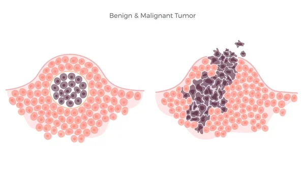
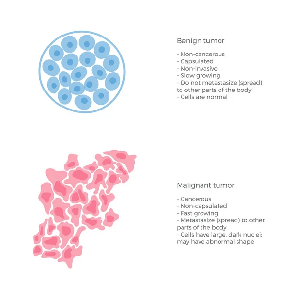
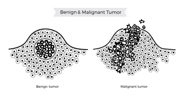
Vector illustration of malignant and benign tumor
Vector  illustration of malignant and benign tumor
Cervical intraepithelial neoplasia. Cervical dysplasia. CIN. Pre-cancerous lesion. Close-up of a cervix. Cancer development from normal cell to Precancerous transformation of cells of the cervix. Vector. Schematic diagram. Detailed poster.
Cervical Intraepithelial Neoplasia Cervical Dysplasia Cin Pre Cancerous Lesion Close Cervix Cancer Development Normal Cell Precancerous Transformation Cells Cervix Vector Schematic Diagram Detailed Poster
Cancer evolution from Mutated cell and Hyperplasia, to Dysplasia and Malignant tumor. Spread cancer cells to other tissues. Vector diagram
Cancer Evolution Mutated Cell Hyperplasia Dysplasia Malignant Tumor Spread Cancer Cells Other Tissues Vector Diagram
Tumor cell and cancer development illustration
Tumor Cell Cancer Development Illustration
Cushing disease endocrinology line icon vector illustration
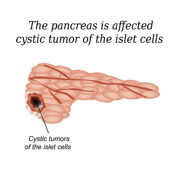
The pancreas is affected cystic tumor of the islet cells. Vector illustration on isolated background
Cervical dysplasia. difference in covering epithelium affected of Cervical Intraepithelial Neoplasia. Cross section of epithelium. Close-up of a normal Squamous cell and Abnormal cells. Cervical cancer evolution. Vector illustration
Cervical Dysplasia Difference Covering Epithelium Affected Cervical Intraepithelial Neoplasia Cross Section Epithelium Close Normal Squamous Cell Abnormal Cells Cervical Cancer Evolution Vector Illustration
Growing Tumor, Tissue Section - Vector Illustration
Medical illustration of the development of breast cancer
Medical Illustration Development Breast Cancer
Cervical cancer. Carcinoma. Malignant neoplasm. Cut-away view of the uterus. female reproductive system. Vector illustration like X-ray image for healthcare design. Reproductive health.
Cervical Cancer Carcinoma Malignant Neoplasm Cut Away View Uterus Female Reproductive System Vector Illustration Ray Image Healthcare Design Reproductive Health
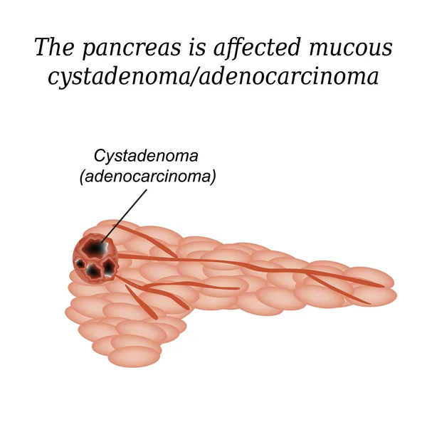
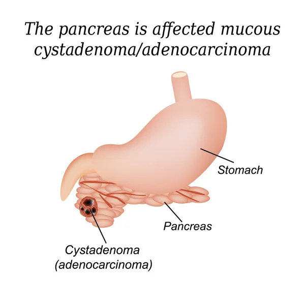
Pancreas mucous cystadenoma, adenocarcinoma. Vector illustration on isolated background
Pleural mesothelioma
Cervical invasive cancer intraepithelial neoplasia Dysplasia 1 2,3 and normal stage cell morphology on pap smear test infographic Female reproductive system Carcinoma speculum view. medical flat style
Cervical Invasive Cancer Intraepithelial Neoplasia Dysplasia Normal Stage Cell Morphology Pap Smear Test Infographic Female Reproductive System Carcinoma Speculum View Medical Flat Style
Growing Tumor, Tissue Section - Vector Illustration
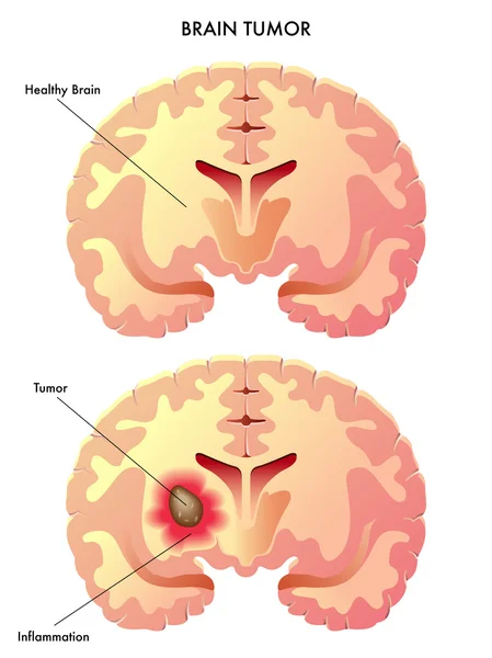
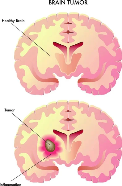
Medical illustration of the effects of the brain tumor
Medical Illustration Effects Brain Tumor
Normal and cancer cell
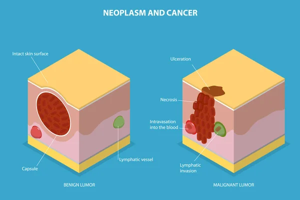
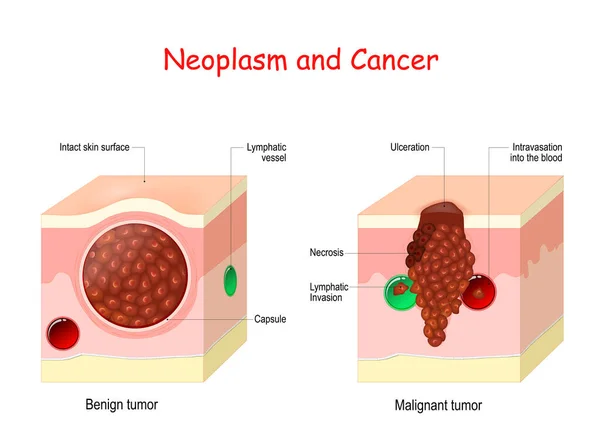
Cancer and Neoplasm. comparison and difference between Malignant and Benign tumor. Benign tumor has a Capsule. Cells of Malignant tumor have Necrosis, Intravasation into the blood vessel, and Lymphatic Invasion.
Cancer Neoplasm Comparison Difference Malignant Benign Tumor Benign Tumor Has Capsule Cells Malignant Tumor Have Necrosis Intravasation Blood Vessel Lymphatic Invasion
Uterine cancer. Cross section of a human uterus with Endometrial tumor. female reproductive system. Vector illustration like X-ray image. Reproductive health.
Uterine Cancer Cross Section Human Uterus Endometrial Tumor Female Reproductive System Vector Illustration Ray Image Reproductive Health
Growing Tumor, Tissue Section - Vector Illustration
Vector illustration of malignant and benign tumor
Vector  illustration of malignant and benign tumor
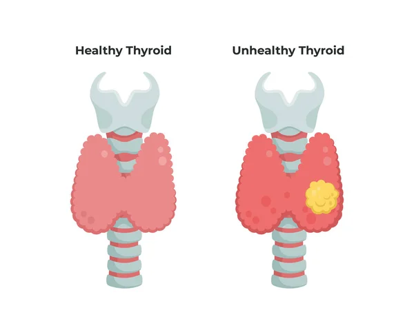
Healthy Thyroid gland and unhealthy thyroid with Inflammation and lump, thyroid cancer concept, flat illustration isolated on white background. Medical infographic elements.
Malignant neoplasm. Cancer and Normal cells. comparison and difference between healthy tissue and tumor. details about chromatin, nucleus and cytoplasm
Malignant Neoplasm Cancer Normal Cells Comparison Difference Healthy Tissue Tumor Details Chromatin Nucleus Cytoplasm
Cancer cells. illustration showing cancer disease development. Healthy tissue and Malignant tumour. Vector diagram for your design, educational, biological, science and medical use
Cancer Cells Illustration Showing Cancer Disease Development Healthy Tissue Malignant Tumour Vector Diagram Your Design Educational Biological Science Medical Use
Line icon Lymphoma icon
Growing Tumor, Tissue Section - Vector Illustration
Pancreas mucous cystadenoma, adenocarcinoma. Vector illustration on isolated background
Vector illustration of malignant and benign tumor
Vector  illustration of malignant and benign tumor
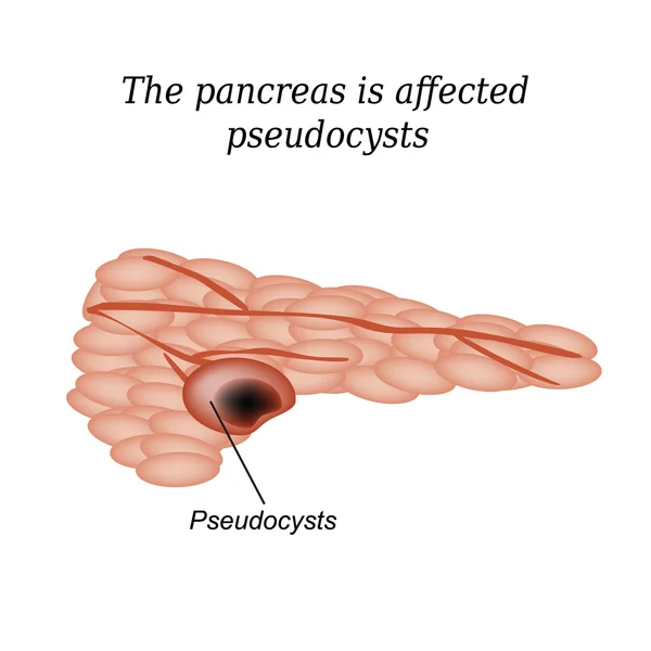
Pancreas pseudocyst affected. Vector illustration on isolated background
Brain tumor
Brain tumor
Growing Tumor, Tissue Section - Vector Illustration
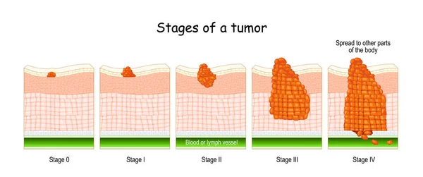
Stages of cancer. Classification of Malignant Tumours (from 0 to 4). system that is most commonly used for the staging process of cancer
Stages Cancer Classification Malignant Tumours System Most Commonly Used Staging Process Cancer
Tumor cell and cancer development illustration
Tumor Cell Cancer Development Illustration
3D Isometric Flat Vector Conceptual Illustration of Neoplasm And Cancer, Tumor Development
Isometric Flat Vector Conceptual Illustration Neoplasm Cancer Tumor Development
Growing Tumor, Tissue Section - Vector Illustration
Pancreatic cancer medical vector illustration
Pancreatic Cancer Medical Vector Illustration
Vector isolated illustration of benign tumor
3D Isometric Flat Vector Illustration of Cancer Cells, Tumor Development
Isometric Flat Vector Illustration Cancer Cells Tumor Development
Medical illustration of symptoms and risk of breast cancer
Medical Illustration Symptoms Risk Breast Cancer
Set of Cervical cancer Dysplasia stages infographic Female reproductive system Carcinoma cells uterus. Front view in a cut. Human anatomy internal organs location scheme flat style icon
Set Cervical Cancer Dysplasia Stages Infographic Female Reproductive System Carcinoma Cells Uterus Front View Cut Human Anatomy Internal Organs Location Scheme Flat Style Icon
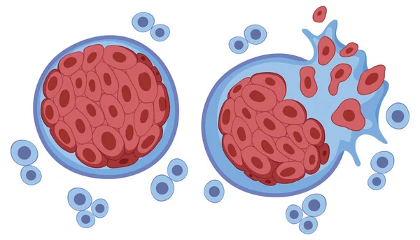
Cancer cells in Benign neoplasm and Malignant tumors. Close-up of carcinocytes that have the ability to metastasis (right). Cells of non-cancerous tumor are surrounded by a fibrous sheath of connective tissue (left). Vector poster
Cancer Cells Benign Neoplasm Malignant Tumors Close Carcinocytes Have Ability Metastasis Right Cells Non Cancerous Tumor Surrounded Fibrous Sheath Connective Tissue Left Vector Poster
Tumor cells concept
Tumor cells concept
Vector illustration of malignant and benign tumor
Vector  illustration of malignant and benign tumor
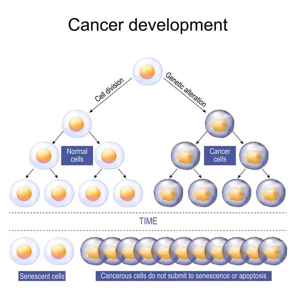
Cancer development. Carcinogenesis or Oncogenesis. Tumor cell initiation. Cell proliferation. Vector illustration
Cancer Development Carcinogenesis Oncogenesis Tumor Cell Initiation Cell Proliferation Vector Illustration
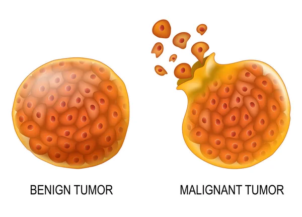
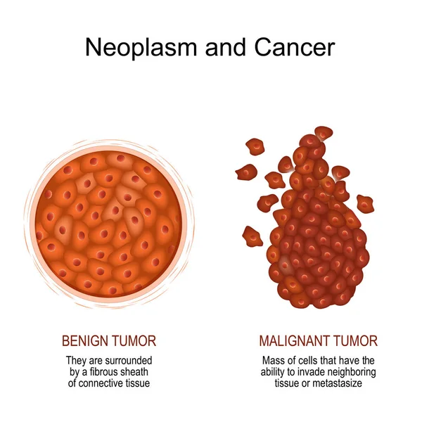
Neoplasm and Cancer. Comparison and difference of a malignant and benign tumor. benign tumor surrounded by a fibrous sheath of connective tissue. malignant Cancer is a Mass of cells that have the ability to invade neighboring tissue or metastasize. v
Neoplasm Cancer Comparison Difference Malignant Benign Tumor Benign Tumor Surrounded Fibrous Sheath Connective Tissue Malignant Cancer Mass Cells Have Ability Invade Neighboring Tissue Metastasize
Tumor cells concept
Tumor cells concept
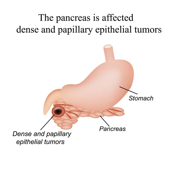
The pancreas is affected dense and papillary epithelial tumors. Vector illustration on isolated background
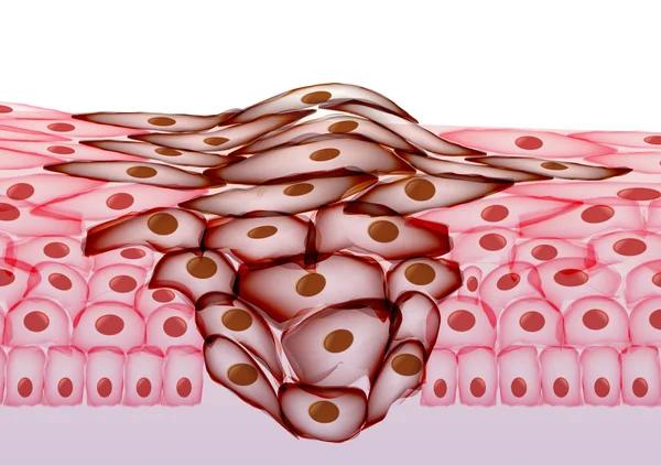
Cervical dysplasia. Cross section of a normal epithelium and Cervical Intraepithelial Neoplasia that caused of Human papillomavirus infection. Close-up of Human papillomavirus. Cervical cancer. Vector illustration
Cervical Dysplasia Cross Section Normal Epithelium Cervical Intraepithelial Neoplasia Caused Human Papillomavirus Infection Close Human Papillomavirus Cervical Cancer Vector Illustration
Structure Hemangeoma. The structure of the red mole on the skin. Birthmark. Infographics. Vector illustration on isolated background
Structure Hemangeoma Structure Red Mole Skin Birthmark Infographics Vector Illustration Isolated Background
Vector isolated illustration of malignant tumor
Cervical dysplasia. Close-up of Normal Squamous epithelial cells and Atypical Precancerous transformation of cells of the cervix. Cervical cancer. Cervical intraepithelial neoplasia. Cytology of CIN. HPV infection. vector illustration isolated on whi
Cervical Dysplasia Close Normal Squamous Epithelial Cells Atypical Precancerous Transformation Cells Cervix Cervical Cancer Cervical Intraepithelial Neoplasia Cytology Cin Hpv Infection Vector Illustration Isolated Whi
Vulvar Cancer Medical Educational Flashcard
Lymphoma icon design
Precocious puberty endocrinology color icon vector illustration
Adaptive cell change. Normal cell, hypertrophy is an increase in the size of cells, and hyperplasia -increase in the number of cells. Vector Poster for education, science, and medical use
Adaptive Cell Change Normal Cell Hypertrophy Increase Size Cells Hyperplasia Increase Number Cells Vector Poster Education Science Medical Use
Human papillomavirus. HPV-related conditions and diseases. Viral infection and associated lesions. Medical illustration. infographic diagram
Human Papillomavirus Hpv Related Conditions Diseases Viral Infection Associated Lesions Medical Illustration Infographic Diagram