Pepsina Stock Vector Images

30 Pepsina vector art & graphics are available under a royalty-free license
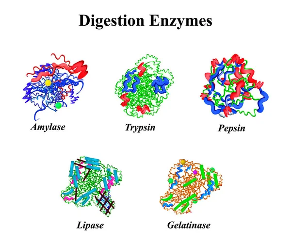
Digestion enzymes set. Chemical molecular formula. Amylase, Trypsin, Gelatinase, Pepsin, Lipase. Infographics. Vector illustration on isolated background.
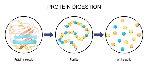
Protein digestion. Protein metabolism. Digestion in the gastrointestinal tract. Infographics. Vector illustration on isolated background.
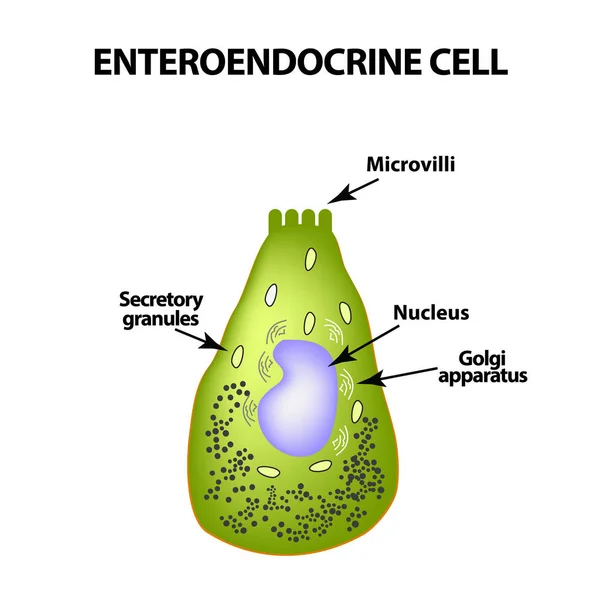
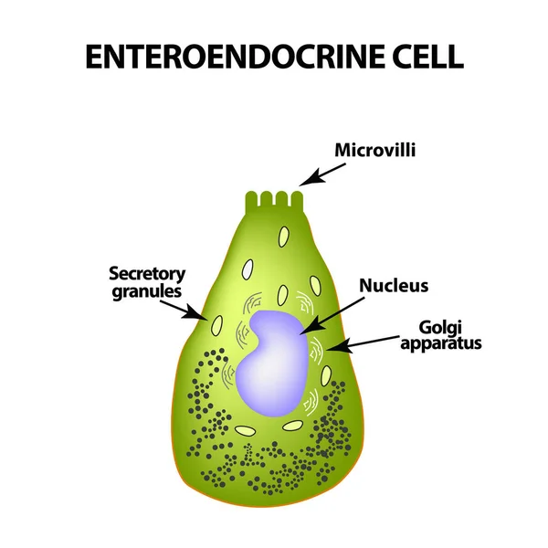
Enteroendocrine cell. Cell of the intestines. Vector illustration on isolated background
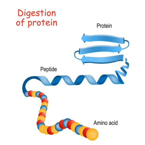
Structure of Protein from amino acid to peptide, and protein. Cl
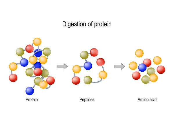
Digestion of Protein. Enzymes (proteases and peptidases), peptid
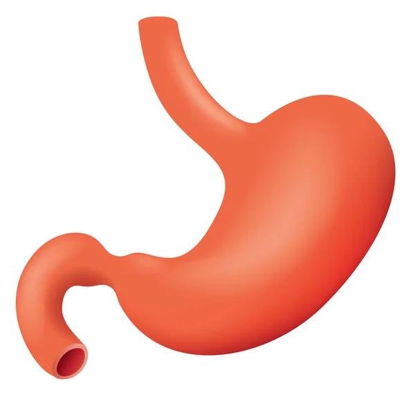
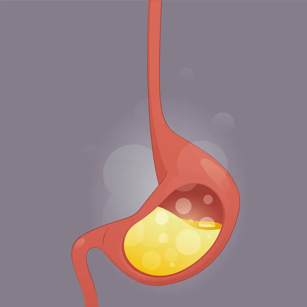
Illustration representing human stomach stomach of the digestive system, anatomy. Ideal for medical and educational materials
Gastric chief cell. Gastric gland cell that releases pepsinogen and gastric lipase. Cell responsible for secretion of chymosin in ruminant animals.
Gastric Chief Cell Gastric Gland Cell Releases Pepsinogen Gastric Lipase Cell Responsible Secretion Chymosin Ruminant Animals
Digestion of protein. Breaking the complex molecule first into peptides then into individual amino acids. The pepsins are enzymes secreted by the stomach that breaks down proteins. Vector illustration
Digestion Protein Breaking Complex Molecule First Peptides Individual Amino Acids Pepsins Enzymes Secreted Stomach Breaks Proteins Vector Illustration
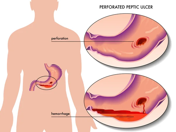
Perforated peptic ulcer
Protein digestion. Enzymes proteases are digestion breaks the protein into single amino acids, which are absorbed into the blood. Vector illustration
Protein Digestion Enzymes Proteases Digestion Breaks Protein Single Amino Acids Which Absorbed Blood Vector Illustration
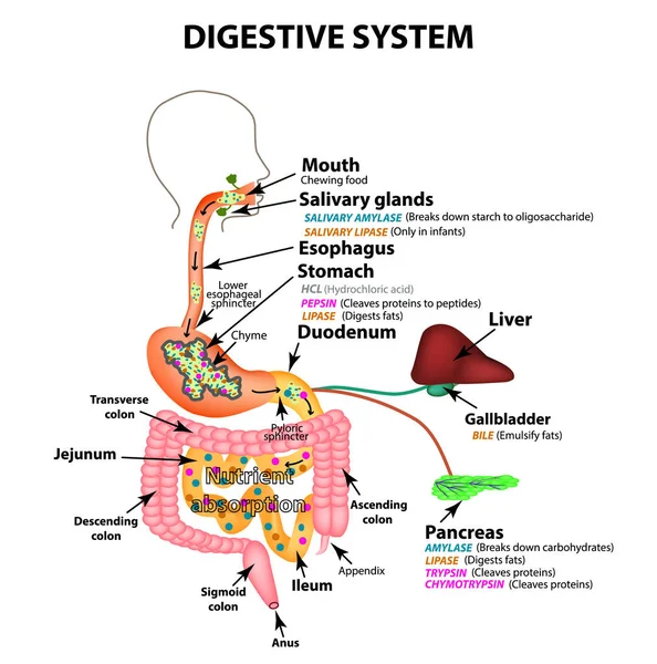
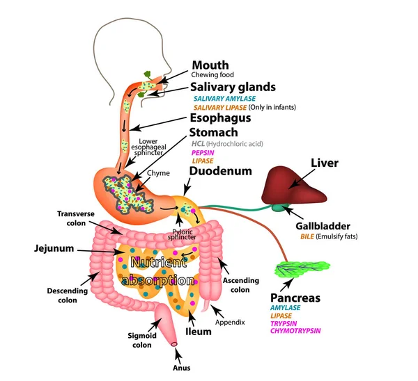
The human digestive system. Anatomical structure. Digestion of carbohydrates, fats and proteins. Enzymes of the gastrointestinal tract, pancreas, liver, gallbladder. Metabolism. Infographics. Vector.
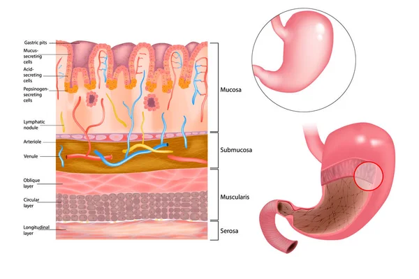
Stomach wall anatomy
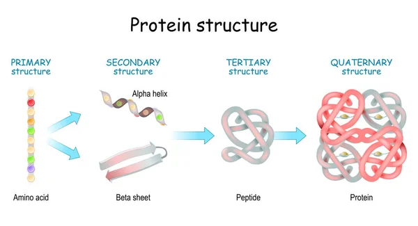
Protein structure levels: Primary, Secondary, Tertiary, and Quaternary. From Amino acid to Alpha helix, Beta sheet, peptide, and protein molecule. concept. Vector illustration.
Protein Structure Levels Primary Secondary Tertiary Quaternary Amino Acid Alpha Helix Beta Sheet Peptide Protein Molecule Concept Vector Illustration
Motilin hormone and internal organs that reaction on polypeptide hormone. Gastrointestinal motility and Intestinal contractions. Migrating motor complex. Human Digestive system. Endocrine system. Vector illustration
Motilin Hormone Internal Organs Reaction Polypeptide Hormone Gastrointestinal Motility Intestinal Contractions Migrating Motor Complex Human Digestive System Endocrine System Vector Illustration
Protein digestion. Protein metabolism. Digestion in the gastrointestinal tract. Infographics. Vector illustration on isolated background.
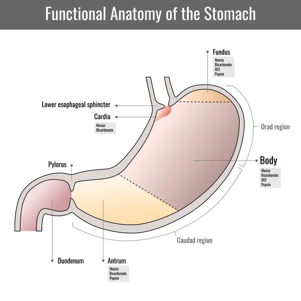
Functional anatomy of the human stomach, internal digestive organ. Parts of the stomach. Stomach wall on white background. Structure and function of Stomach Anatomy system
Functional Anatomy Human Stomach Internal Digestive Organ Parts Stomach Stomach Wall White Background Structure Function Stomach Anatomy System
Diagram of the alkaline mucous layer in the stomach. Diagram of the histological cross-section of the stomach Layers of the Stomach.
Diagram Alkaline Mucous Layer Stomach Diagram Histological Cross Section Stomach Layers Stomach
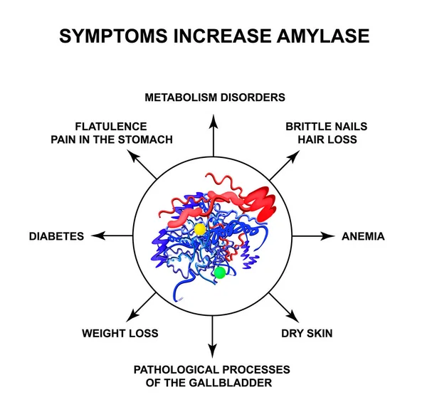
Symptoms of increased amylase. The enzyme amylase. Infographics. Vector illustration on isolated background.
The human digestive system. Anatomical structure. Digestion of carbohydrates, fats and proteins. Enzymes of the gastrointestinal tract, pancreas, liver, gallbladder. Metabolism. Infographics. Vector.
Molecules, bold line icons. The illustrations are a vector, editable stroke, 48x48 pixel perfect files. Crafted with precision and eye for quality.
Molecules Bold Line Icons Illustrations Vector Editable Stroke 48X48 Pixel Perfect Files Crafted Precision Eye Quality
Three black and white icons representing human stomach with different levels of fullness, useful for explaining digestion process
Three Black White Icons Representing Human Stomach Different Levels Fullness Useful Explaining Digestion Process
Protein structure. Amino acids, Alpha helix, Polypeptide chains, and Complex of protein molecule. Protein is a polymer (polypeptide) that formed from sequences of amino acids. Isometric Flat vector illustration.
Protein Structure Amino Acids Alpha Helix Polypeptide Chains Complex Protein Molecule Protein Polymer Polypeptide Formed Sequences Amino Acids Isometric Flat Vector Illustration
Gastric acid anatomy. Cross section of human stomach and molecule HCl hydrochloric acid. Gastric juice. Vector illustration.
Gastric Acid Anatomy Cross Section Human Stomach Molecule Hcl Hydrochloric Acid Gastric Juice Vector Illustration
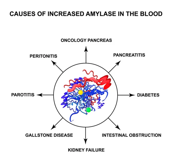
Causes of increased amylase in the blood. The enzyme amylase. Infographics. Vector illustration on isolated background
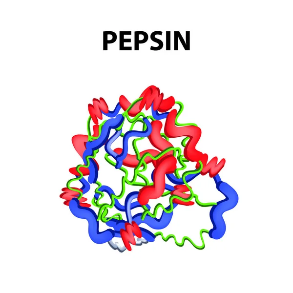
Pepsin is a molecular chemical formula. Enzyme of the stomach. Infographics. Vector illustration on an isolated background.
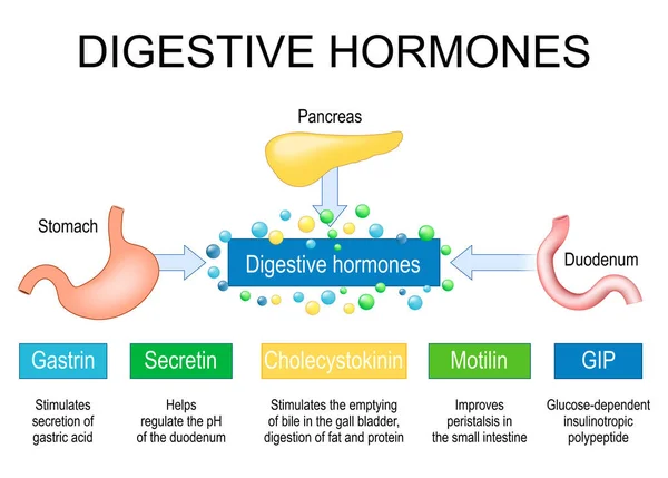
Digestive hormones. Human internal organs Pancreas, Stomach and Duodenum like part of endocrine system. Gastrointestinal hormones gastrin, cholecystokinin, secretin, Gastric inhibitory peptide and motilin. Vector illustration
Digestive Hormones Human Internal Organs Pancreas Stomach Duodenum Part Endocrine System Gastrointestinal Hormones Gastrin Cholecystokinin Secretin Gastric Inhibitory Peptide Motilin Vector Illustration
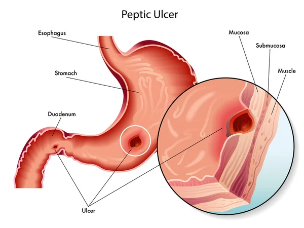
Peptic ulcer scheme
Peptic ulcer scheme
Acid reflux
Acid reflux
Protein Digestion. Enzymes proteases and peptidases are digestion breaks the protein into smaller peptide chains and into single amino acids, which are absorbed into the blood. Vector illustration
Protein Digestion Enzymes Proteases Peptidases Digestion Breaks Protein Smaller Peptide Chains Single Amino Acids Which Absorbed Blood Vector Illustration
Enteroendocrine cell. Cell of the intestines. Vector illustration on isolated background