Temporal bone Stock Vector Images

81 Temporal bone vector art & graphics are available under a royalty-free license
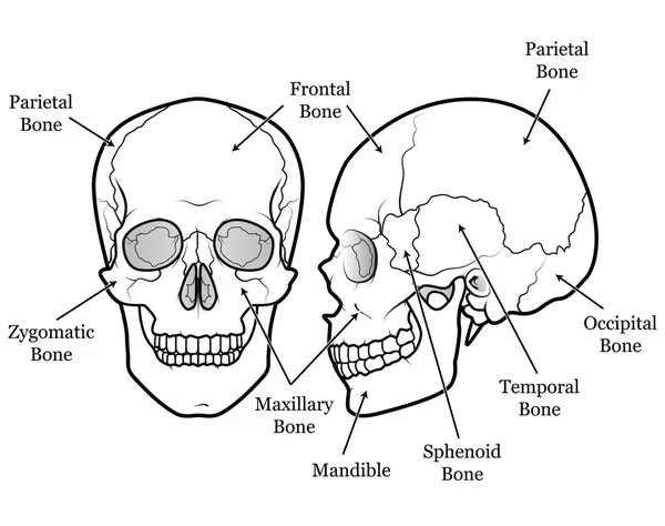
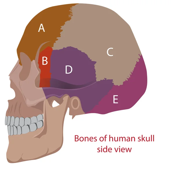
Human Skull Diagram Illustration
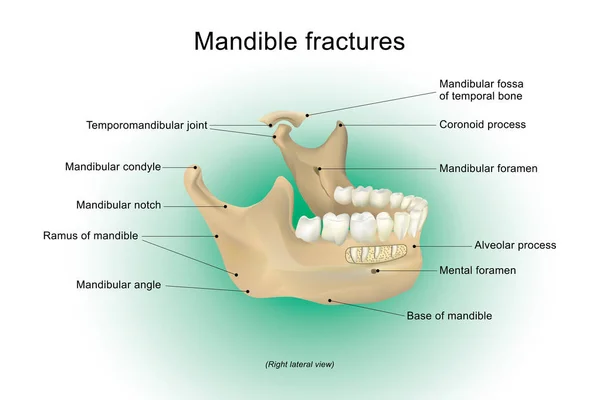
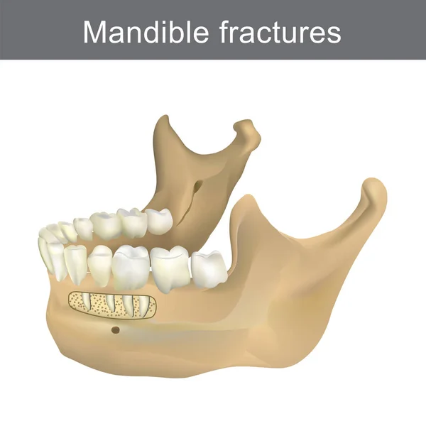
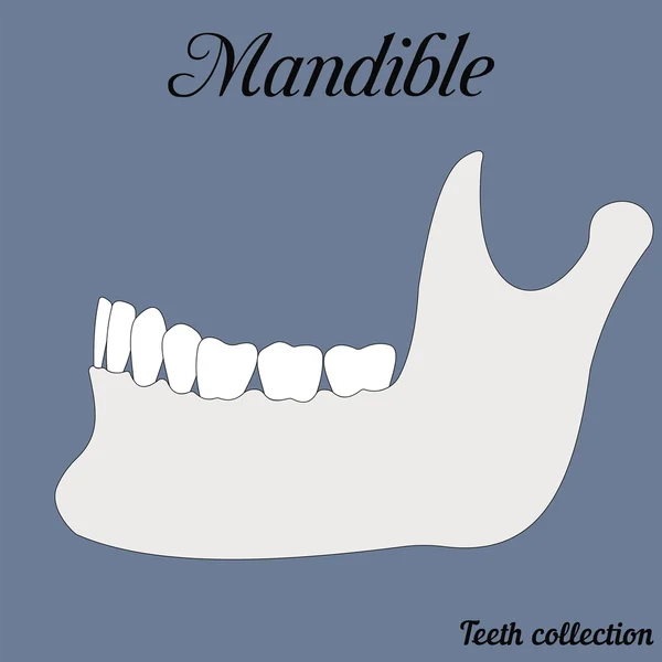
Mandible fractures. Anatomy Vector design, Illustration.
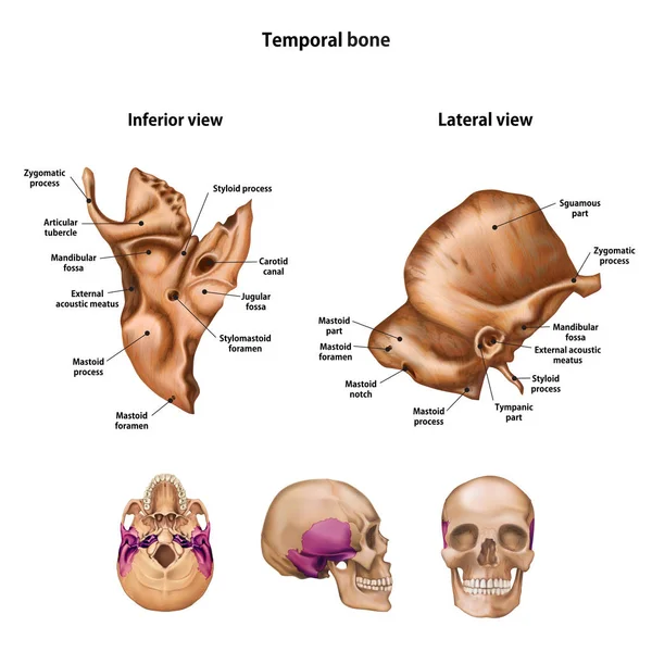
Temporal bone. With the name and description of all sites.
Temporal Bone Name Description All Sites
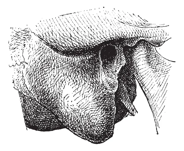
Human Mastoid Process, vintage engraving
Skull Chart
Skull Chart
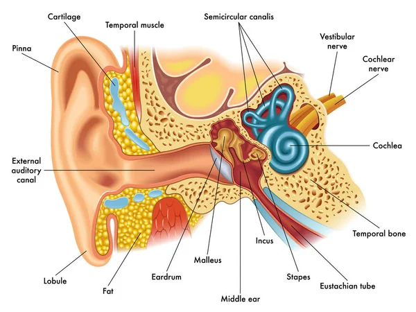
Ear anatomylustration of ear anatomy
Illustration of skull sutures diagram
Illustration Skull Sutures Diagram
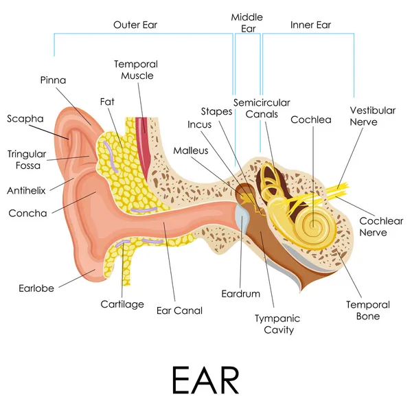
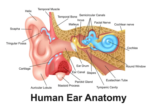
Human Ear Anatomy
Human Ear Anatomy
Mandible fractures is the largest, strongest and lowest bone in
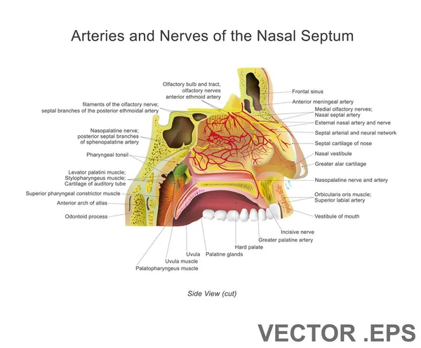
Arteries and Nerves of the Nasal Septum. Anatomy Vector design, Illustration.
Human vs Chimpanzee Skull Anatomy Comparison Diagram
Human Chimpanzee Skull Anatomy Comparison Diagram
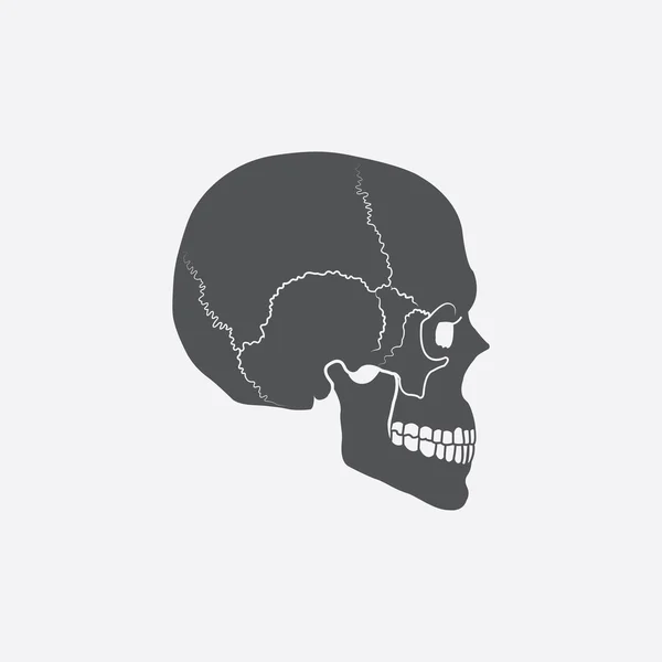
Side view of Human Skull
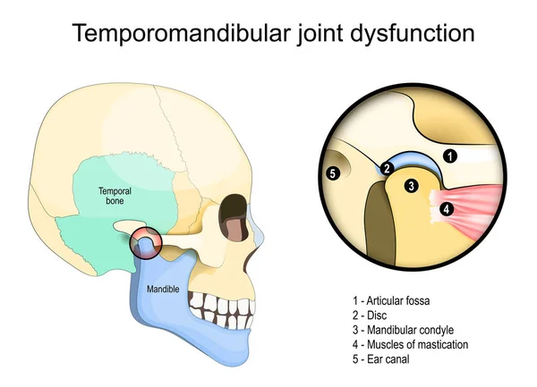
Temporomandibular joint dysfunction syndrome. Side view of a Human skull with Temporal bone. Close-up of Muscles of mastication, and joint that connect the mandible to the skull. Vector illustration
Temporomandibular Joint Dysfunction Syndrome Side View Human Skull Temporal Bone Close Muscles Mastication Joint Connect Mandible Skull Vector Illustration
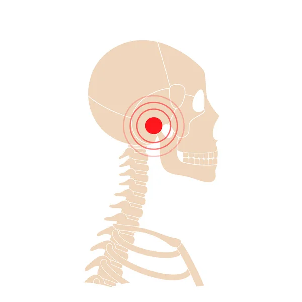
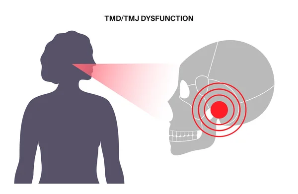
Temporomandibular joint disorder. TMD or TMJ dysfunction. Pain in the jaw joint, temporal bone locking or displaced disc. Transcutaneous electrical nerve stimulation. Human skull and mandible vector
Temporomandibular Joint Disorder Tmd Tmj Dysfunction Pain Jaw Joint Temporal Bone Locking Displaced Disc Transcutaneous Electrical Nerve Stimulation Human Skull Mandible Vector
Zygomatic bone. With the name and description of all sites.
Zygomatic Bone Name Description All Sites
Human skull bones structure diagram schematic vector illustration. Medical science educational illustration
Human Skull Bones Structure Diagram Schematic Vector Illustration Medical Science Educational Illustration
Zygomatic bone. With the name and description of all sites.
Zygomatic Bone Name Description All Sites
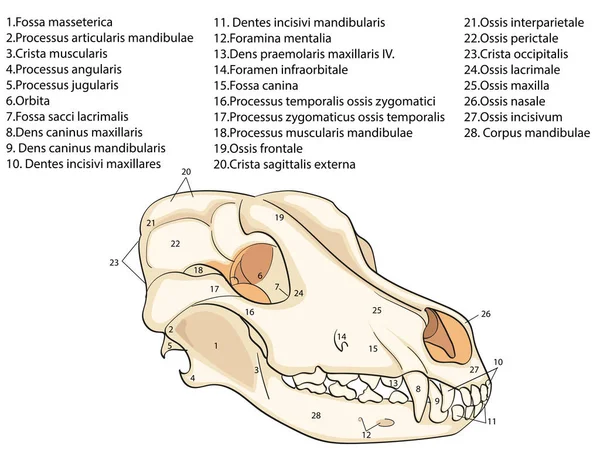
The skull of a dog. Structure of the bones of the head, anatomical design. In Latin
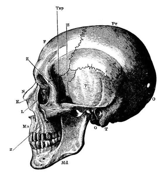
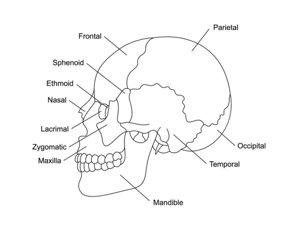
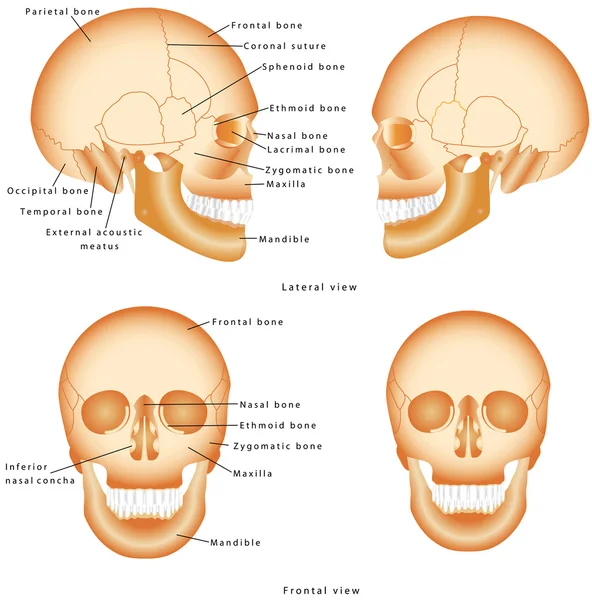
The side view of the human skull, with the parts labelled as, 'O', occipital bone; 'T', temporal bone; 'Pr', parietal bone; 'F', frontal bone; S'', sphenoid; 'Z', malar; 'Mx', maxilla; 'N', nasal; 'E', ethmoid; 'L', lachrymal; 'Md', inferior maxilla.
Side View Human Skull Parts Labelled Occipital Bone Temporal Bone Parietal Bone Frontal Bone Sphenoid Malar Maxilla Nasal Ethmoid Lachrymal Inferior Maxilla
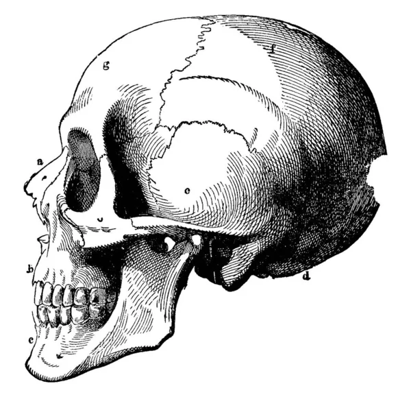
A typical representation of the human skull, with its parts labelled as a, b, c, d, e, f and g, representing, nasal bone, superior maxillary, inferior maxillary, occipital, temporal, parietal and frontal bone, respectively, vintage line drawing or en
Typical Representation Human Skull Its Parts Labelled Representing Nasal Bone Superior Maxillary Inferior Maxillary Occipital Temporal Parietal Frontal Bone Respectively Vintage Line Drawing
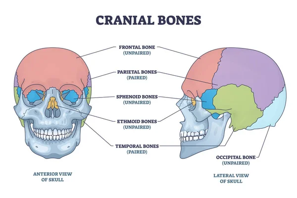
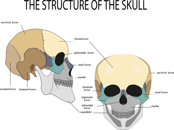
Cranial bones anatomy and skull skeleton medical division outline diagram. Labeled educational frontal, parietal, sphenoid, ethmoid and temporal bone vector illustration. Paired and unpaired examples.
Cranial Bones Anatomy Skull Skeleton Medical Division Outline Diagram Labeled Educational Frontal Parietal Sphenoid Ethmoid Temporal Bone Vector Illustration Paired Unpaired Examples
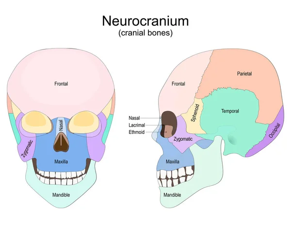
Human skull from the front and side view. Neurocranium. cranial bones. Vector poster for medical, and education use
Human Skull Front Side View Neurocranium Cranial Bones Vector Poster Medical Education Use
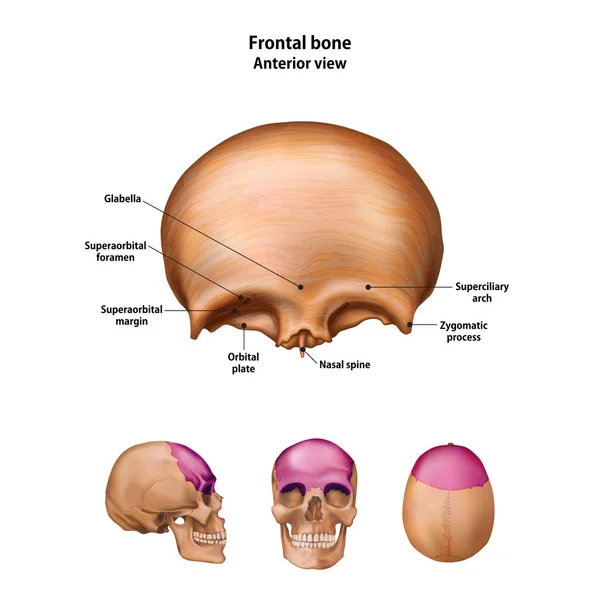
Frontal bone. With the name and description of all sites.
Frontal Bone Name Description All Sites
Human skull
Human skull
A diagram in which complete structure of human skull which is defined clearly, vintage line drawing or engraving illustration.
Diagram Which Complete Structure Human Skull Which Defined Clearly Vintage Line Drawing Engraving Illustration
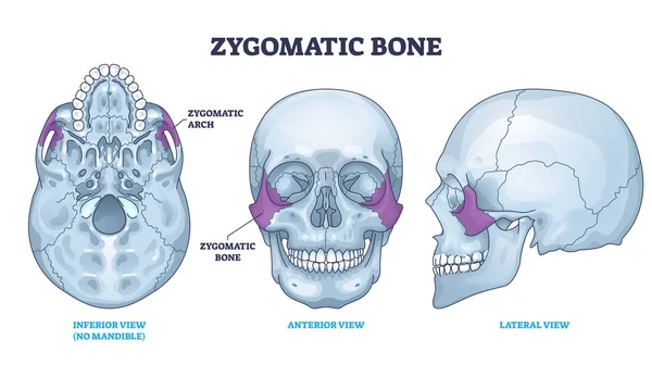
Zygomatic bone location with human skull skeleton anatomy outline diagram. Labeled educational scheme with cheekbone from inferior, anterior and lateral view vector illustration. Head parts structure.
Zygomatic Bone Location Human Skull Skeleton Anatomy Outline Diagram Labeled Educational Scheme Cheekbone Inferior Anterior Lateral View Vector Illustration Head Parts Structure
The skull of a dog. Structure of the bones of the head, anatomical design. In Latin
The temporal Bone of the human skull, situated at the sides and base of the skull, with its parts labelled, vintage line drawing or engraving illustration.
Temporal Bone Human Skull Situated Sides Base Skull Its Parts Labelled Vintage Line Drawing Engraving Illustration
Skeleton of the human head, vintage engraved illustration. La Vie dans la nature, 1890.
Skeleton Human Head Vintage Engraved Illustration Vie Dans Nature 1890
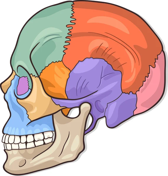
Human skull bones anatomy. Cranial colored parts structure. Outline sutures. Zygomatic, nasal, maxilla, temporal lacrimal mandible frontal parietal occipital. Brain protection. Vector illustration.
Human Skull Bones Anatomy Cranial Colored Parts Structure Outline Sutures Zygomatic Nasal Maxilla Temporal Lacrimal Mandible Frontal Parietal Occipital Brain Protection Vector Illustration
Human anatomy
Human anatomy
Human Ear Detailed Anatomy
Human skull bones anatomy with descriptions. Cranial parts structure. Human internal organ illustration. Zygomatic, nasal, maxilla, temporal, frontal head parts.
Human Skull Bones Anatomy Descriptions Cranial Parts Structure Human Internal Organ Illustration Zygomatic Nasal Maxilla Temporal Frontal Head Parts
Human skull anatomy,
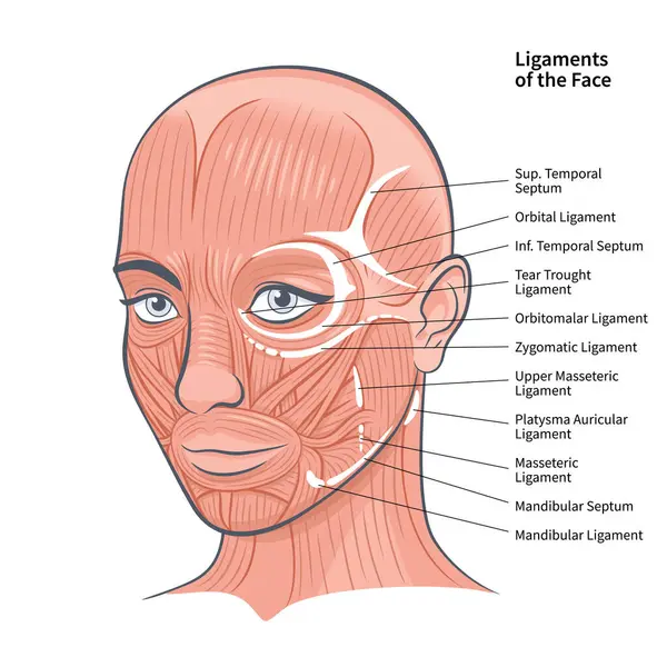
The Functional Anatomy of the Woman Face Ligaments Muscles Scheme Portrait Vector Illustration
Functional Anatomy Woman Face Ligaments Muscles Scheme Portrait Vector Illustration
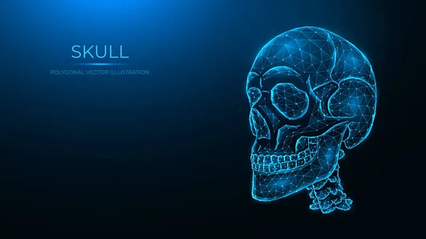
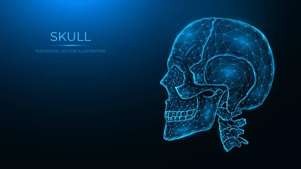
Polygonal vector illustration of a human skull, side view. Low poly anatomical model of the skull and cervical spine on dark blue background.
Polygonal Vector Illustration Human Skull Side View Low Poly Anatomical Model Skull Cervical Spine Dark Blue Background
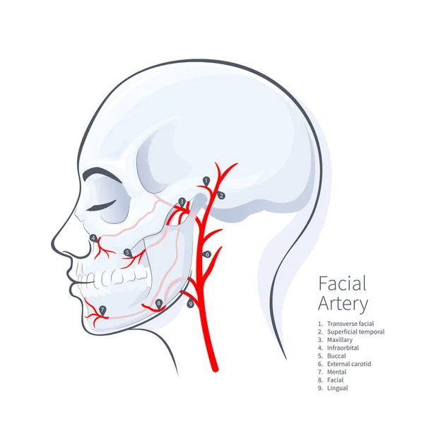
Woman head scull faceial artery scheme vector illustration on white backgound
Woman Head Scull Faceial Artery Scheme Vector Illustration White Backgound
Sutures of the skull as human head bone medical division outline diagram. Labeled educational anatomical scheme with squamous, coronal, lamboid, sagittal and occipitomastoid parts vector illustration.
Sutures Skull Human Head Bone Medical Division Outline Diagram Labeled Educational Anatomical Scheme Squamous Coronal Lamboid Sagittal Occipitomastoid Parts Vector Illustration
Polygonal vector illustration of a human skull, side view. Anatomical model of the skull and cervical spine on dark blue background.
Polygonal Vector Illustration Human Skull Side View Anatomical Model Skull Cervical Spine Dark Blue Background
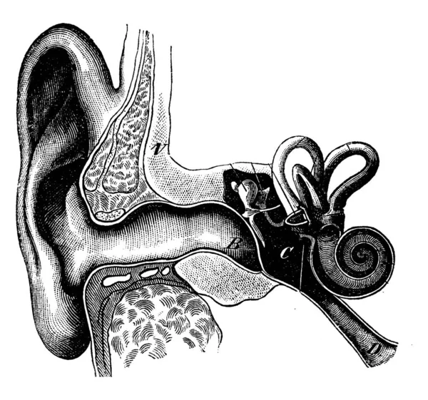
Section showing the organization of the ear, vintage engraved illustration. La Vie dans la nature, 1890.
Section Showing Organization Ear Vintage Engraved Illustration Vie Dans Nature 1890
Human skull low poly art. Polygonal skull on a white background.
Human Skull Low Poly Art Polygonal Skull White Background
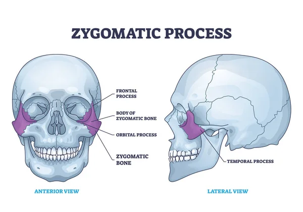
Zygomatic process as human cheek bone skeleton anatomy outline diagram. Labeled educational cheekbone location and skull parts structure vector illustration. Frontal, orbital and temporal process.
Zygomatic Process Human Cheek Bone Skeleton Anatomy Outline Diagram Labeled Educational Cheekbone Location Skull Parts Structure Vector Illustration Frontal Orbital Temporal Process
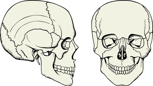
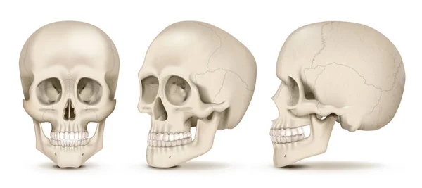
Realistic human skulls front and side views set isolated on white background vector illustration
Realistic Human Skulls Front Side Views Set Isolated White Background Vector Illustration
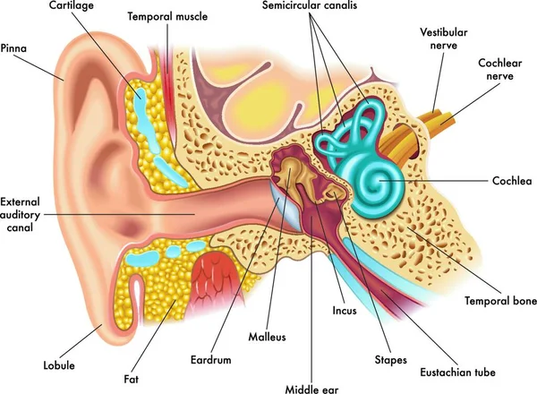
Medical illustration of ear anatomy
Medical Illustration Ear Anatomy
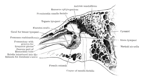
The section through the left temporal bone, showing inner wall of tympanic cavity, with its parts labeled, vintage line drawing or engraving illustration.
Section Left Temporal Bone Showing Inner Wall Tympanic Cavity Its Parts Labeled Vintage Line Drawing Engraving Illustration
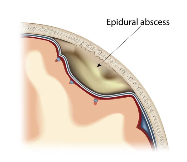
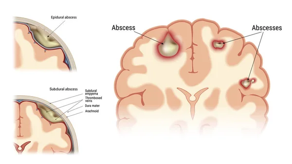
Epidural abscess vector
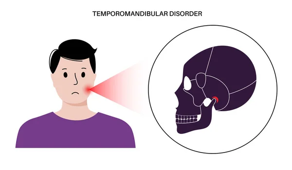
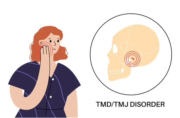
Temporomandibular joint disorder. TMD or TMJ dysfunction. Pain in the jaw joint, temporal bone locking or displaced disc. Transcutaneous electrical nerve stimulation. Human skull and mandible vector
Temporomandibular Joint Disorder Tmd Tmj Dysfunction Pain Jaw Joint Temporal Bone Locking Displaced Disc Transcutaneous Electrical Nerve Stimulation Human Skull Mandible Vector
TMJ disorder vector illustration. Labeled jaw condition educational scheme.
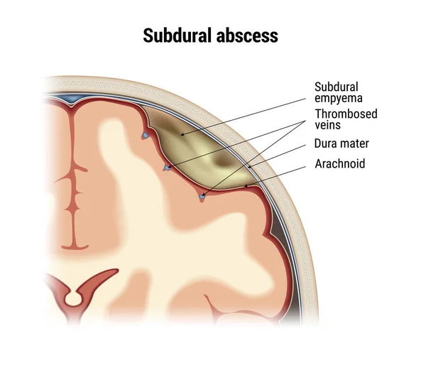
Subdural abscess vector
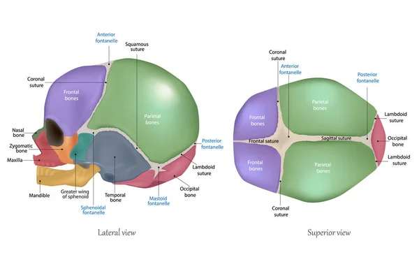
Anatomy of the Newborn Skull. Cranial sutures and Fontanelle. Lateral and superior view
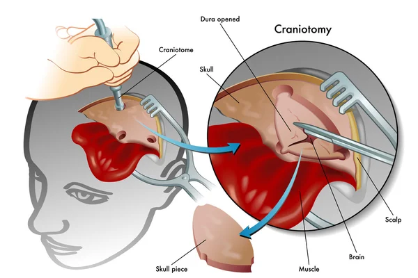
Medical illustration showing a surgical craniotomy
Jugular foramen anatomy shows skull base passage, internal jugular vein, glossopharyngeal and vagus nerves clarify pars nervosa vs vascularis compartments. Outline diagram
Jugular Foramen Anatomy Shows Skull Base Passage Internal Jugular Vein Glossopharyngeal Vagus Nerves Clarify Pars Nervosa Vascularis Compartments Outline Diagram
Vector illustration of cat skull anatomy. veterinary science
Vector Illustration Cat Skull Anatomy Veterinary Science
Pain, injury or inflammation of the skull bones polygonal vector illustration. Low poly model of a human skull
Pain Injury Inflammation Skull Bones Polygonal Vector Illustration Low Poly Model Human Skull
Human anatomy
Human anatomy
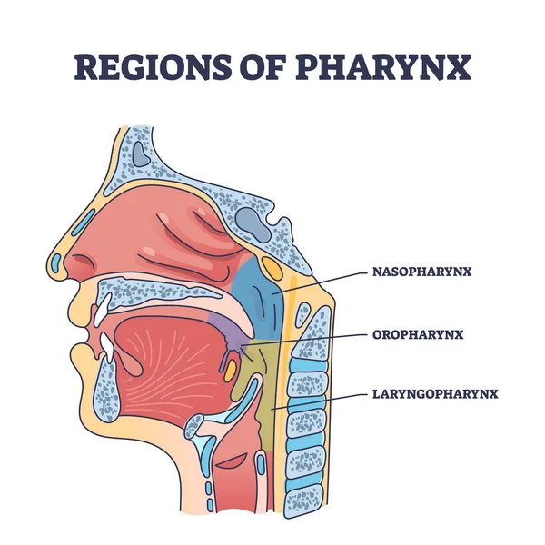
Regions of pharynx and throat parts division from cavity side view outline diagram. Labeled educational scheme with nasopharynx, oropharynx and laryngopharynx location anatomy vector illustration.
Regions Pharynx Throat Parts Division Cavity Side View Outline Diagram Labeled Educational Scheme Nasopharynx Oropharynx Laryngopharynx Location Anatomy Vector Illustration
Zygomatic acrh with skull cheekbone skeletal parts anatomy outline diagram. Labeled educational medical scheme with temporal process of bone location from inferior and lateral view vector illustration
Zygomatic Acrh Skull Cheekbone Skeletal Parts Anatomy Outline Diagram Labeled Educational Medical Scheme Temporal Process Bone Location Inferior Lateral View Vector Illustration
Human Skull structure
Human head bones
Human head bones
Human skull anatomy.
Skull icon of vector illustration for web and mobile
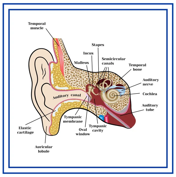
The structure of the human ear.
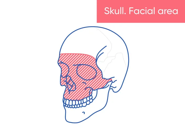
Human skull anatomy - facial area vector illusttation flat medicine poster
Pharynx or throat anatomical and medical oral structure outline diagram. Labeled educational detailed description with tongue, epiglottis, esophagus, cavity and palatini body parts vector illustration
Pharynx Throat Anatomical Medical Oral Structure Outline Diagram Labeled Educational Detailed Description Tongue Epiglottis Esophagus Cavity Palatini Body Parts Vector Illustration
Head bones
Head bones
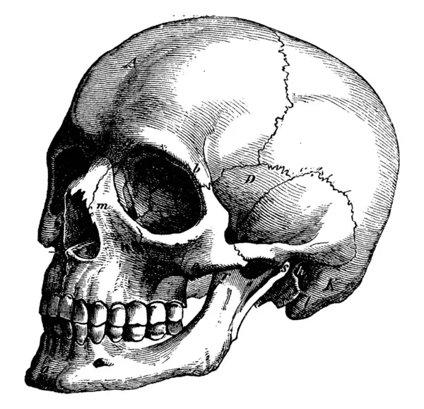
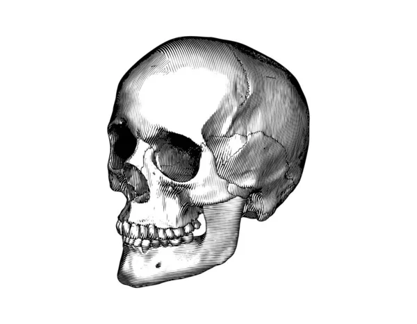
Monochrome black vintage engraved drawing human skull close jaw perspective side view vector biology illustration isolated on white background
Monochrome Black Vintage Engraved Drawing Human Skull Close Jaw Perspective Side View Vector Biology Illustration Isolated White Background
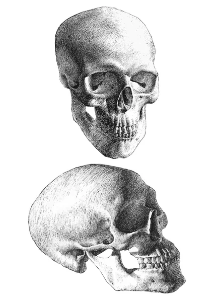
Illustration with two anatomical skulls
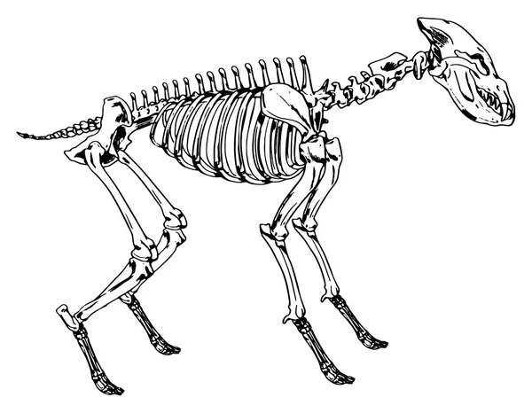
Skeleton of a hyena
Skeleton of a hyena
Bones of head
Bones of head
Human craniotomy scheme
Closure of teeth
Closure of teeth
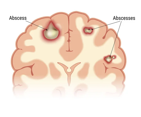
Intracerebral abscess vector
Polygonal vector illustration of human skulls, front, side, and back views. Set of low poly models of skulls with cervical spine isolated on dark blue background. Head bone anatomy concept art.
Polygonal Vector Illustration Human Skulls Front Side Back Views Set Low Poly Models Skulls Cervical Spine Isolated Dark Blue Background Head Bone Anatomy Concept Art
Abscess set vector
Abscess set vector
Temporomandibular joint disorder. TMD or TMJ dysfunction.Consultation with doctor. Pain in the jaw joint, temporal bone locking. Transcutaneous electrical nerve stimulation. Human skull flat vector
Temporomandibular Joint Disorder Tmd Tmj Dysfunction Consultation Doctor Pain Jaw Joint Temporal Bone Locking Transcutaneous Electrical Nerve Stimulation Human Skull Flat Vector
Temporomandibular joint disorder. TMD or TMJ dysfunction. Pain in the jaw joint, temporal bone locking or displaced disc. Transcutaneous electrical nerve stimulation. Human skull and mandible vector
Temporomandibular Joint Disorder Tmd Tmj Dysfunction Pain Jaw Joint Temporal Bone Locking Displaced Disc Transcutaneous Electrical Nerve Stimulation Human Skull Mandible Vector
Temporomandibular joint disorder. TMD or TMJ dysfunction. Pain in the jaw joint, temporal bone locking or displaced disc. Transcutaneous electrical nerve stimulation. Human skull and mandible vector
Temporomandibular Joint Disorder Tmd Tmj Dysfunction Pain Jaw Joint Temporal Bone Locking Displaced Disc Transcutaneous Electrical Nerve Stimulation Human Skull Mandible Vector
Temporomandibular joint disorder. TMD or TMJ dysfunction.Consultation with doctor online. Pain in the jaw joint, temporal bone locking. Transcutaneous electrical nerve stimulation. Human skull vector
Temporomandibular Joint Disorder Tmd Tmj Dysfunction Consultation Doctor Online Pain Jaw Joint Temporal Bone Locking Transcutaneous Electrical Nerve Stimulation Human Skull Vector
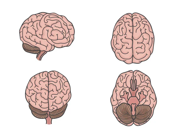
Anatomy of brain
Anatomy of brain
Temporomandibular joint disorder. TMD or TMJ dysfunction. Pain in the jaw joint, temporal bone locking or displaced disc. Transcutaneous electrical nerve stimulation. Human skull and mandible vector
Temporomandibular Joint Disorder Tmd Tmj Dysfunction Pain Jaw Joint Temporal Bone Locking Displaced Disc Transcutaneous Electrical Nerve Stimulation Human Skull Mandible Vector
Human brain
Human brain