Trocantere Stock Vector Images

32 Trocantere vector art & graphics are available under a royalty-free license
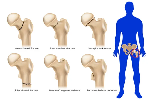
Femoral neck fracture. Types of hip fractures. Subtrochanteric, Intertrochanteric, Transcervical and Subcapital neck fracture, Fracture of the greater and lesser trochanter.
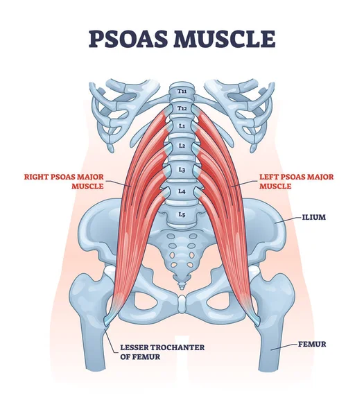
Psoas muscle as deep body muscular system for spine health outline diagram
Quadratus lumborum muscle or QL for strong and healthy spine outline diagram
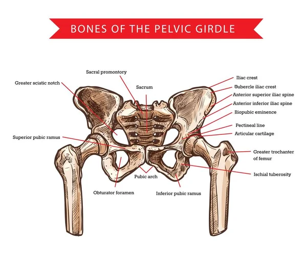
Pelvis bones of pelvic girdle, vector sketch of human anatomy and medicine. Bones and joints structure of skeleton hips, sacrum, femur and coccyx, sacral promontory, pubic arch and iliac spine
Pelvis Bones Pelvic Girdle Vector Sketch Human Anatomy Medicine Bones Joints Structure Skeleton Hips Sacrum Femur Coccyx Sacral Promontory Pubic Arch Iliac Spine
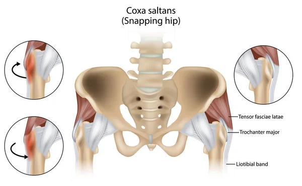
Coxa Saltans or Snapping Hip Snapping Hip Syndrome also referred to as dancer hip. Trochanter major, Tensor fasciae latae and Liotibial band
Coxa Saltans Snapping Hip Snapping Hip Syndrome Also Referred Dancer Hip Trochanter Major Tensor Fasciae Latae Liotibial Band
Bursitis inflammation concept
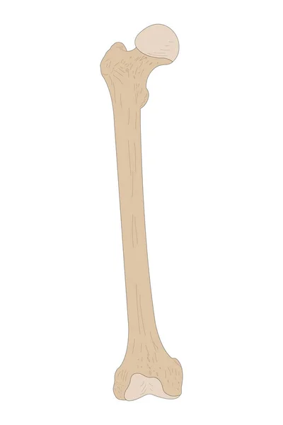
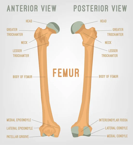
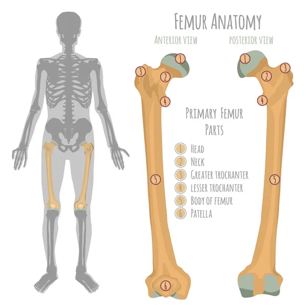
Right Femur. Anterior (ventral) view.
Right Femur Anterior Ventral View
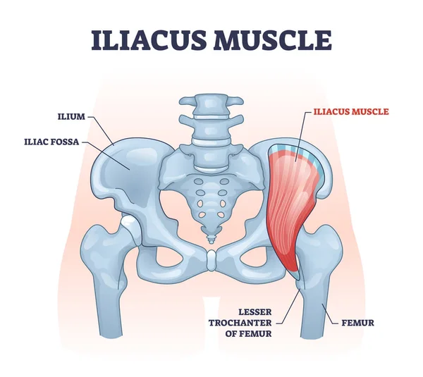
Iliacus muscle with hip or groin muscular, skeletal anatomy outline diagram
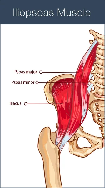
Vector illustration of a iliopsoas muscle
Vector Illustration Iliopsoas Muscle
Hip labrum tear, Hip joint medical illustration flashcard
Hip Labrum Tear Hip Joint Medical Illustration Flashcard
Gluteus medius muscle with human hip and groin anatomy outline diagram
Iliopsoas biomechanics contrasts relaxed vs active contraction, showing the psoas-iliacus powering hip flexion, key objects, iliopsoas muscle, pelvis, lumbar spine. Outline diagram
Iliopsoas Biomechanics Contrasts Relaxed Active Contraction Showing Psoas Iliacus Powering Hip Flexion Key Objects Iliopsoas Muscle Pelvis Lumbar Spine Outline Diagram
Ants
Ants
A praying mantis
A praying mantis
Human femur bones
Human femur bones
Gluteus medius trigger points map referred pain patterns across hip and pelvis, showing muscle fibers, X-marked TrPs, and shaded pain zones for clinical guidance. Outline diagram
Gluteus Medius Trigger Points Map Referred Pain Patterns Hip Pelvis Showing Muscle Fibers Marked Trps Shaded Pain Zones Clinical Guidance Outline Diagram
Gluteal tendinopathy anatomy compares healthy and pathological hip, highlighting gluteus medius, tendon insertion, and IT band with pain referral area. Outline diagram
Gluteal Tendinopathy Anatomy Compares Healthy Pathological Hip Highlighting Gluteus Medius Tendon Insertion Band Pain Referral Area Outline Diagram
The Ant
The Ant
Gluteus minimus muscle with hips muscular system and bones outline concept
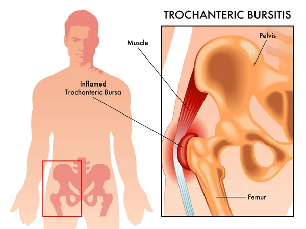
Trochanteric bursitis as bursa inflammation located in hip outline diagram
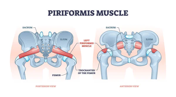
Piriformis muscle with hip skeleton and muscular system outline diagram
Male hip bone anatomy
Trochanteric bursitis vector illustration on white background
Trochanteric Bursitis Vector Illustration White Background
Snapping hip extra-articular mechanics, iliopsoas tendon and IT band snap over pelvic bone, focus on iliopsoas, iliotibial band, greater trochanter. Outline diagram
Snapping Hip Extra Articular Mechanics Iliopsoas Tendon Band Snap Pelvic Bone Focus Iliopsoas Iliotibial Band Greater Trochanter Outline Diagram
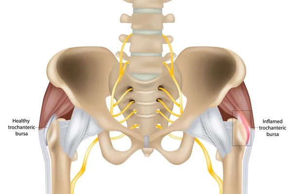
Hip Trochanteric bursitis is inflammation of the bursa. Illustration of the Healthy and inflamed trochanteric bursa. Greater trochanteric pain syndrome.
Hip Trochanteric Bursitis Inflammation Bursa Illustration Healthy Inflamed Trochanteric Bursa Greater Trochanteric Pain Syndrome
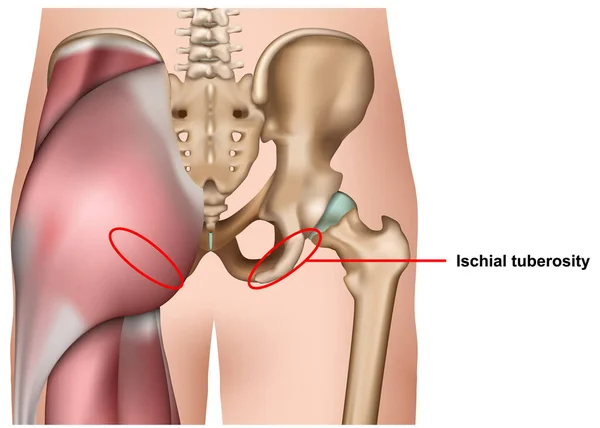
Ischial tuberosity 3d medical vector illustration isolated on white background infographic
Ischial Tuberosity Medical Vector Illustration Isolated White Background Infographic
Piriformis trigger points and referred pain mapped on the posterior pelvis, showing piriformis muscle, sciatic nerve, and hip region zones. Outline diagram
Piriformis Trigger Points Referred Pain Mapped Posterior Pelvis Showing Piriformis Muscle Sciatic Nerve Hip Region Zones Outline Diagram
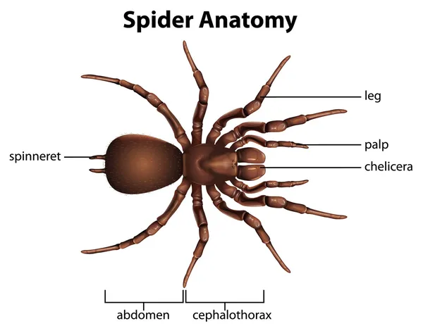
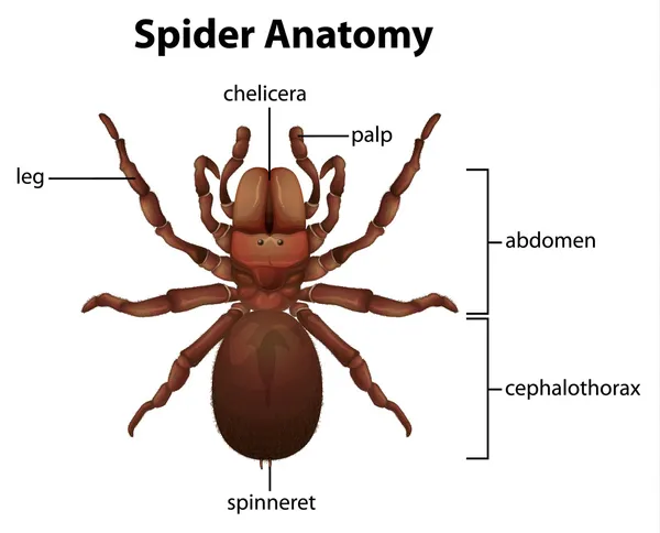
Anatomy of a spider
Anatomy of a spider
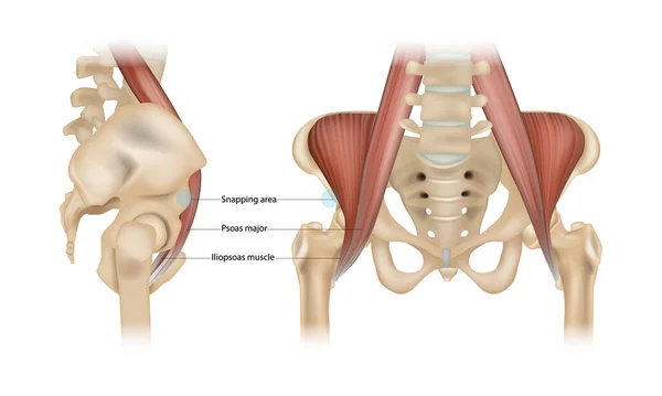
Internal snapping hip Syndrome. Psoas major, Iliopsoas muscle and Snapping area. Vector
Internal Snapping Hip Syndrome Psoas Major Iliopsoas Muscle Snapping Area Vector
Hip flexor anatomy primary vs secondary shows labeled muscles for psoas major, iliacus, and rectus femoris on the pelvis, clarifying function and grouping. Outline diagram
Hip Flexor Anatomy Primary Secondary Shows Labeled Muscles Psoas Major Iliacus Rectus Femoris Pelvis Clarifying Function Grouping Outline Diagram
Spider anatomy
Spider anatomy
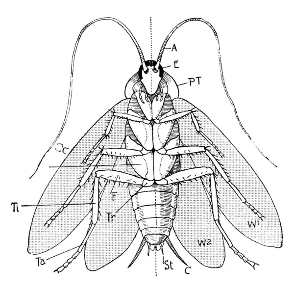
Labels: A, antennae; E, eye; P.T, prothorax; W1, first pair of wings; W2, second pair of wings; C, cercus; St, style; Co, coxa; Tr, trochanter; and other, vintage line drawing or engraving illustration.
Labels Antennae Eye Prothorax First Pair Wings Second Pair Wings Cercus Style Coxa Trochanter Other Vintage Line Drawing Engraving Illustration