Current page requires JavaScript, this web browser either does not support JavaScript, or scripts are being blocked.
Please turn on JavaScript or use different browser.
Photos
Vectors
Illustrations
Free Pictures
Videos
Music & SFX
AI Assistant
AI Image Generator
Free Background Remover
Free Image Upscaler
Reverse Image Search
AI training data
Pricing
Illustrations
Log In
Sign Up
Beine knochen Stock Illustrations
31,982,171 Beine knochen illustrations, Beine knochen clipart illustrations & paintings
Create with AI
Watercolor beine knochen
Landscape
Happiness
Nature
Lifestyle
Science
Animals
Business
Food
Architecture
Sport
Best Match
Fresh
Popular
Concept of orthopedic medical technology, graphic of a man running with skeleton x-ray scan
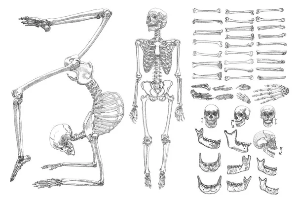
Human bones skeleton collection set
Feet
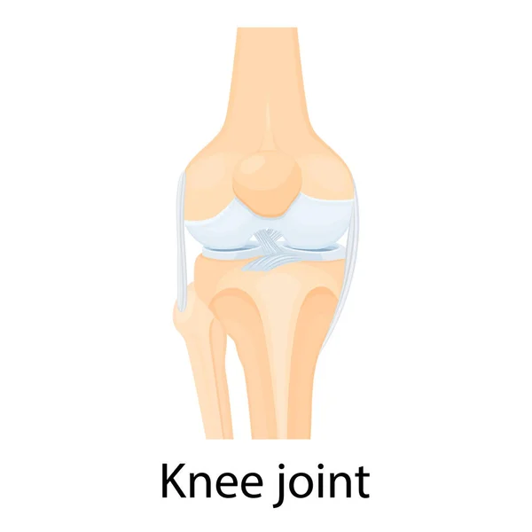
Knee joint anatomy isolated on white
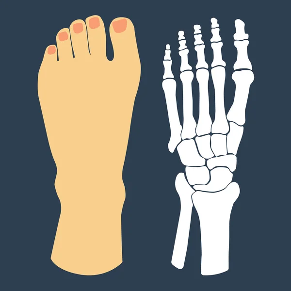
The flat design of the foot and the foot skeleton. Vector illustration.
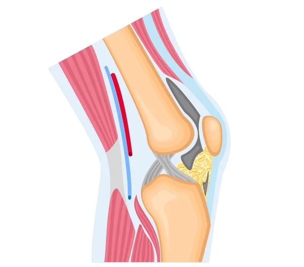
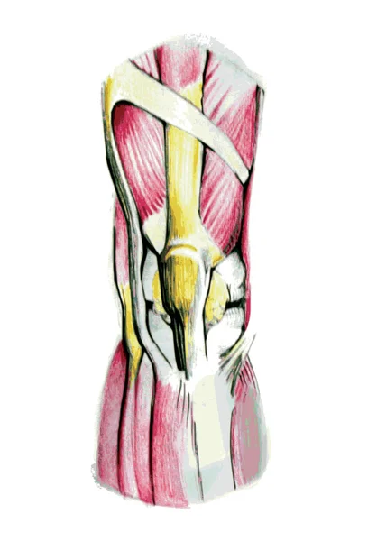
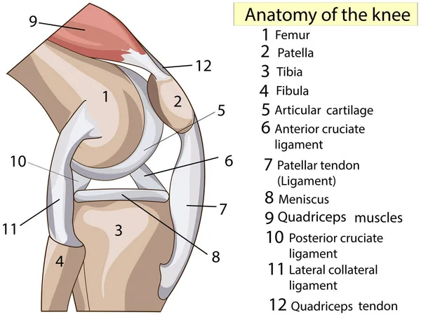
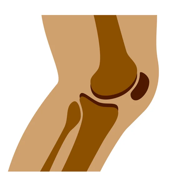
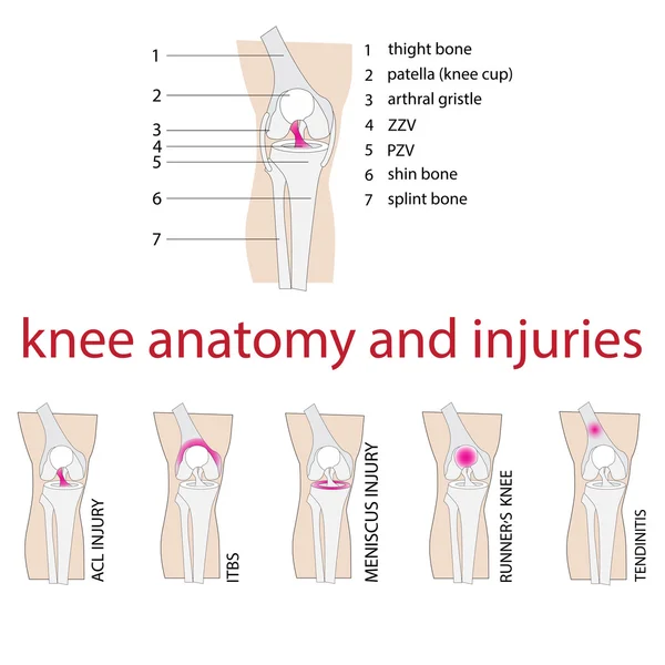
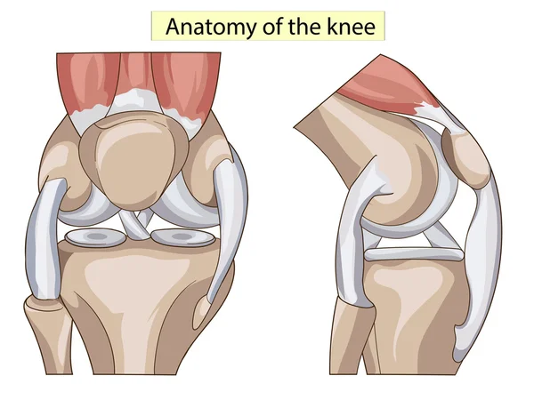
Simplified anatomy of the knee
Cartoon illustration of a businessman who specializes in reusing license plates
Temporarily disabled sad girl with broken leg with bandage is sitting.Crutches are around. Unhappy injured young woman. Ankle bone fracture or sprain with gypsum.Vector illustration in trendy colors
First aid.Arm and leg bone fractures with gypsum or orthopedic cast. Temporarily disabled sad child with broken limbs with bandage. Nurse is working.Unhappy injured girl is sitting on medical couch.
Set of three young women with broken leg.Girl with orthopedic cast walking using crutches.Ankle bone fracture or sprain with gypsum.Unhappy injured lady with bandage is sitting.Trendy vector poster
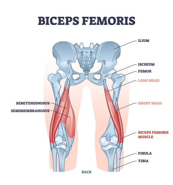
Biceps femoris muscle with human leg and thigh structure outline diagram
Human knee joint
Cartoon old people fracture
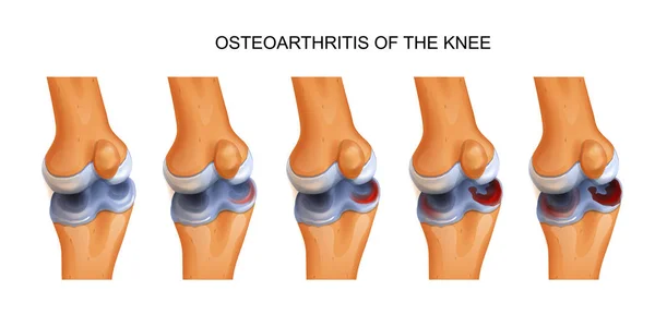
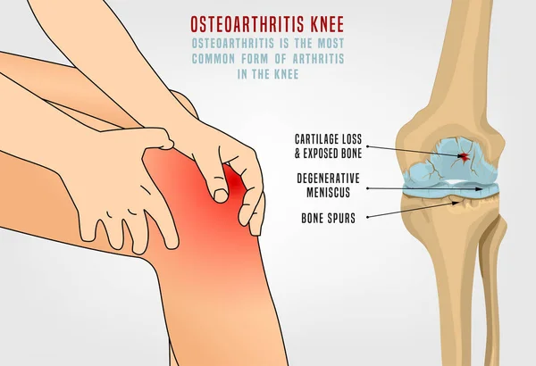
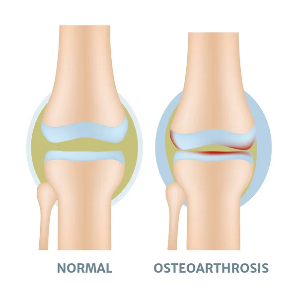
Vector illustration of osteoarthritis of the knee
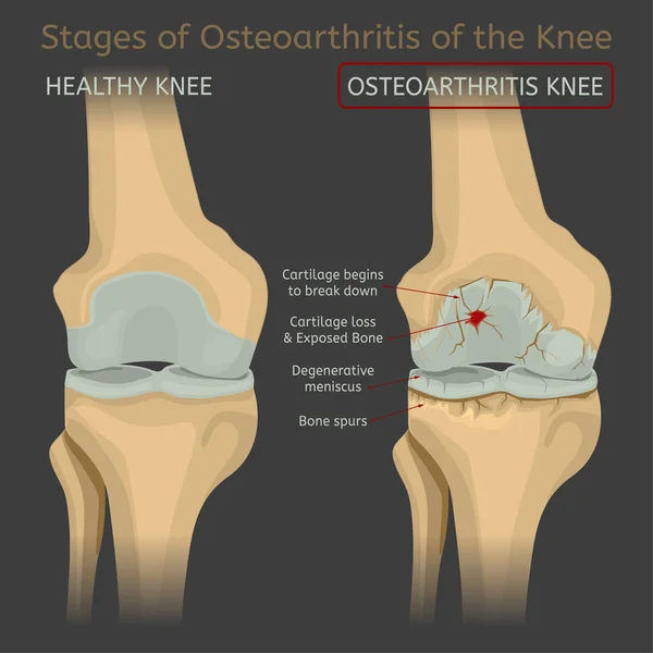
Stages of osteoarthritis of the knee
Flexor hallucis longus muscle with human leg and foot bones outline diagram
Knee Pain vector logo icon illustrator
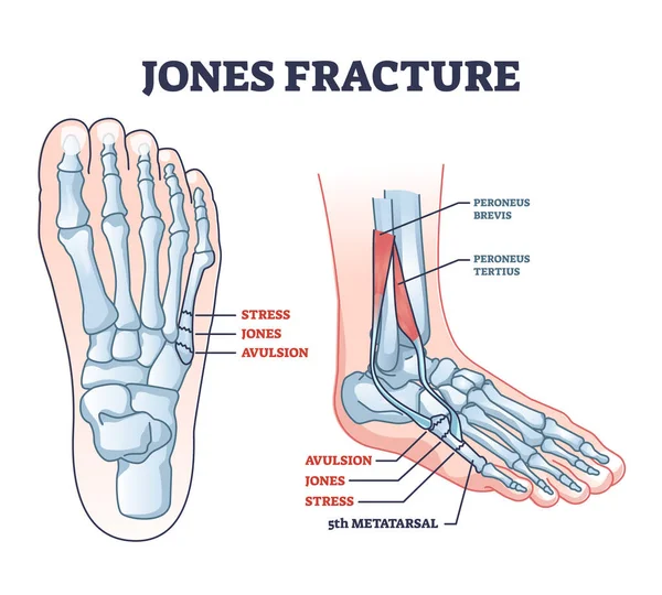
Jones fracture and foot pinky finger bone broken damage outline diagram. Labeled educational scheme with bone stress or avulsion sections vetor illustration. Peroneus brevis and tertius muscle anatomy
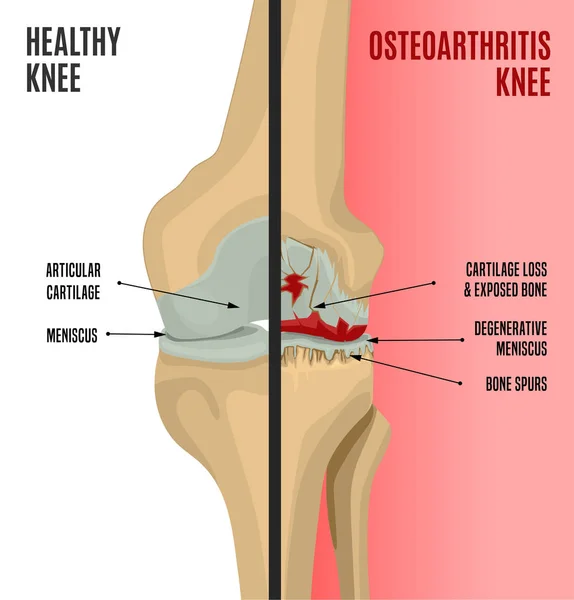
Osteoarthritis Knee Poster
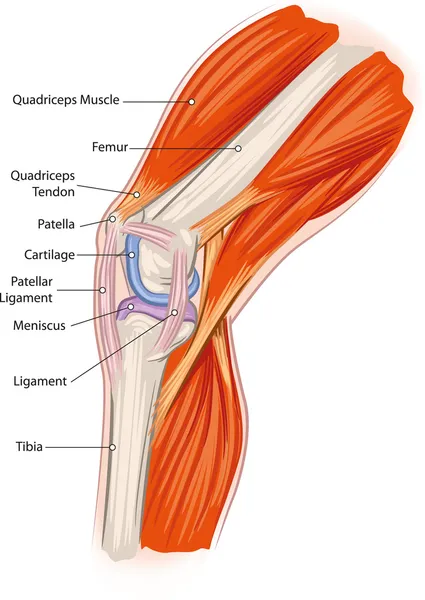
Human Knee joint anatomy. Knee Tendons, anatomical diagram
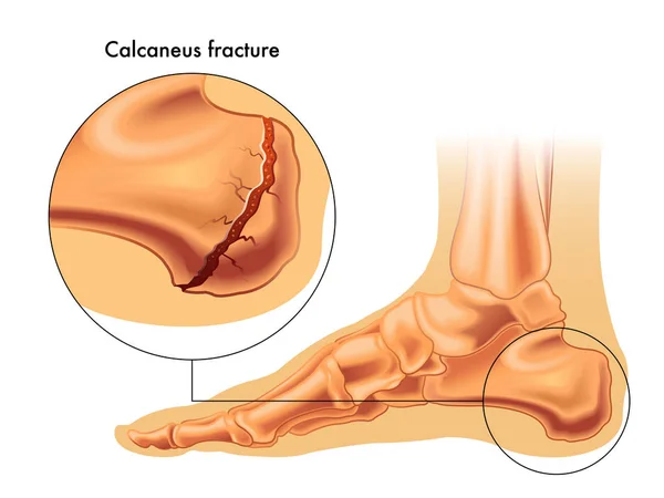
Calcaneus fracture illustration
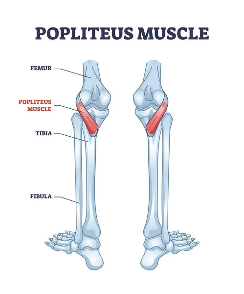
Popliteus muscle as leg and knee muscular joint anatomy outline diagram
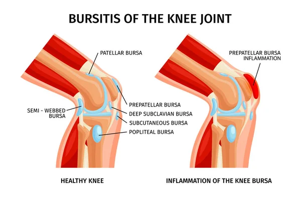
Bursitis of knee bursa realistic infographics with anatomy of healthy and unhealthy joints vector illustration
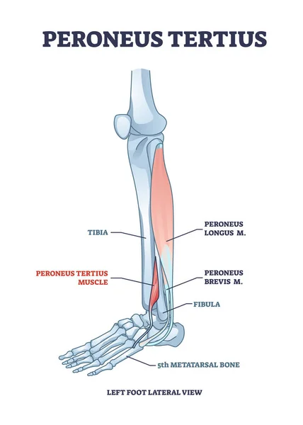
Peroneus tertius leg muscle with longus and brevis location outline diagram
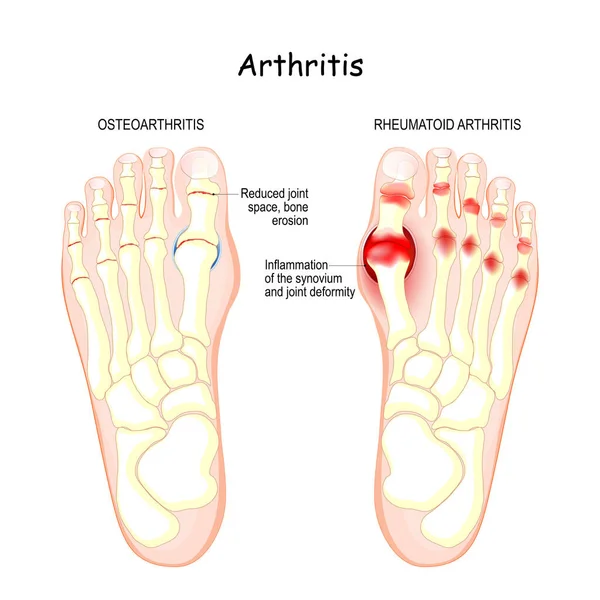
Osteoarthritis, rheumatoid arthritis, and posttraumatic arthritis. comparison of human foot with different forms of arthritis. inflamed synovium and joint deformity
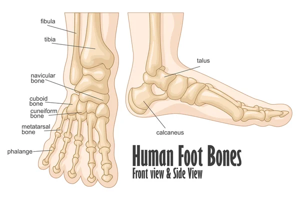
Human foot bones front and side view anatomy
Tibialis posterior muscle with human leg skeletal structure outline diagram
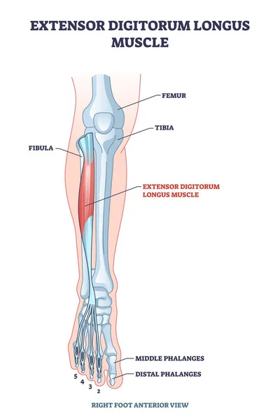
Extensor digitorum longus muscle with foot skeletal system outline diagram
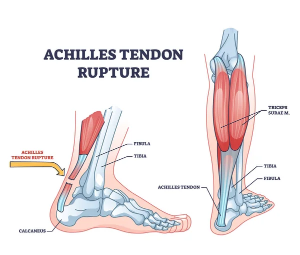
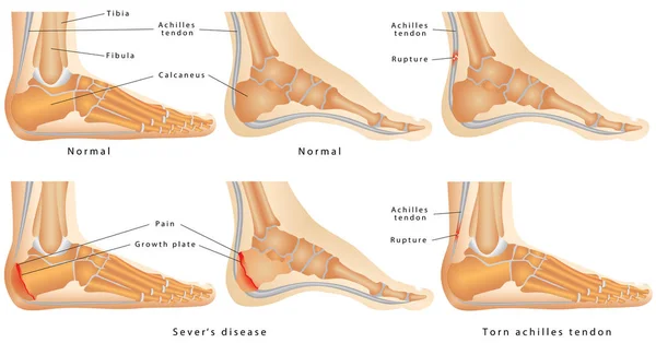
Achilles tendon rupture as painful injury and leg trauma outline diagram. Labeled educational anatomical scheme with orthopedic problem explanation vector illustration. Medical body muscle condition.
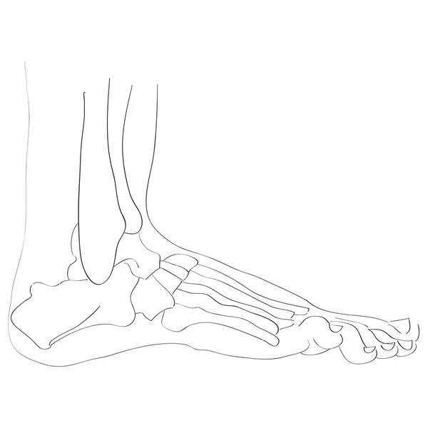
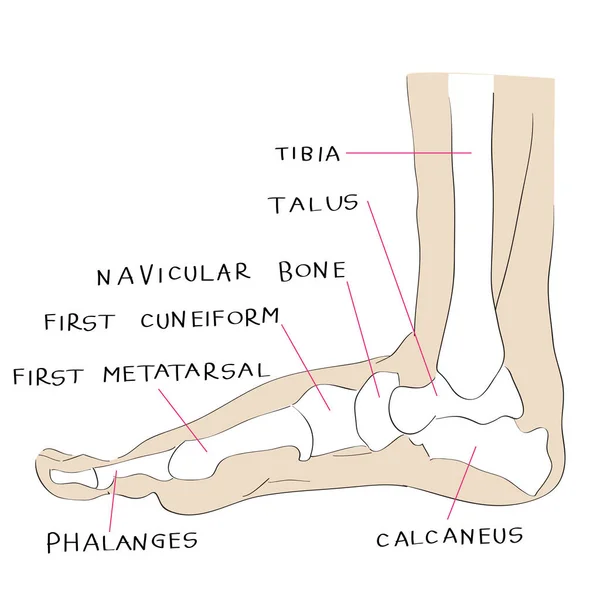
Lateral view foot bones
Heel Spur.
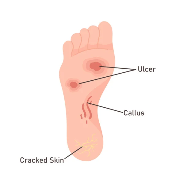
Human Foot Diseases Vector Illustration
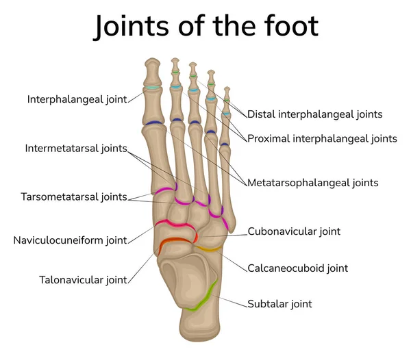
Illustration of the joints of the foot. The bones of the foot are depicted and the joints between them are schematically shown.
Human Knee Osteoarthritis diagram illustration
Human leg and joints. Symbol. Vector illustration.
Stages of osteoarthritis of the knee
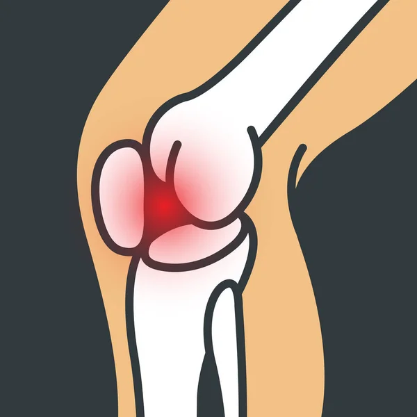
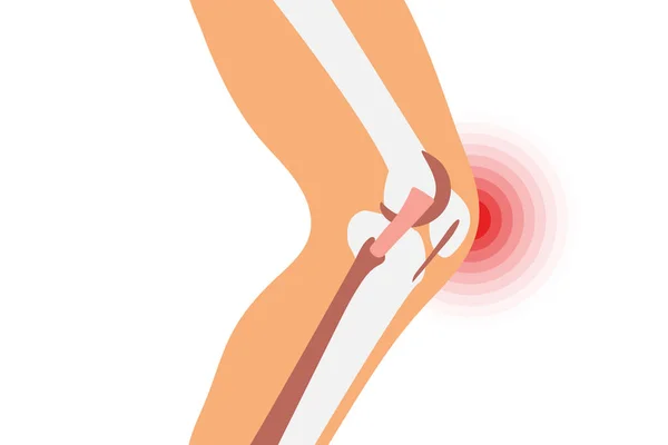
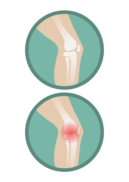
Knee pain illustration. Pain circle on a knee joint. Medicine concept.
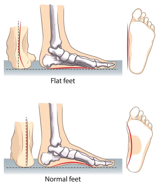
Pronated foot, arch deformation, bottom and back view . Foot weight distribution.
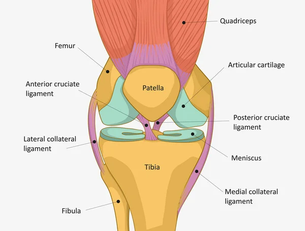
Knee joint isolated vector illustration, flat design. Ligaments of the knee. Anterior and Posterior cruciate ligaments, Patellar and Quadriceps, tendons, Medial and Lateral collateral ligaments.
Anatomy. Subscribe. Structure knee joint raster Basic Medical Education
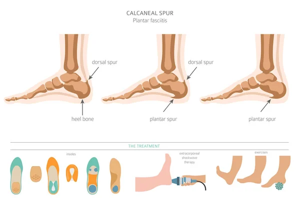
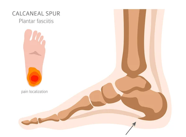
Calcaneal spur. Plantar fasciitis set. Medical desease infograph
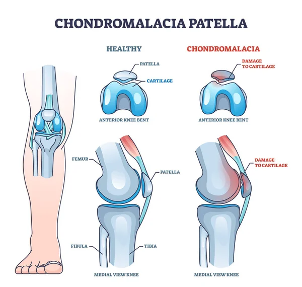
Chondromalacia patella knee breakdown compared with healthy outline diagram. Labeled educational kneecap tissue damage with cartilage problem and anatomical leg joint structure vector illustration.
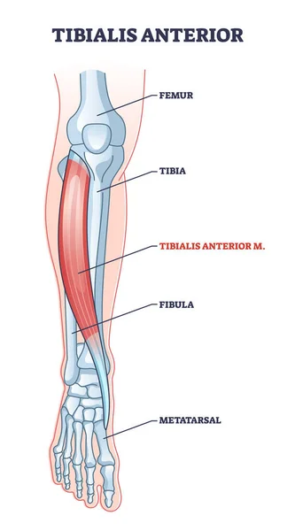
Tibialis anterior muscle with human leg skeletal structure outline diagram
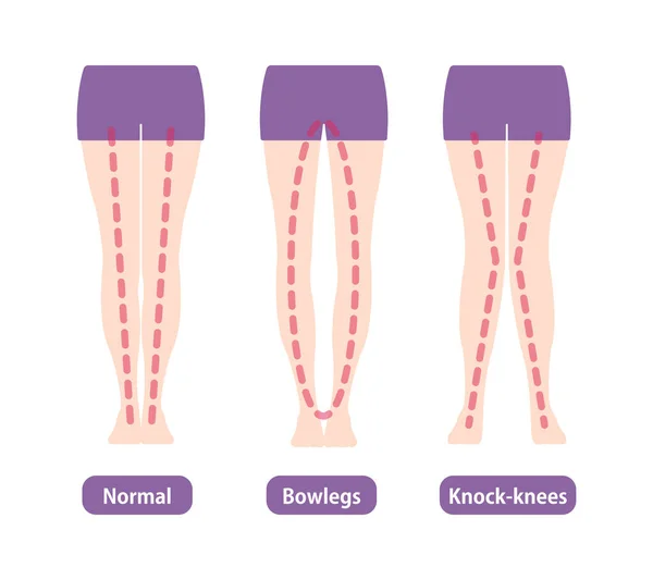
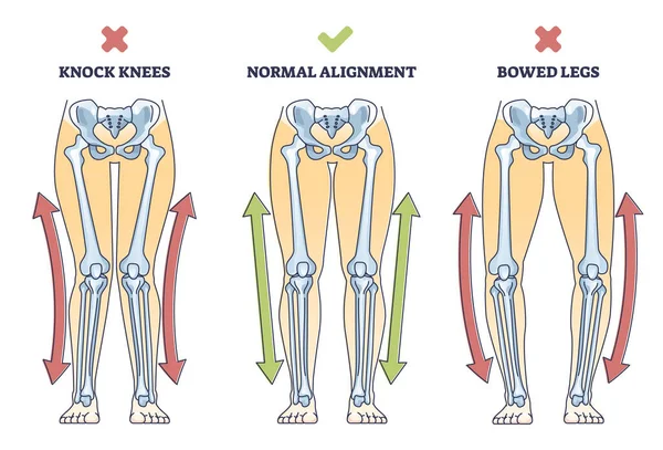
Difference types of legs angles and knees vector illustration
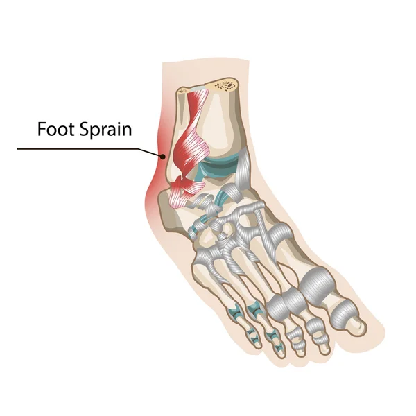
Foot Sprain
Human foot structure, anatomical medical teaching poster, bones of the heel, phalanges, joint, ankle. Healthcare mockup, vector illustration.
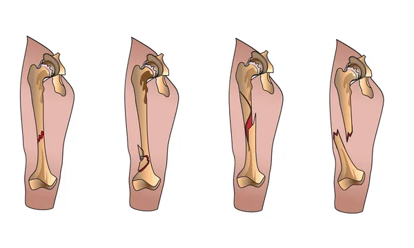
Types of bone fractures medical educational vector
Knee bone anatomy
The anatomy of a group of muscles of the human thigh, their structure on a white background. Vector 3D illustration
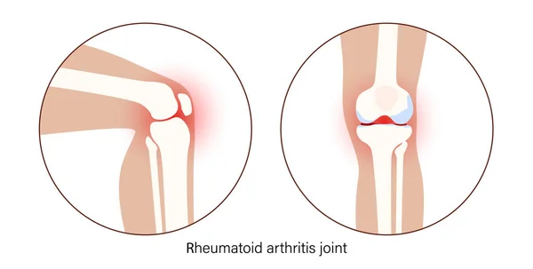
Arthritis in knee joint
Soleus muscle with anatomical leg bones skeletal structure outline diagram
Vastus lateralis muscle location and hip or leg skeletal bone outline diagram
Extensor hallucis longus muscle with foot skeletal system outline diagram
Bowed legs syndrome with normal and knock legs comparison outline diagram. Labeled educational physical defect with outward bending and knees does not touch vector illustration. Anatomical explanation
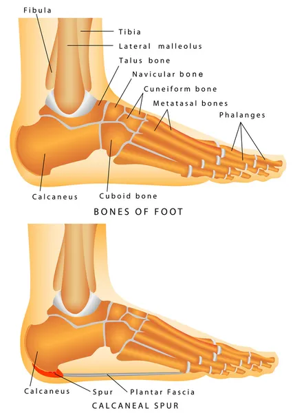
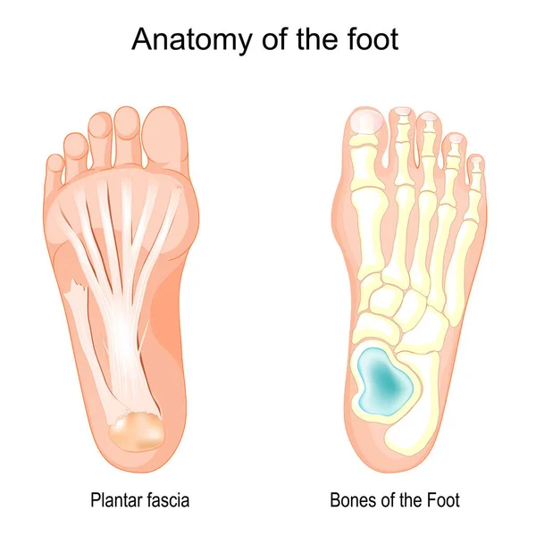
Anatomy of the foot. Bones of the Foot and Plantar fascia. Vector illustration
Knee Tendons medical vector illustration scheme, anatomical diagram.
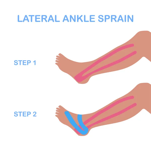
Lateral ankle sprain. Correct kinesiology taping.
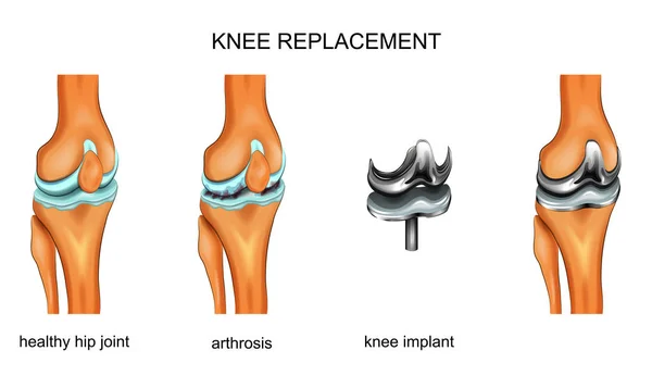
Vector illustration of a total knee replacement
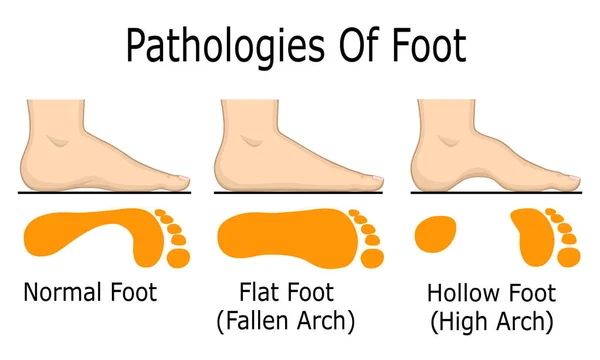
Pathologies of foot
Osteoarthritis Knee Poster
Puncture synovitis of the knee joint
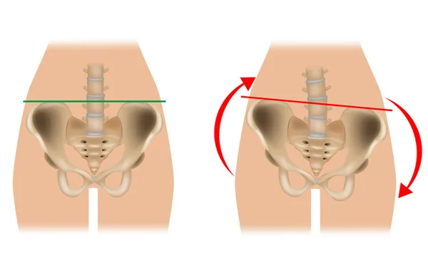
Comparison of pelvic distortion. Normal pelvis and distorted pelvis. A Twisted Pelvis, also called Pelvic Torsion
Calcaneal spur. Plantar fasciitis set. Medical desease infograph
Human knee joint
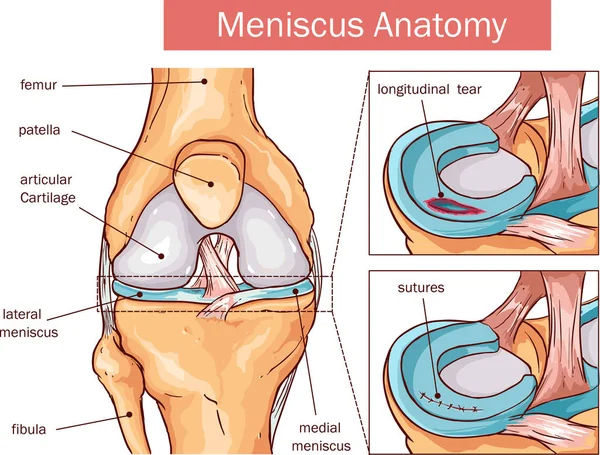
Vector illustration of a Meniscus tear and surgery
Vector human knee
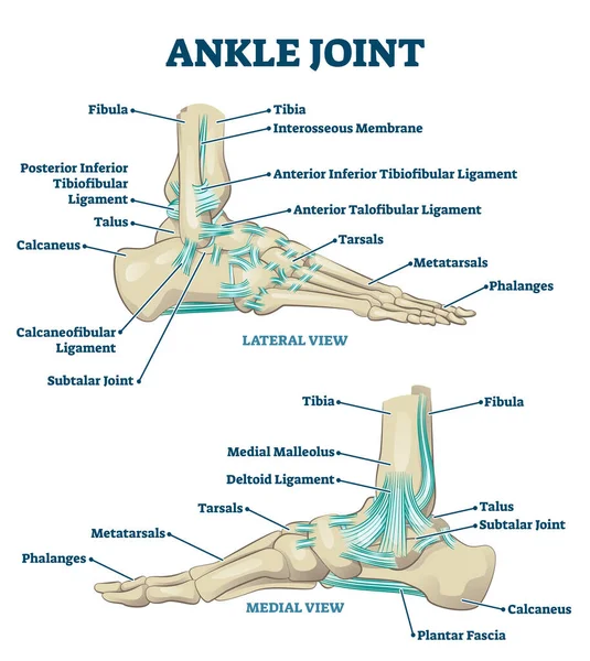
Ankle joint vector illustration. Labeled educational leg structure scheme.
Illustration of a knee joint
Realistic 3d Illustration Knee with Torn Miniscus
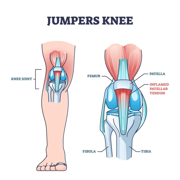
Jumpers knee or patellar tendonitis tendon bone inflammation outline diagram
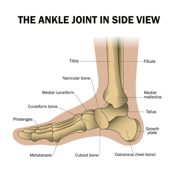
Ankle(side view)
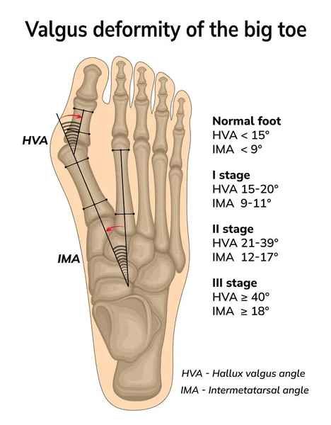
Illustration of valgus deformity of the big toe. The angles of bone displacement during deformation are shown
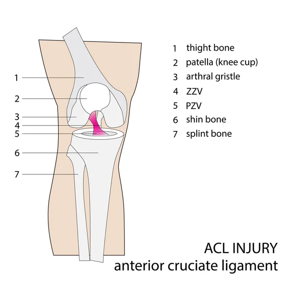
ACL injury
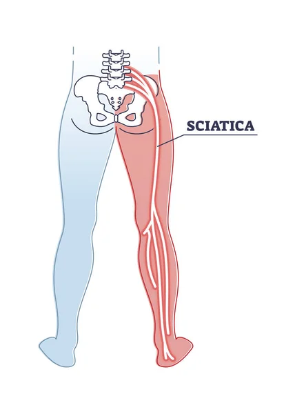
Sciatica pain or nerve weakness as leg lumbar radiculopathy outline diagram
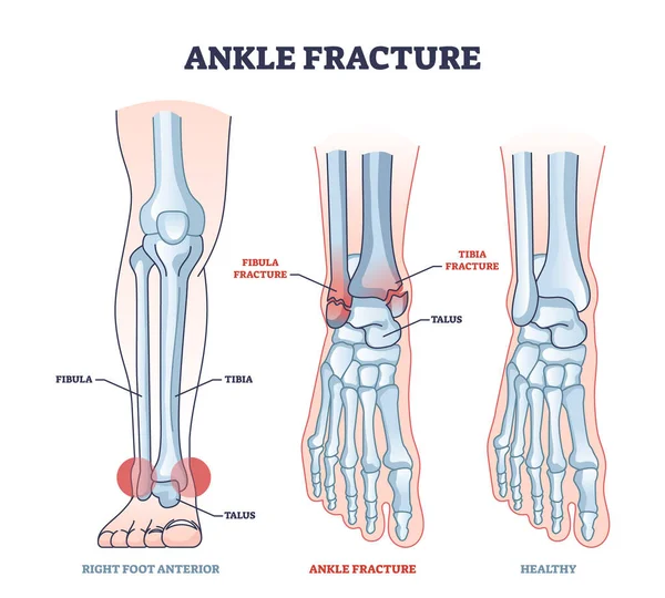
Ankle fracture and right foot anterior trauma medical view outline diagram. Labeled educational scheme with anatomical comparison for healthy and fractured bones vector illustration. Leg tibia injury
Vector hand drawn flat illustration plantar fasciitis infographic for medical studies. Isolated on black background.
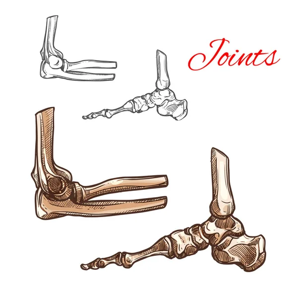
Bone and joint sketch of human foot, elbow, ankle
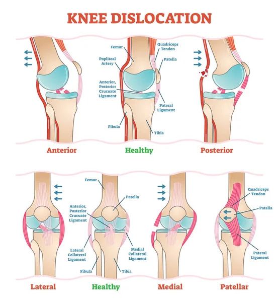
Knee Dislocations - medical vector illustration diagrams. Anatomical knee injury types scheme.
Human foot bones anatomy with descriptions. Colored leg base parts structure. Educational diagram with human internal organ illustration. Talus, calcaneus, tarsals, cuboid, metatarsals foot parts.
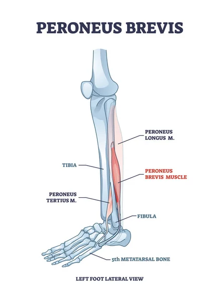
Peroneus brevis leg muscle with longus and tertius location outline diagram
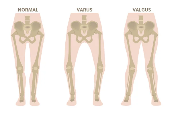
Valgus and varus leg deformities. Diagram showing the deformed bones of the lower extremities. Cosmetic pathology. Vector illustration.
Knee anatomy
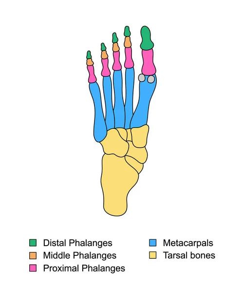
Foot bones anatomy with descriptions. Colored leg base parts structure. Educational diagram with internal organ illustration. Distal, proximal and middle phalanges, metacarpal, tarsal bones foot parts
Orthopedic medical icons
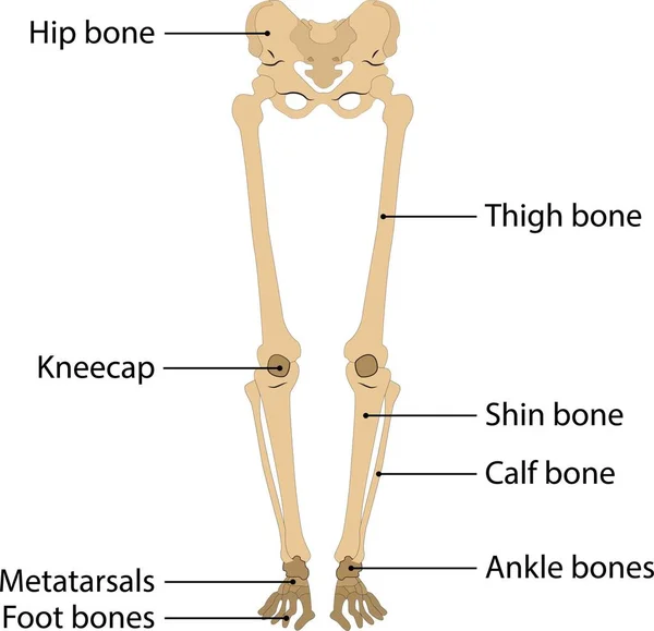
Human body leg skeleton. vector illustration. on white background. human leg bone
Knee anatomy
Arthritis in knee joint
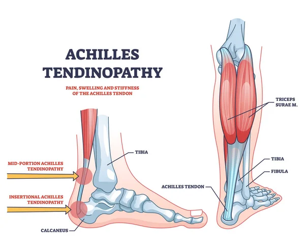
Achilles tendinopathy as injury to tendon in heel outline diagram. Labeled educational scheme with anatomical leg and foot skeleton and muscles vector illustration.Trauma and band of tissue problem.
Types of fractures
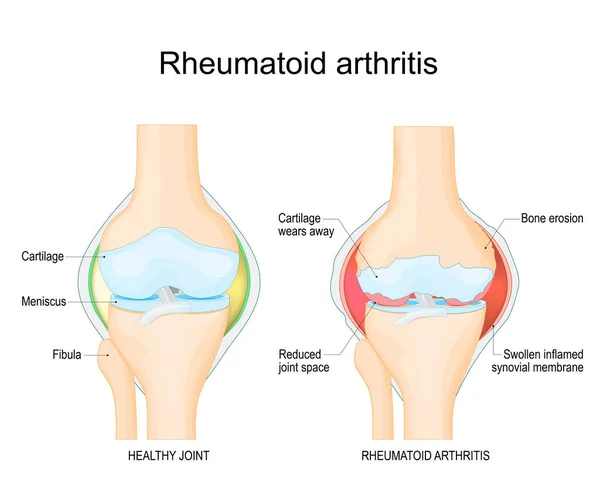
Rheumatoid arthritis. A comparison between a healthy knee and joint with Bone erosion, Cartilage wears, Reduced joint space and Swollen inflamed synovial membrane. Vector illustration with labels. diagnostic images to aid patient and doctor
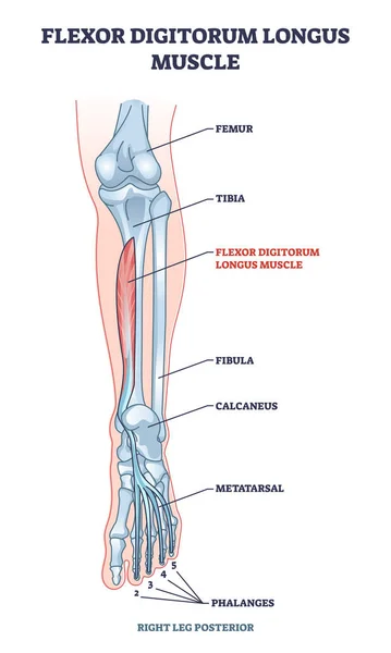
Flexor digitorum longus muscle with human leg and foot bones outline diagram
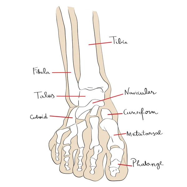
Front view foot bones text
Anatomy of human knee vector sketch of leg bones and joint, medicine design. Side and front view of knee bones, hand drawn femur, patella, tibia and fibula, tibial plateau and lateral condyle
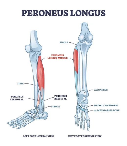
Peroneus longus muscle with leg muscular and skeletal system outline diagram
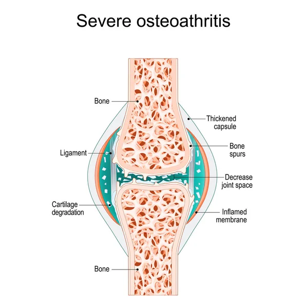
Severe osteoarthritis. Synovial joint with bone Spurs, Inflamed membrane, Decreased joint space, and Cartilage degradation. vector illustration
Internal lateral view foot bones color
Anatomy. Structure knee joint vector
Knee joints with osteoparosis. Vector flat illustration
Fracture of the metatarsal bones in the foot. Anatomical structure of the foot. Skeleton. Broken bones. Vector illustration on isolated background
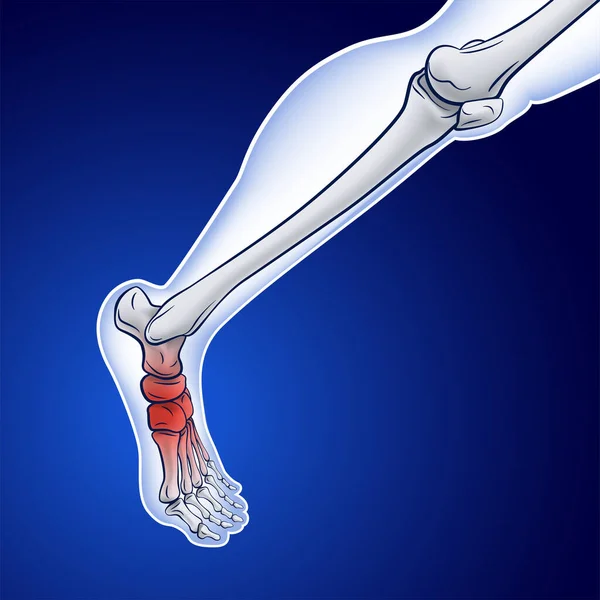
An illustration of a running skeletal line showing a red dot indicating an injury.
Next Page
1
2
3
4
5
...
100
Page
Go
Home
Illustrations
beine knochen illustrations & clipart