Current page requires JavaScript, this web browser either does not support JavaScript, or scripts are being blocked.
Please turn on JavaScript or use different browser.
Photos
Vectors
Illustrations
Free Pictures
Videos
Music & SFX
AI Assistant
AI Image Generator
Free Background Remover
Free Image Upscaler
Reverse Image Search
AI training data
Pricing
Illustrations
Log In
Sign Up
Celulas Stock Illustrations
47,618,971 Celulas illustrations, Celulas clipart illustrations & paintings
Create with AI
Watercolor celulas
Photos celulas
Landscape
Happiness
Nature
Lifestyle
Science
Animals
Business
Food
Architecture
Sport
Best Match
Fresh
Popular
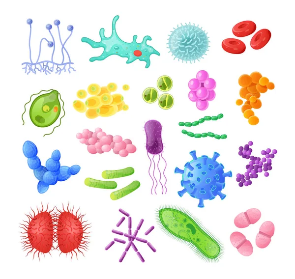
Microorganism, bacteria, virus cell, bacillus, disease bacterium and fungi cells.
Blood Cells Mascots
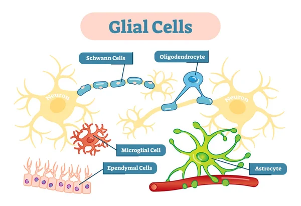
Nervous system Glial cells vector illustration schematic diagram.
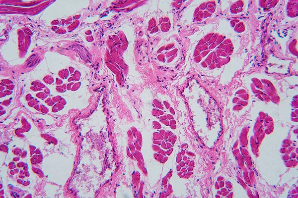
Human tongue, longitudinal section, organic background
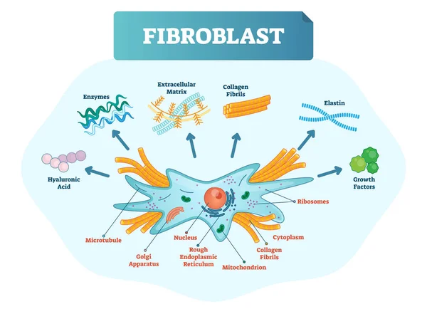
Fibroblast vector illustration. Scheme with extracellular, collagen fibrils, elastin, hyaluronic acid, microtubule, golgi apparatus, nucleus and ribosomes.
Pancreatic gland cells
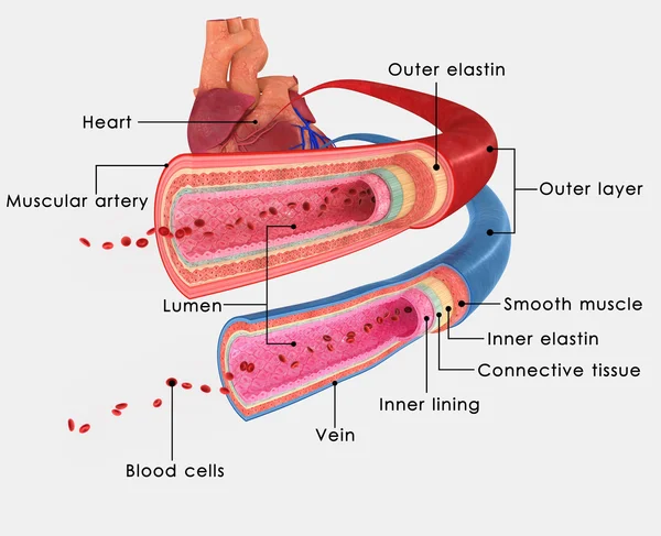
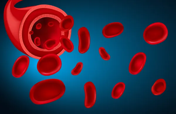
BLOOD VESSELS
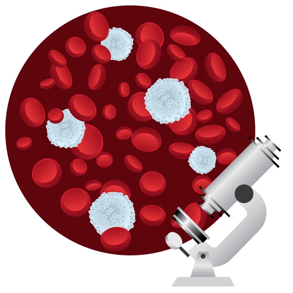
Cells under microscope
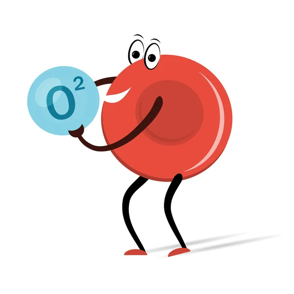
Red Blood Cell with Oxygen Cartoon
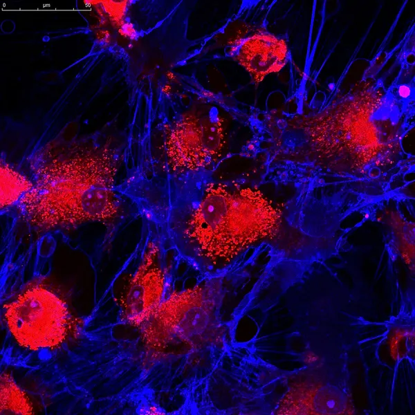
Mesenchymal stem cells labeled with fluorescent molecules
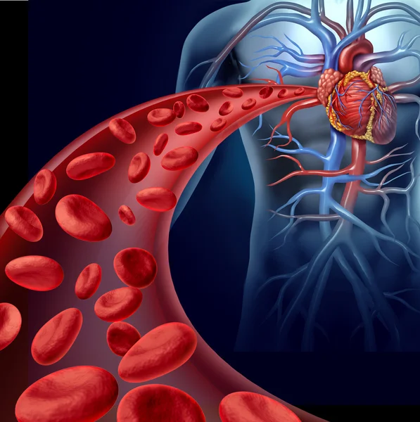
Heart Blood Health
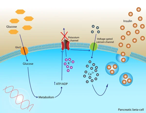
Insulin secretion
Cartoon of Blood Cell - Erythrocytes, Leukocytes, Thrombocytes
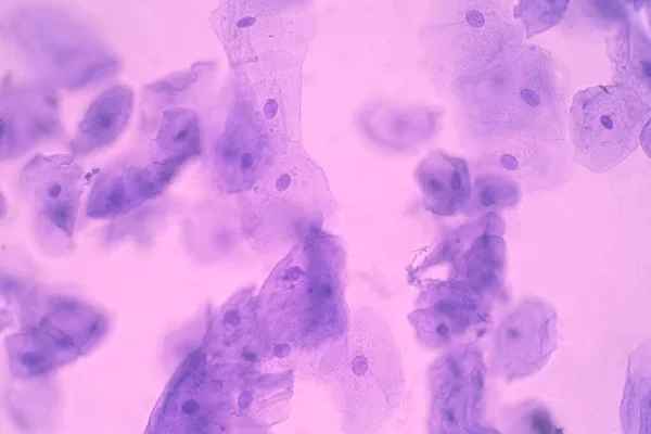
Human Cheek Epithelial Cells. The tissue that lines the inside of the mouth is known as the basal mucosa and is composed of squamous epithelial cells. Education pathology.
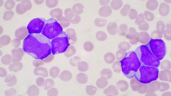
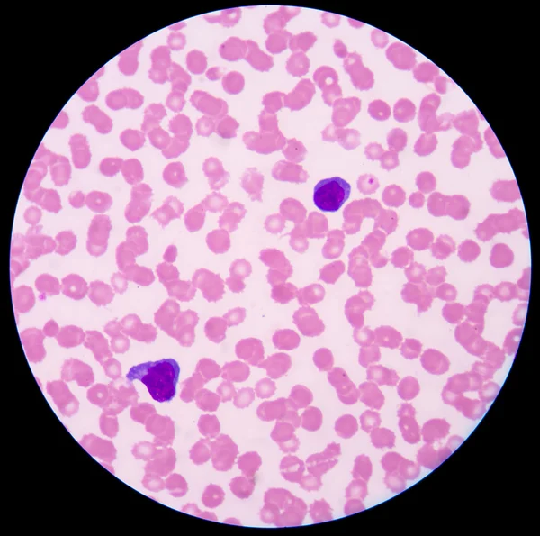
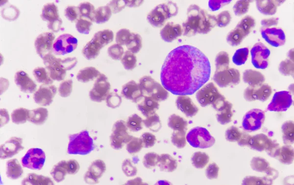
Blast cell in leukemia pateins in blood smear.
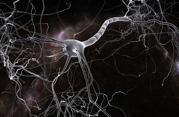
Neuron cell
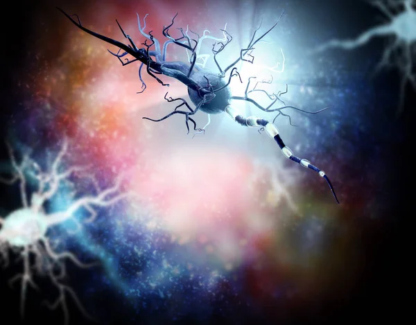
Damaged nerve cells, concept for neurodegenerative and neurological disease, tumors, brain surgery.
Biology. Round concept vector hand drawn elements in doodle style.
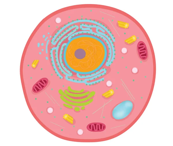
Animal cells are the basic unit of life.
Human genome research. Scientist working with a dna helix, genome or gene structure. Human genome project. Flat style vector illustration.
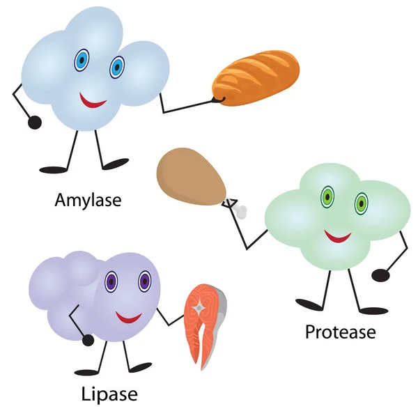
How enzymes work. Fermentation and disgestion food
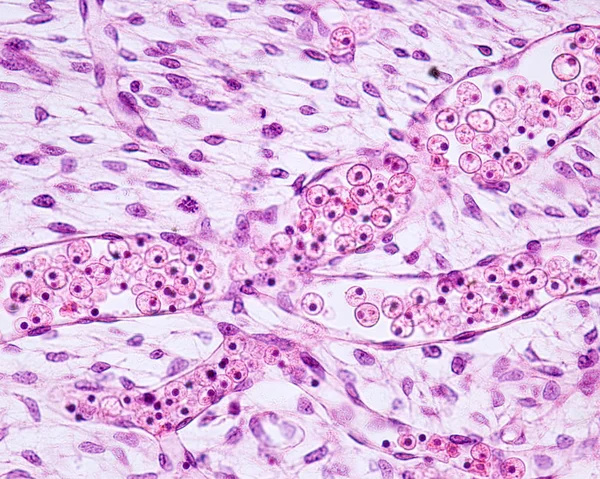
Embryonic blood vessels
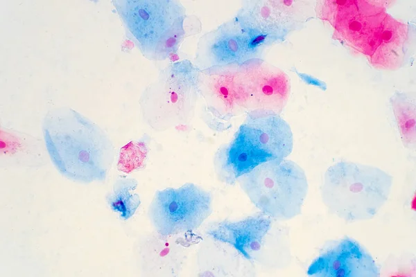
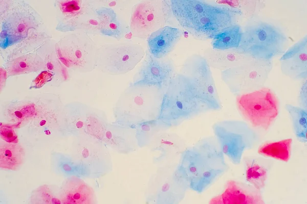
Squamous epithelial cells of human cervix under the microscope view. Pap smear test is a procedure to test for cervical cancer in women.
Process of metastases cancer stages illustration isolated on white background.
Eosinophilic cationic protein is incorporated into the membranes of helminth cells and disrupts their integrity. Eosinophil is a blood cell. Vector illustration on isolated background
Microscopic section of human kidney tissue
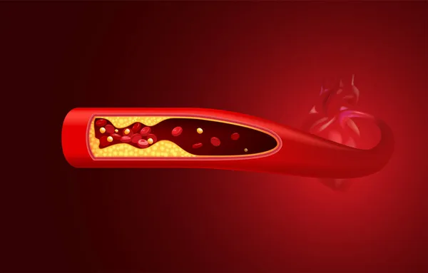
Red blood cells and cholesterol clog before the heart.
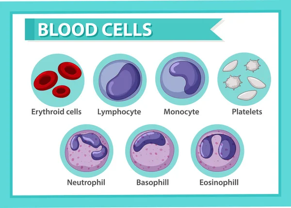
Type of blood cells illustration
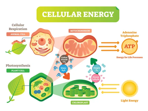
Animal and plant cell energy cycle vector illustration diagram with mitochondrion and chloroplast.
Blood Cells Circulation
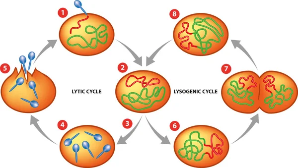
Lytic and Lysogenic cycle
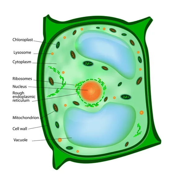
Plant cell
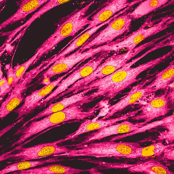
Real fluorescence microscopic view of human skin cells in culture. Nucleus are in yellow, cell membranes were labeled with pink
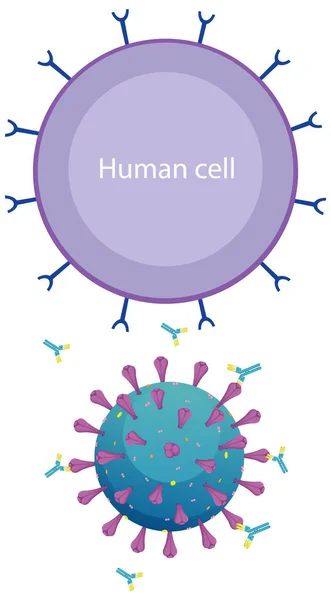
Informative human cell and coronavirus diagram illustration
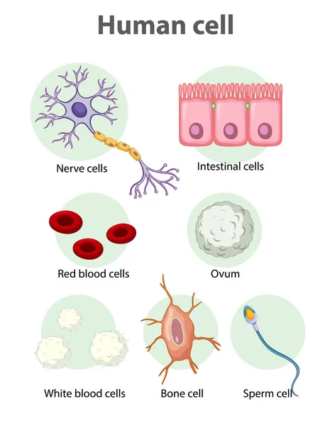
Information poster on human cells illustration
Melanocytes are specialized cells that produce melanin pigments.
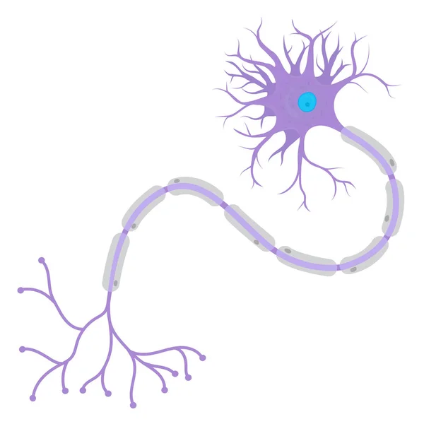
Structure of a Neurons in the human body.
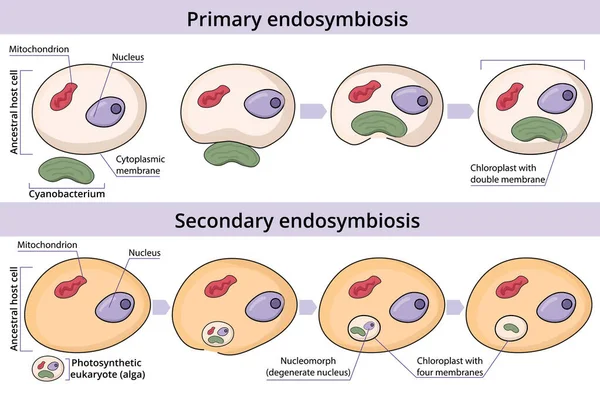
Process of primary and secondary endosymbiosis. Cell engulfs and absorbs a prokaryotic cell. Secondary endosymbiosis occurs when a eukaryotic cell engulfs and absorbs another eukaryotic cell.
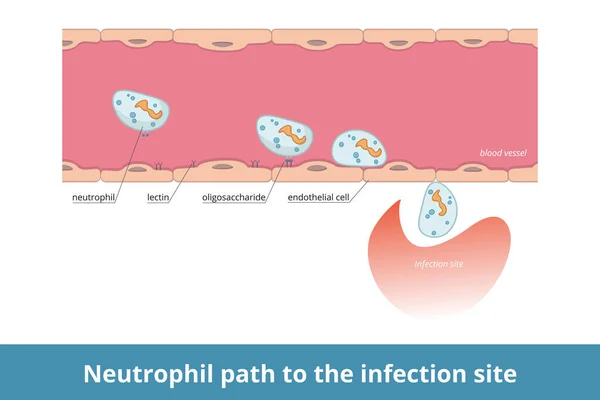
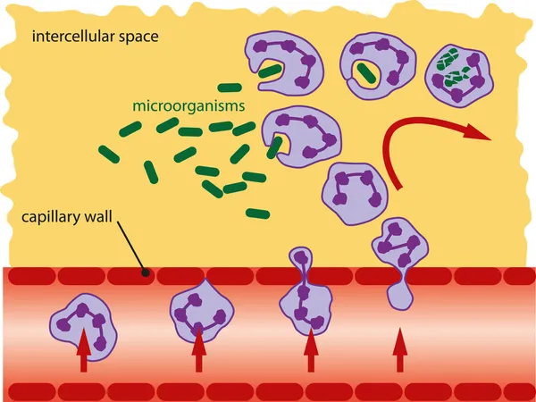
Neutrophil path to the infection site. Cell-surface carbohydrate are recognized by lectins that allows them to migrate from blood to infection site.
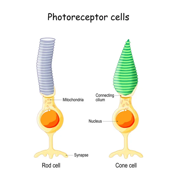
Anatomy of Photoreceptor. cell of a retina in the eye. Cone cells in respond to color vision and send signals to brain. Rod cells are used in peripheral vision
Blood corpuscles magnified about 400 diameters and the red corpuscles have arranged themselves in rouleaux, vintage line drawing or engraving illustration.
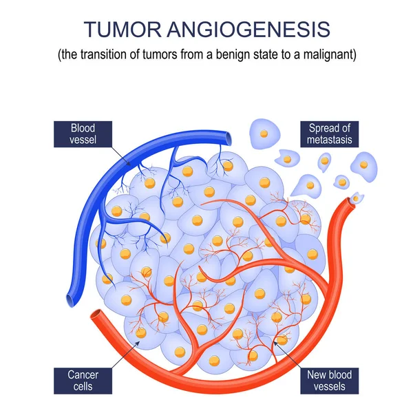
Tumor angiogenesis. transition of tumors from a benign state to a malignant. tumor grow. Cancer cells and Spread of metastasis. Vector poster
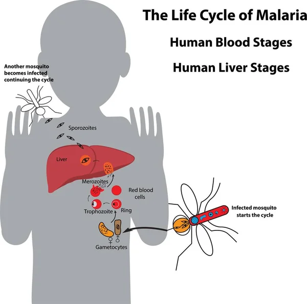
Life cycle of malaria parasite, human blood stages and human liver stages
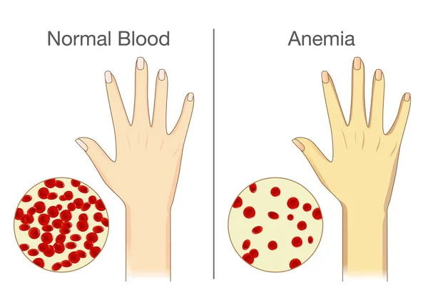
The effect of Anemia on skin blood flow in human.
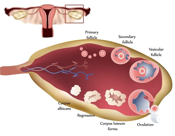
Normal ovary, follicular development and ovulation
Tissue of Small intestine (Duodenum), Large intestine Human and Stomach Human under the microscope in Lab.
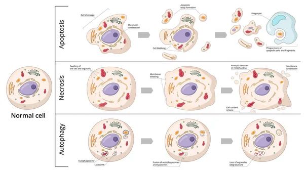
Three basic forms of cell death: apoptosis (chromosome condensation, nuclear fragmentation), autophagy (autophagosome formation), necrosis (membrane rupture, organelles swelling).
Cell structure
Human tongue longitudinal section organic background
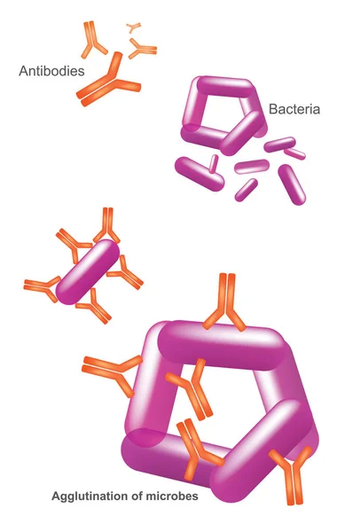
Antibody and Bacteria. Illustration Health care and medical infographic.
Yeast cells with epithelial tissue
Blood
Red Blood Cells
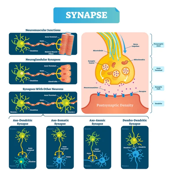
Synapse vector illustration. Labeled diagram with neuromuscular example.
Type of Muscle Cells for health education Infographic illustration
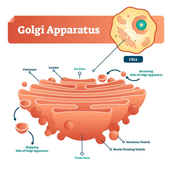
Golgi apparatus vector illustration. Labeled microscopic scheme with cisternae, lumen, secretory and newly forming vesicle. Diagram with receiving and shipping side.
Health and biochemistry laboratory of nanotechnology. Molecule helix of dna, genome or gene evolution. Vector blue science genome clone sequence concept with characters. Flat isometric illustration.
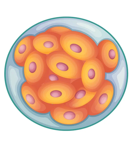
Morula
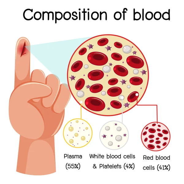
Composition of blood diagram illustration
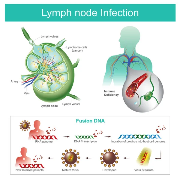
The Lymph nodes are infected Failure of the immune system, the b
Great image of red cells under the microscope
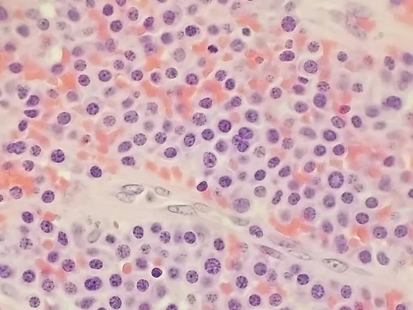
Salamander testicular tissue under the microscope 100x
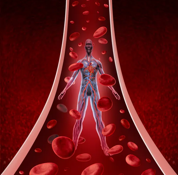
Human Circulation Health
Smaller red to pink normal red blood cells or erythrocytes.
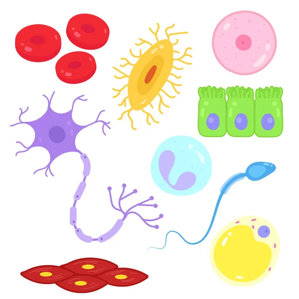
Types of cells in the human body.
Squamous epithelial cells of human cervix under the microscope view. Pap smear test is a procedure to test for cervical cancer in women.
DNA molecule structure isolated in white. 3d illustration
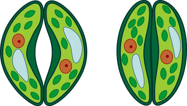
Guard cells of stoma
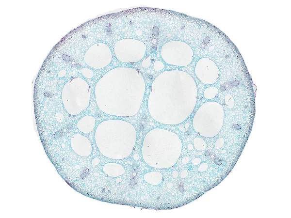
Cross section cut slice of plant stem under the microscope microscopic view of plant cells for botanic education high quality
Blood cells.
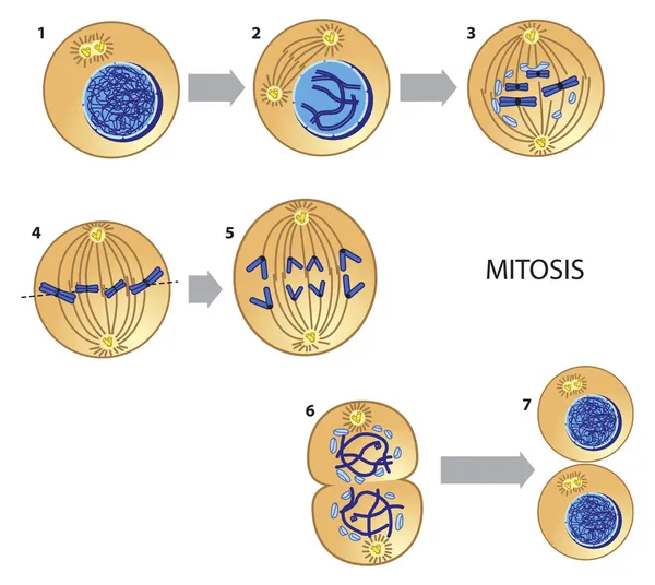
Mitosis
Leucocytes
Ovary and ovulation process
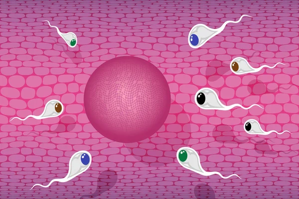
Sperm race
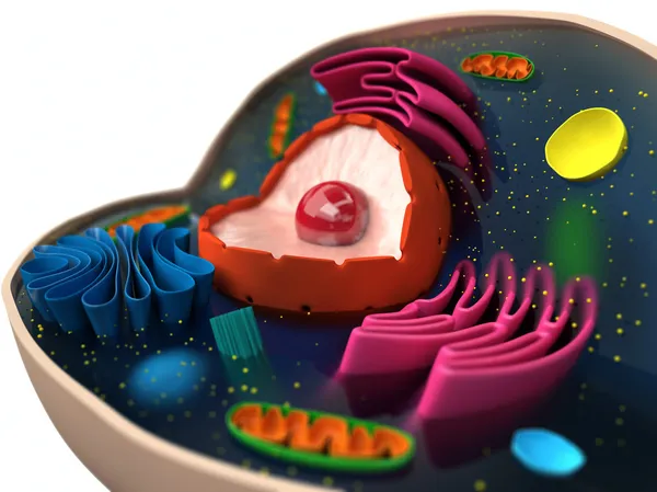
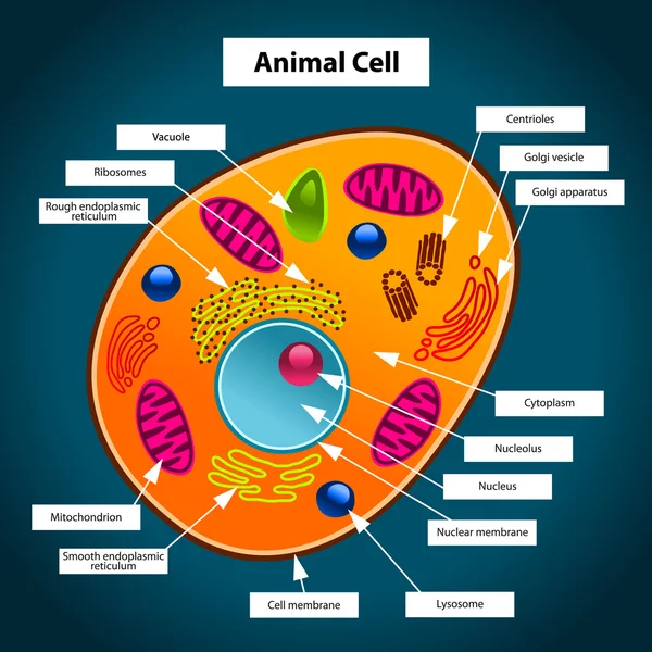
Animal Cell
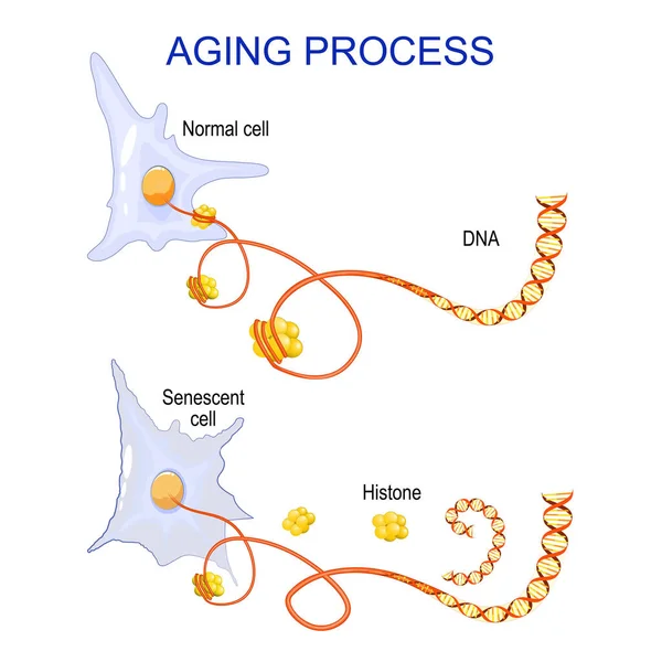
Aging process into cells. chromatin, DNA and histones change in ageing and senescent cells. Vector
Malaria
Slime molds, as a group, are polyphyletic under the microscope for education.
Host cells with spores (mold) are inside wood under the microscope for education.
Human gas exchange process with oxygen cycle explanation outline diagram
Neuron, nerve cell anatomy
Type of Muscle Cells for health education Infographic illustration
Cartoon picture of contagious corona virus showing anxious face. Vector illustration
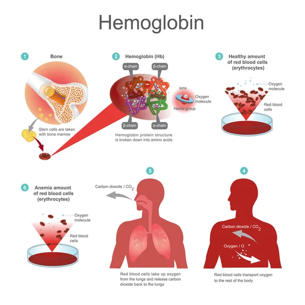
Red blood cells (erythrocytes) begins in the bone marrow. Red bl
Vector illustration of human somatic cells
Petri dish with mold culture in the hand of a scientist
Biology. Round concept of vector hand drawn elements.
Virus infection, antibodies and blood cells
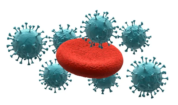
Blood cells on virus (Isolated with clipping path)
Health and biochemistry laboratory of nanotechnology. Molecule helix of dna, genome or gene evolution. Vector blue science genome clone sequence concept with characters. Flat isometric illustration.
Real fluorescence microscopic view of human skin cells
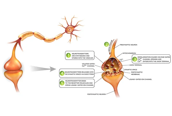
Neurons and closeup of synapse detailed anatomy
Serologic Test (Antibody) by Immune Cells - Illustration as EPS 10 File
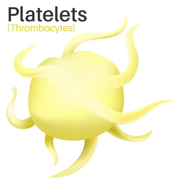
Platelets are tiny blood cells.
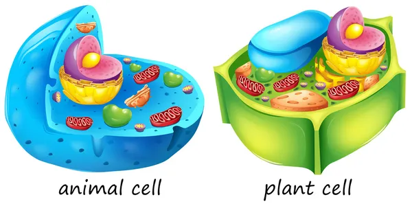
Animal and plant cells
Plasmablastic anaplastic Multiple Myeloma - Plasmacytoma Biopsy Specimen
Immature blood cells in leukemia.
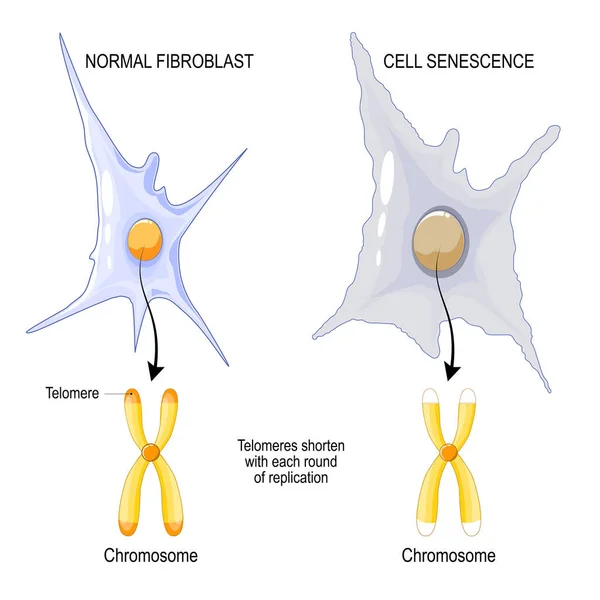
Chromosome of a Normal fibroblast and Cell senescence. Telomeres shorten with each round of replication. aging process. Vector poster
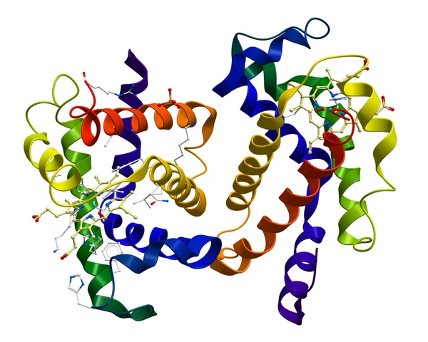
Human oxyhemoglobin molecular structure
Blood vessels with artery and vein internal structure vector illustration
Next Page
1
2
3
4
5
...
100
Page
Go
Home
Illustrations
celulas illustrations & clipart
Related searches
white blood cell
,
cells
,
immune system
,
cell cartoon
,
mean virus