Current page requires JavaScript, this web browser either does not support JavaScript, or scripts are being blocked.
Please turn on JavaScript or use different browser.
Photos
Vectors
Illustrations
Free Pictures
Videos
Music & SFX
AI Assistant
AI Image Generator
Free Background Remover
Free Image Upscaler
Reverse Image Search
AI training data
Pricing
All Images
Log In
Sign Up
Histology Images
Create with AI
Images histology
Vectors histology
pathology
human cells
microscopy
cells
radiation therapy
biopsy
micrograph
pathology microscope
skin cells
microscopic
microscopic tissue
clinical research
pathologist
microscope slide
Best Match
Fresh
Popular
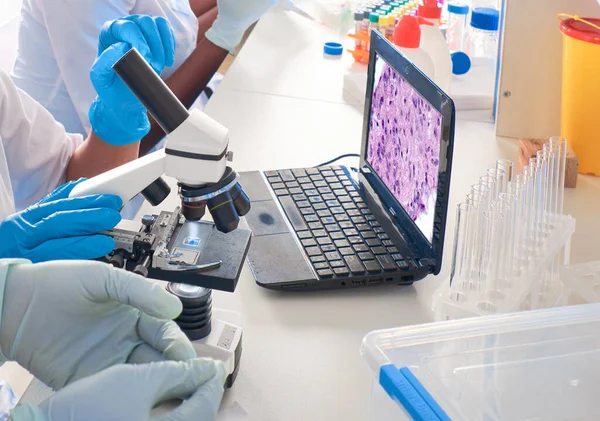
African female tech, scientists white coat and gloves do pcr test, work with microscope in modern laboratory or research facility. Analysis of lung tissue from COVID-19 patients, clinical cases.
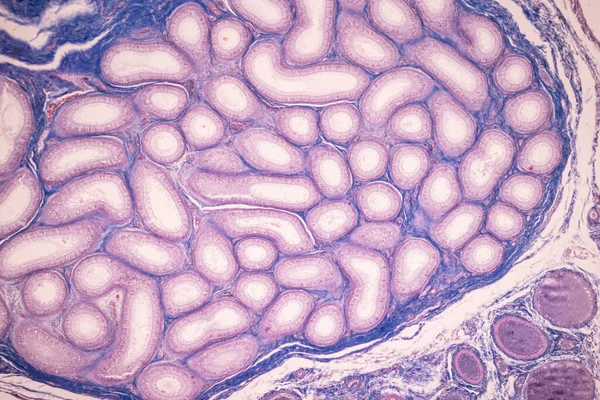
Anatomy and Histological Ovary and Testis human cells under microscope.
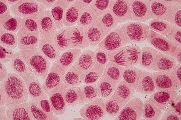
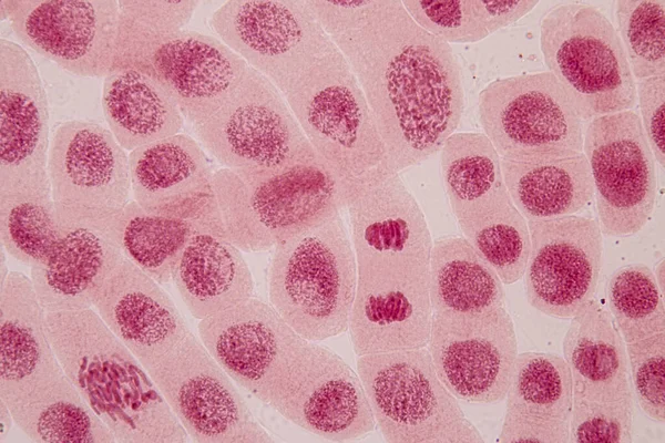
Root tip of Onion and Mitosis cell in the Root tip of Onion under a microscope.
Root tip of Onion and Mitosis cell in the Root tip of Onion under a microscope.
Tissue of Small intestine (Duodenum) and Vermiform appendix Human under the microscope in Lab.
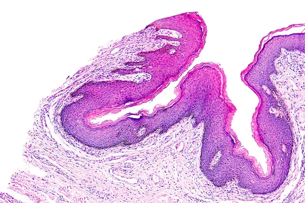
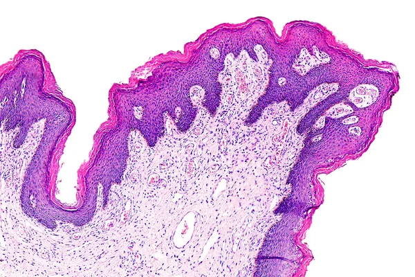
Skin papilloma of a human
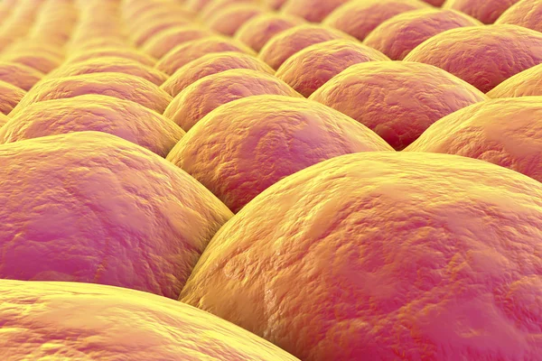
Adipose tissue
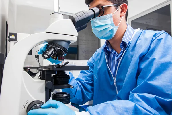
Modern microscope station in research facility
Biological cell isolated on whithe background microscope
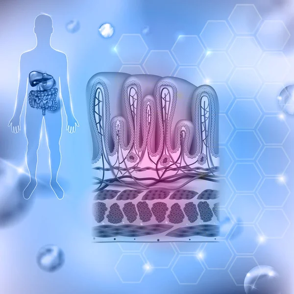
Stomach wall layers detailed anatomy, beautiful colorful drawing
Cancer cell in the moment that divides, 3d illustration
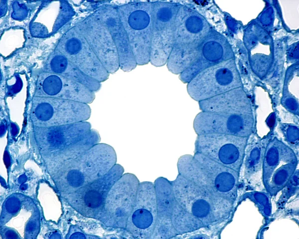
Kidney. Collecting duct
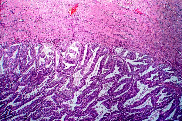
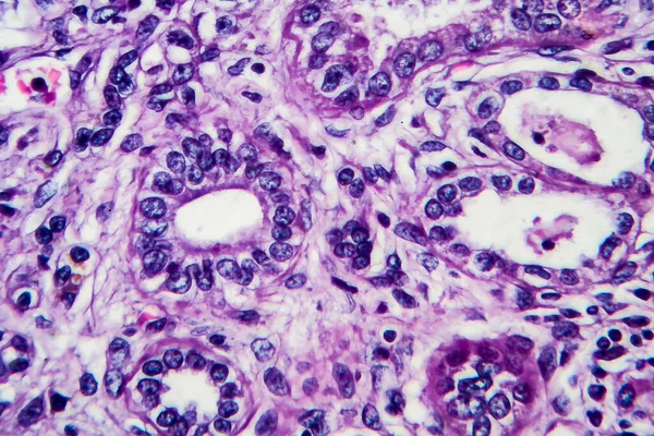
Endometrial adenocarcinoma, light micrograph, photo under microscope
Brain astrocytoma of a human
Fibrosarcoma of a human
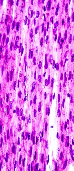
Heart muscle, light micrograph
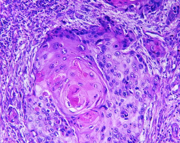
Squamous cell carcinoma of a human
Pancreas section organic background
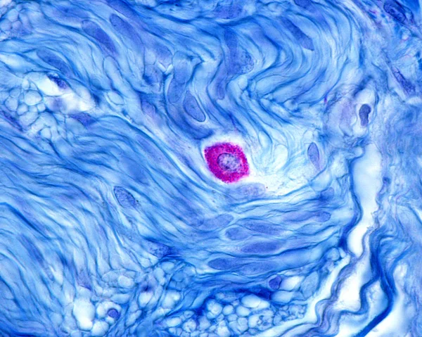
Mast cell
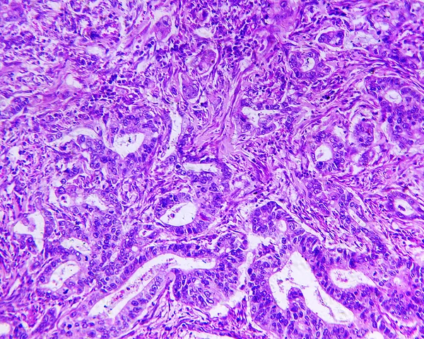
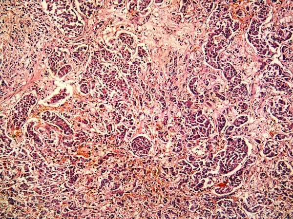
Gastric adenocarcinoma of a human
Ovarian follicle
Medical student in anatomy dissecting a kidney
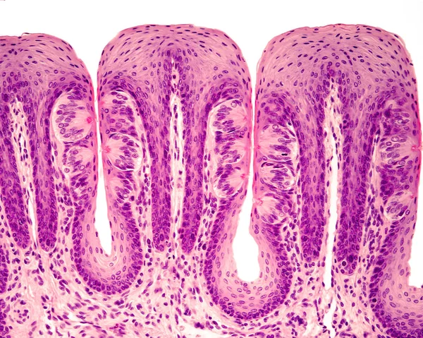
Taste buds
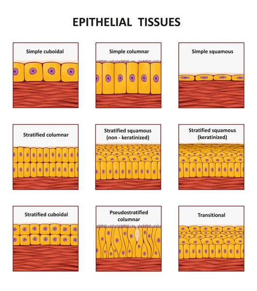
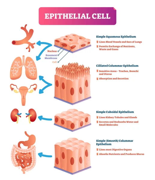
Different types of epithelial tissue collection.
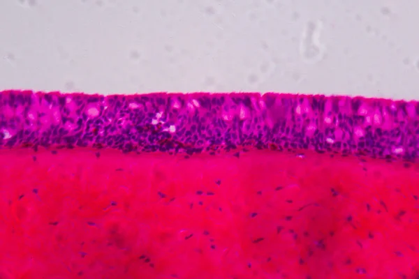
Respiratory epithelium
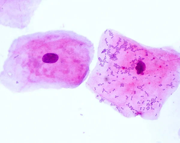
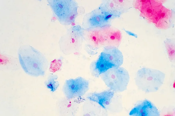
Squamous epithelial cells
Cell division background
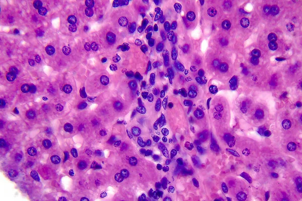
Coccidiosis, coccidia in liver, light micrograph, photo under microscope
Skin papilloma of a human
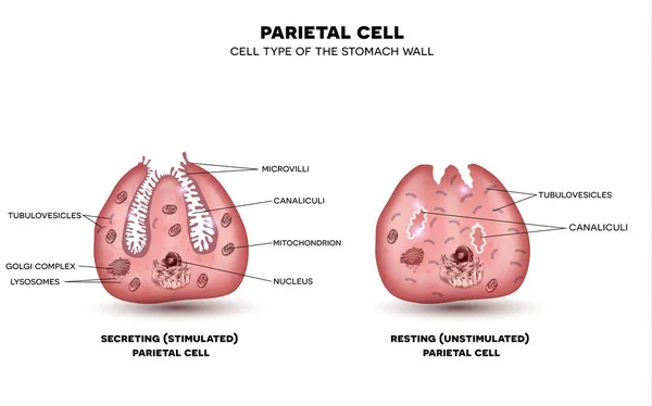
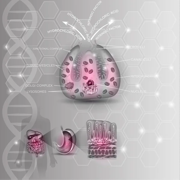
Parietal cell of stomach wall, located in the gastric glands sec
Epithelial cells vector illustration. Medical location and meaning diagram.
The words CANCER DIAGNOSIS written on a white notepad on a blue background near a stethoscope, syringe, electronic thermometer and pills. Medical concept
Tissue sections, battery of jars with fixed tissue samples on gl
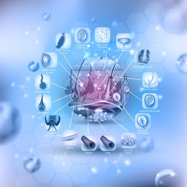
Skin anatomy round shape, detailed illustration
Chronic nephritis, light micrograph, photo under microscope. High magnification
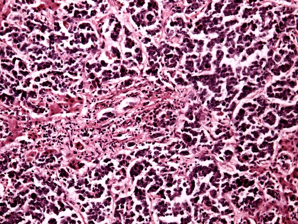
Small-cell lung cancer of a human
Light micrograph of a kidney
Gastric adenocarcinoma, light micrograph
Male scientist looking at slides under the microscope
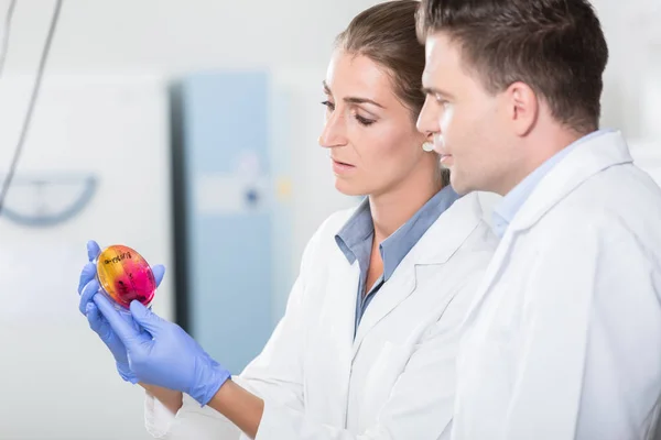
Scientists in laboratory regarding sample in petri dish
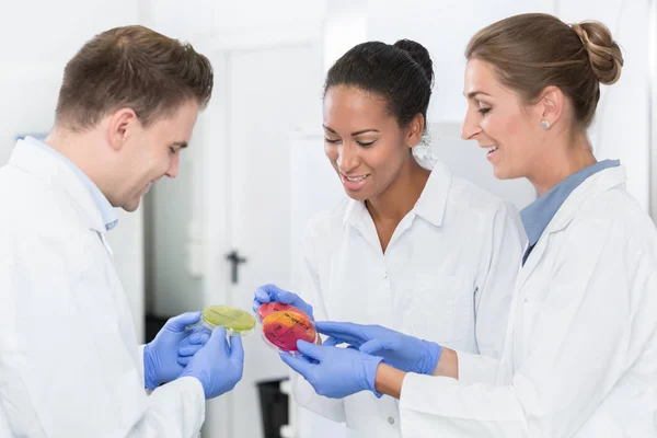
Group of food laboratory researchers comparing bacteria cultures
Micrograph plant root tip tissue cell
Large intestine of a rat
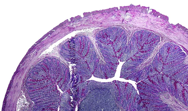
This is a histological photograph of the human small intestine. Magnify 40x.
Histopathology of diffuse sclerosing glomerulonephritis, light micrograph, photo under microscope
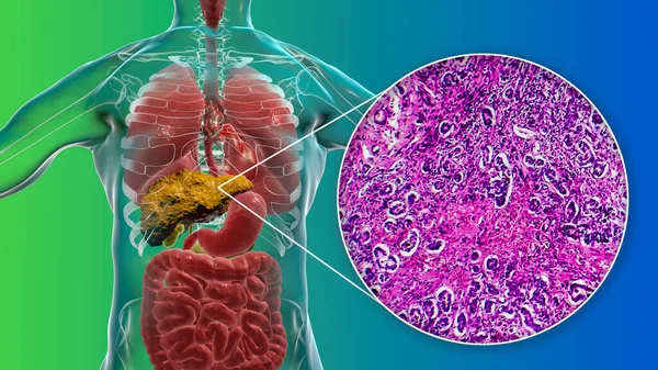
Liver with cirrhosis inside human body. 3D illustration and light micrograph of biliary cirrhosis
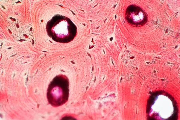
Histology of human compact bone tissue under microscope view for education, muscle bone connection and connective tissue
Parietal cell of stomach wall, located in the gastric glands
Photomicrograph showing histology of a atheroma cyst on the thorax in the anatomical pathology laboratory
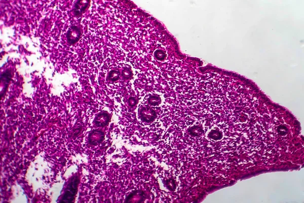
Endometrial hyperplasia, light micrograph, photo under microscope
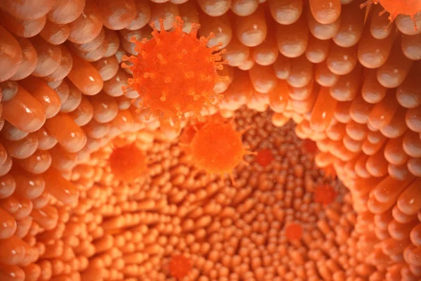
3d illustration Intestinal villi. Intestine lining. Microscopic villi and capillary. Human intestine. Viral infection causing chronic disease. Hepatitis viruses, influenza virus, cell infect organism.
Male scientist or tech with dark hair and brown eyes works in sc
Liver cancer of a human
Hepatocellular cancer of liver of a human
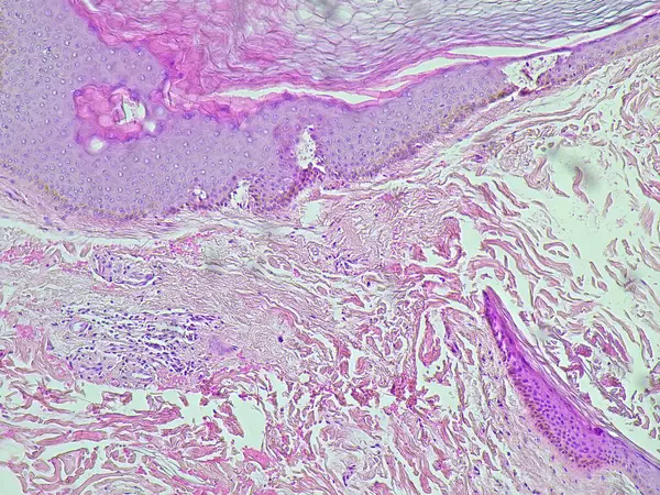
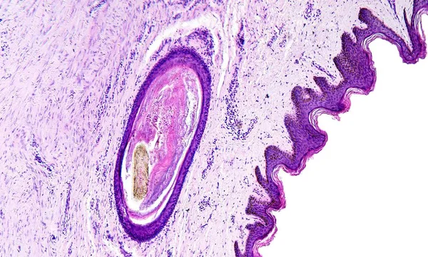
Histology of human tissue, show skin with hair follicles as seen under the microscope, 10x zoom
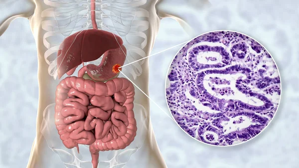
Stomach adenocarcinoma, gastric cancer, illustration and light micrograph
Colon biopsy from the colonoscopy. The pathology report describes normal colonic mucosa fragment with colic glands. Haematoxylin and eosin staining technique slide.
Endometrial adenocarcinoma, light micrograph
Spinal cord, cross-section. 3D illustration which shows the white and the grey matter with dorsal and ventral horns
Histopathology of interstitial nephritis, light micrograph, photo under microscope
Photomicrograph of lobar pneumonia during the hemorrhagic edema period, displaying lung tissue inflammation with hemorrhagic changes.
Lab technician examines scrapings on glass slides
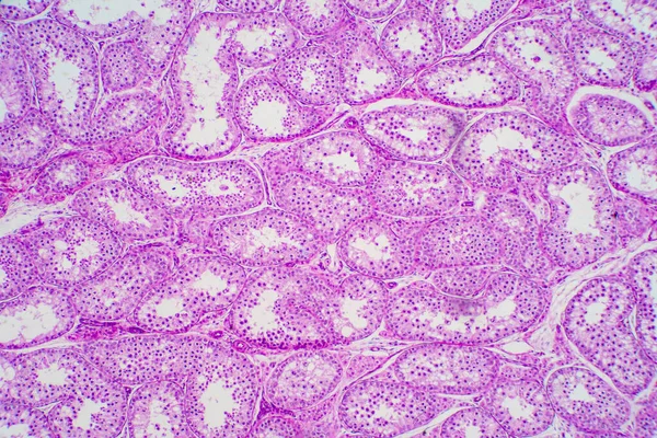
Cross section Human testis under microscope view. Shows spermatogonia, spermatocytes in meiosis, spermatids, and spermatozoa
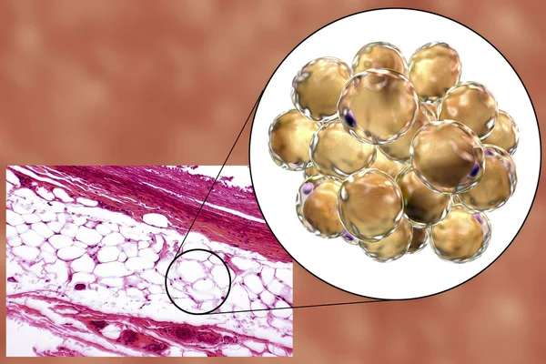
Fat cells, micrograph and 3D illustration
Human cells, micrograph and 3D illustration
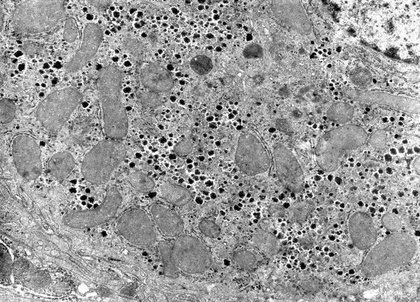
Cytoplasm. Cell organelles, TEM
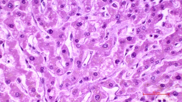
Light micrograph of a human liver stained with hematoxylin and eosin. The hepatocytes are arranged in cords separated by clear areas where hepatic sinusoids showing red blood cells are located.
Layer of human cells
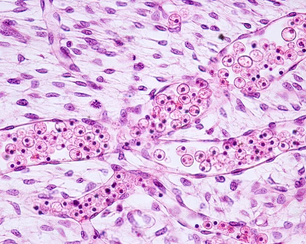
Embryonic blood vessels
Squamous epithelial cells of human cervix under the microscope view. Pap smear test is a procedure to test for cervical cancer in women.
Under high magnification microscopic view of vagina
Under high magnification microscopic view of tooth
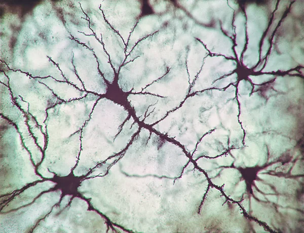
2D illustration, neurons and their connections. Microscopic photo simulation of the human nervous system.
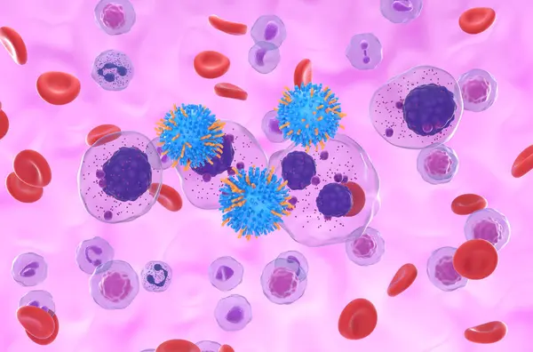
CAR T cell therapy in Multiple myeloma (MM) - isometric view 3d illustration
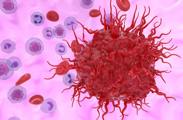
Metastatic neuroendocrine tumor cell in the blood flow - 3d illustration closeup view
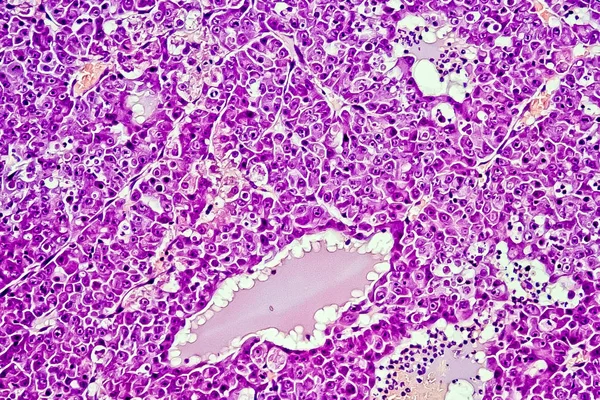
Hepatocellular carcinoma - liver cancer in rat, 60x zoom
Cells of the bark from a cork tree.
Intestinal villi. Intestine lining. Microscopic capillary. Human intestine. Concept of a healthy or diseased intestinal. Viruses or bacteria, cell infected organism, decreased immunity, 3D Rendering
Micrograph of ovary showing primordial, primary and secondary follicles isolated on white background.
Root tip of Onion show Mitosis cell in the Root tip under the microscope view.
Microscopic section of human kidney tissue
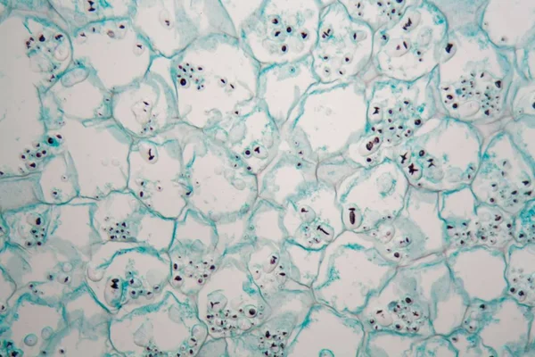
Lillium Anther under Optical Microscope
Developing long bone. Growth plate
Light micrograph of a liver
Elastic cartilage of human outer ear
Osteon, highlighting osteocytes, Haversian canal, and the lacunocanalicular network in bone tissue structure diagram hand drawn schematic raster illustration. Medical science educational illustration
Human sperm in the testis morphology under microscope. Micrograph showing spermatozoon for pathology education.
3D illustration of a Caucasian man with transparent skin, revealing a fatty liver, highlighting liver anatomy and fat accumulation, alongside with light micrograph of liver steatosis.
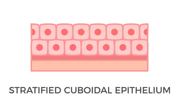
Stratified cuboidal epithelium. Epithelial tissue types. Multiple layers of cube-like cells. Occurs in the excretory ducts of sweat glands and salivary glands. Medical illustration. Vector.
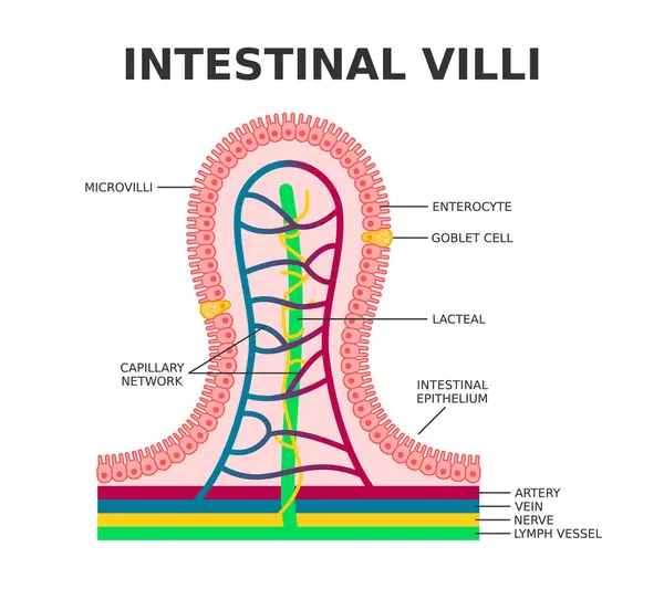
Intestinal villi. Microvilli. Intestinal epithelium. Villi absorb nutrients from the food. Intestinal epithelial cells with capillary network. Enterocyte and Goblet cell. Vector illustration.
Human prostate. Corpora amylacea
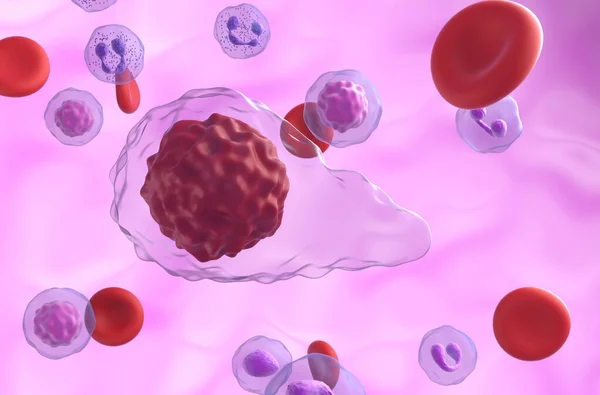
Acute myeloid leukemia (AML) cells in blood flow - section view 3d illustration
Primary myelofibrosis (PMF) cells in blood flow - closeup view 3d illustration
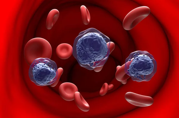
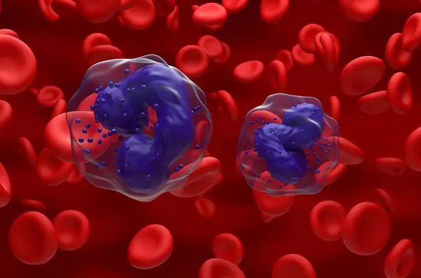
Chronic Myelogenous Leukemia (CML) cells in the blood flow - closeup view 3d illustration
Epithelial Tissue with Goblet Cells. Goblet Cell Structure Illustration. Mucus Secreting. Human Goblet Cell Anatomy Educational Chart
Human intestine with intestinal bacteria, 3D illustration
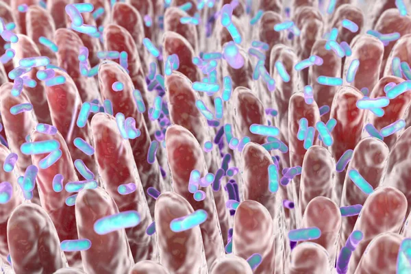
Intestinal villi and bacteria. Human intestine. 3d illustration.
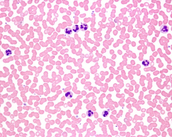
Human blood smear. Leukocytosis in an acute infection. Most of the neutrophil leukocytes exhibit nuclear hypersegmentation.
Anodonta gills ciliated epithelium under the microscope - Abstra
Male scientist looking at slides under the microscope
Next Page
1
2
3
4
5
...
100
Page
Go
Home
Photos
histology photos & images
Related searches
anatomic pathology
,
hematoxylin
,
tissue microscopic image
,
cell tissue
,
cancer pathology
,
liver cell