Actine Stock Vector Images

75 Actine vector art & graphics are available under a royalty-free license
Colorful structure skeletal muscle scheme on white background
ATP muscle contraction cycle vector illustration labeled educational scheme
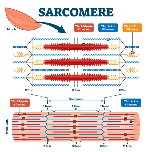
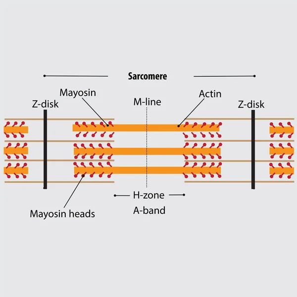
Sarcomere muscular biology scheme vector illustration
Actin vector illustration. Labeled diagram with protein structure.
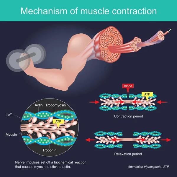
The muscle contraction as a result of Nerve impulses set off a biochemical reaction that causes myosin to stick to actin. Human body infographic.
Muscle Contraction Result Nerve Impulses Set Biochemical Reaction Causes Myosin Stick Actin Human Body Infographic
Smooth muscle tissue structure. Walls of internal organs and blood vessels in the human body. Spindle shaped myocyte. Intestine anatomy, digestive system diagram, medical flat vector illustration.
Smooth Muscle Tissue Structure Walls Internal Organs Blood Vessels Human Body Spindle Shaped Myocyte Intestine Anatomy Digestive System Diagram Medical Flat Vector Illustration
The women with freckles and melasma on skin face vector illustration isolated on white background. Types of pigmentation, Freckles, Ephelides, Solar Lentigines, Actinic lentigines and melasma.
Women Freckles Melasma Skin Face Vector Illustration Isolated White Background Types Pigmentation Freckles Ephelides Solar Lentigines Actinic Lentigines Melasma
Deep sea blue background with actin and corals
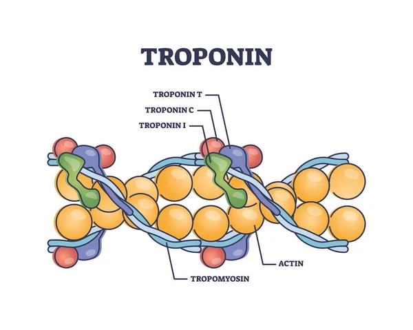
Troponin complex and three regulatory proteins structure outline diagram. Labeled educational scheme with tropomyosin and actin for muscle contraction vector illustration. Myocardial infarction marker
Troponin Complex Three Regulatory Proteins Structure Outline Diagram Labeled Educational Scheme Tropomyosin Actin Muscle Contraction Vector Illustration Myocardial Infarction Marker
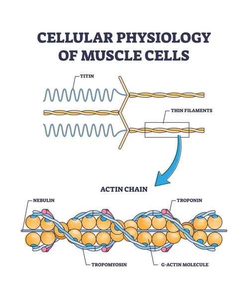
Cellular physiology of muscle cells with closeup structure outline diagram
3 types of muscle ( white, mixed and red muscle ) vector illustration set.
Types Muscle White Mixed Red Muscle Vector Illustration Set
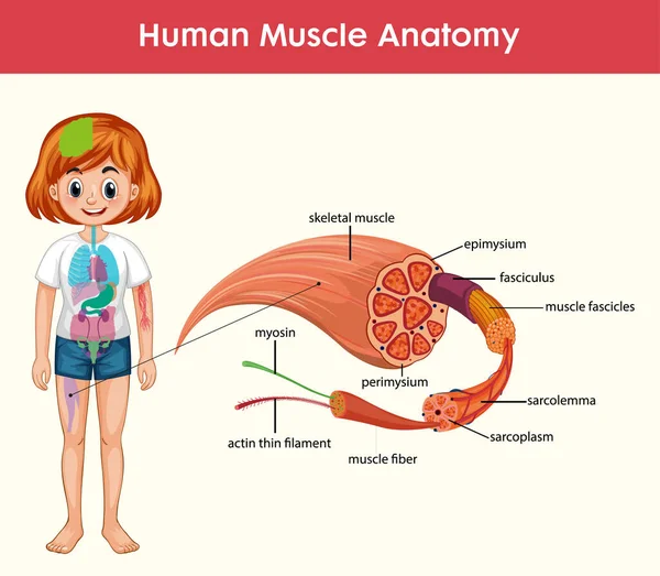
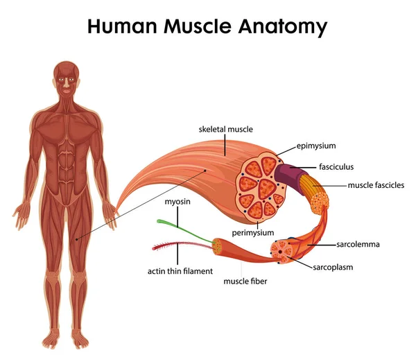
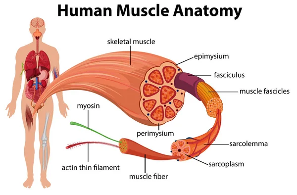
Human Muscle Anatomy Infographic With Body Anatomy illustration
Human Muscle Anatomy Infographic Body Anatomy Illustration
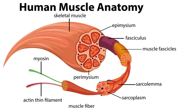
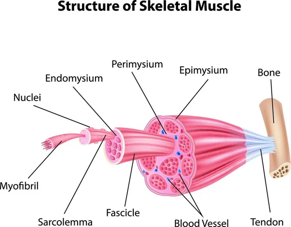
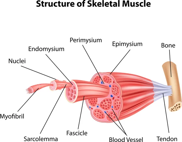
Structure of skeletal muscle vector illustration
Structure Skeletal Muscle Vector Illustration
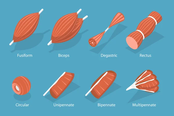
3D Isometric Flat Vector Set of Skeletal Fiber Muscle, Types of Tissue
Isometric Flat Vector Set Skeletal Fiber Muscle Types Tissue
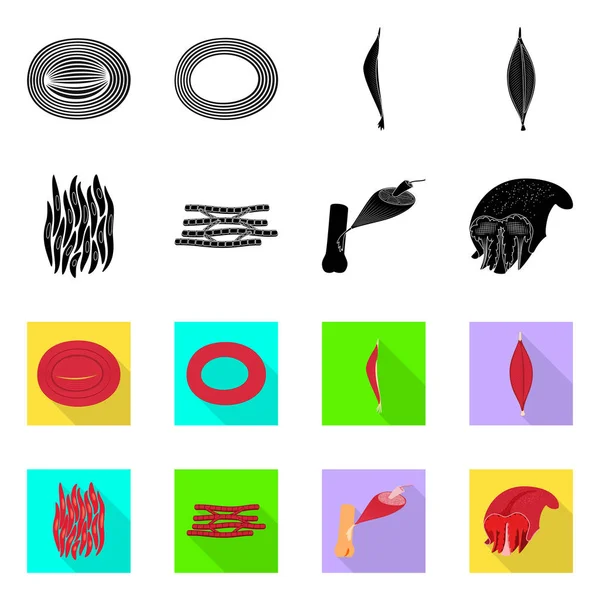
Vector illustration of muscle and cells symbol. Set of muscle and anatomy vector icon for stock.
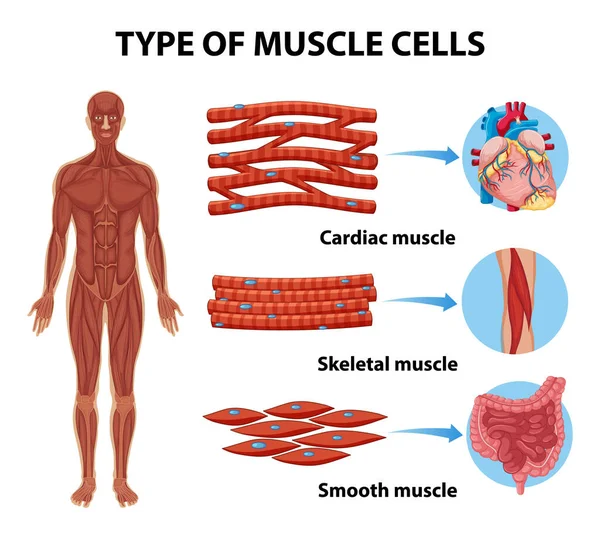
Type of Muscle Cells for health education Infographic illustration
Type Muscle Cells Health Education Infographic Illustration
Platelets structure thrombocytes anatomy diagram. Biology educational poster. Labeled mitochondria, dense body, granules, glycogen, myosin, lysosome, actin, canalicular system, microtubules.
Platelets Structure Thrombocytes Anatomy Diagram Biology Educational Poster Labeled Mitochondria Dense Body Granules Glycogen Myosin Lysosome Actin Canalicular System Microtubules
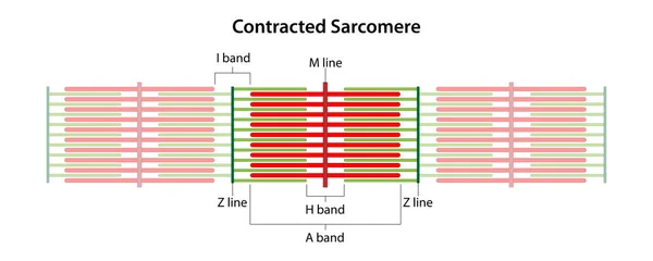
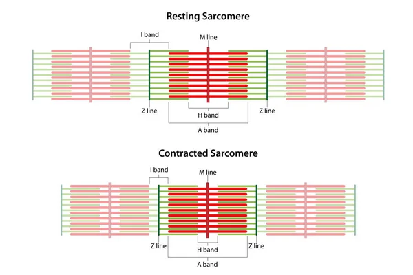
Contracted Sarcomere. Location of the I band, A band, H band, M line, and Z lines.
Contracted Sarcomere Location Band Band Band Line Lines
Type of Muscle Cells for health education Infographic illustration
Type Muscle Cells Health Education Infographic Illustration
Vector illustration of muscle and cells icon. Set of muscle and anatomy vector icon for stock.
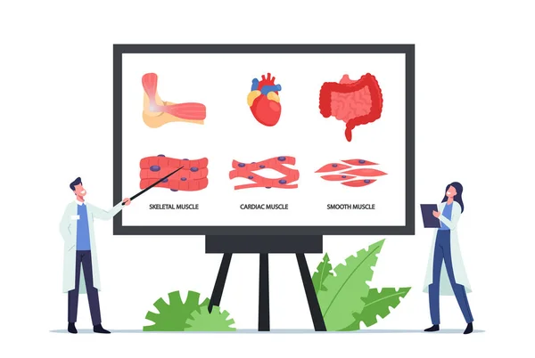
Types of Muscles Landing Page Template. Tiny Doctors Characters at Huge Board with Infographics Presenting Muscles
Vector design of fiber and muscular sign. Collection of fiber and body stock vector illustration.
Measurement tape icon outline vector. Muscle arm. Bicep power
Measurement Tape Icon Outline Vector Muscle Arm Bicep Power
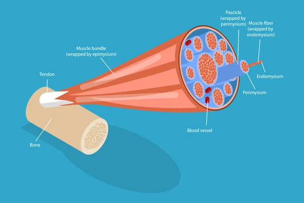
3D Isometric Flat Vector Conceptual Illustration of Skeletal Muscles, Anatomical Structure
Isometric Flat Vector Conceptual Illustration Skeletal Muscles Anatomical Structure
Chronic cheilitis. Dry chapped lips. Ripped skin. Disease of the mouth. Flaking skin flakes. Vector illustration
Chronic Cheilitis Dry Chapped Lips Ripped Skin Disease Mouth Flaking Skin Flakes Vector Illustration
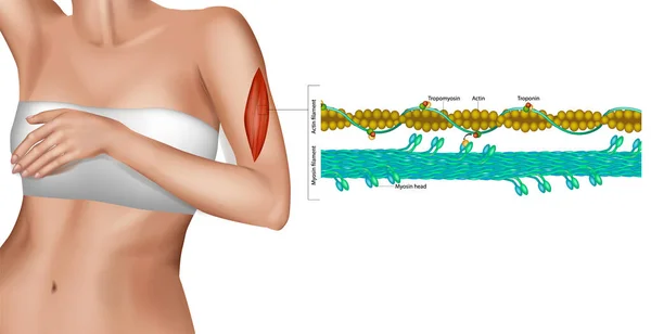
Actin filament and Myosin filament. Structure Myosin. Muscle Actin myosin interaction. Troponin or troponin complex.
Actin Filament Myosin Filament Structure Myosin Muscle Actin Myosin Interaction Troponin Troponin Complex
Muscular Health Concept. Tiny Doctors at Huge Board with Infographics Presenting Skeletal, Cardiac and Smooth Muscles
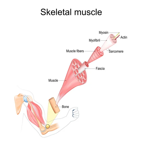
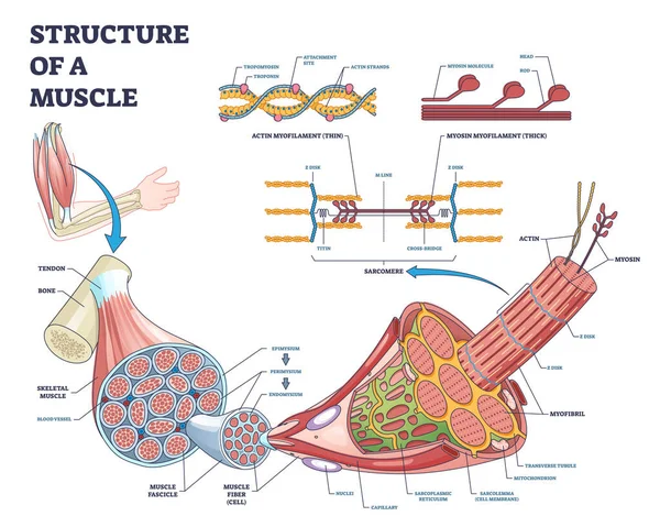
Skeletal Muscle anatomy. structure of Muscle fibers from Fascia and Tendon to Actin and Myosin. Vector poster
Skeletal Muscle Anatomy Structure Muscle Fibers Fascia Tendon Actin Myosin Vector Poster
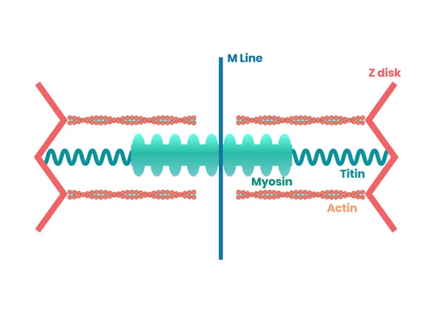
Heart muscle proteins and philament structure. Myosin, actin and titin illustration
Skeletal muscle anatomy outline diagram. Structure with anatomical inner layers. Labeled educational medical vector illustration.
Skeletal Muscle Anatomy Outline Diagram Structure Anatomical Inner Layers Labeled Educational Medical Vector Illustration
Human Muscle Anatomy Landing Page Template. Characters at Board with Infographics Skeletal, Cardiac and Smooth Muscles
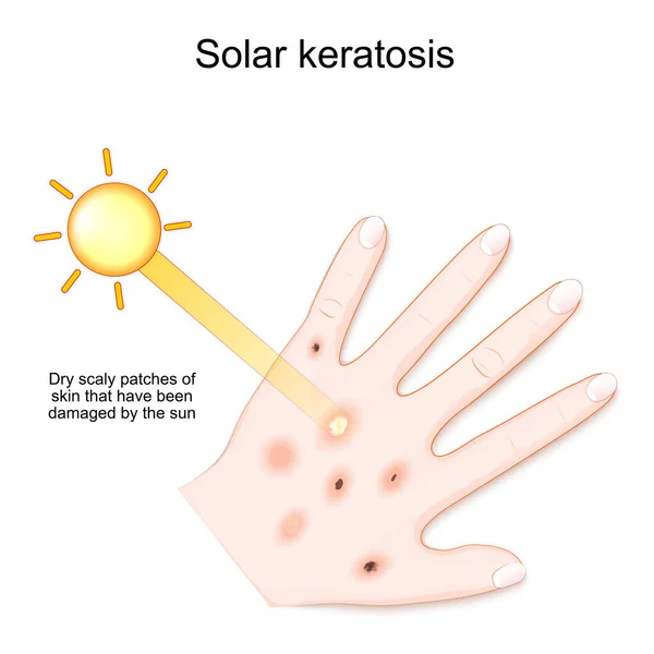
Actinic keratosis are dry scaly patches of skin that have been damaged by the sun. humans hand with solar keratosis. vector illustration
Actinic Keratosis Dry Scaly Patches Skin Have Been Damaged Sun Humans Hand Solar Keratosis Vector Illustration
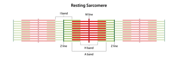
Resting Sarcomere. Location of the I band, A band, H band, M line, and Z lines.
Resting Sarcomere Location Band Band Band Line Lines
Simple Black Muscle Fiber Shape Icon Illustration
Vector illustration of actinic keratosis. malignization. cancer
Vector Illustration Actinic Keratosis Malignization Cancer
Solar keratosis. Dry scaly patches of skin that have been damaged by the sun. Human's palm with rash after sun. vector illustration
Solar Keratosis Dry Scaly Patches Skin Have Been Damaged Sun Human Palm Rash Sun Vector Illustration
The muscle contraction as a result of Nerve impulses set off a biochemical reaction that causes myosin to stick to actin. Human body infographic.
Muscle Contraction Result Nerve Impulses Set Biochemical Reaction Causes Myosin Stick Actin Human Body Infographic
Structure of muscle with isolated myosin and actin closeup outline diagram
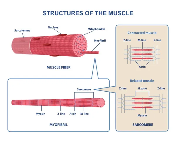
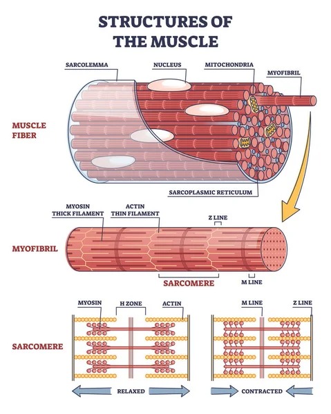
Structures of muscle with fiber, myofibril and sarcomere outline diagram
Human Muscle Anatomy Diagram illustration
Human Muscle Anatomy Diagram Illustration
Vector illustration of fiber and muscular logo. Collection of fiber and body stock symbol for web.
Structure of skeletal muscle vector illustration
Structure Skeletal Muscle Vector Illustration
Protein jar icon outline vector. Arm muscle. Gym man
Protein Jar Icon Outline Vector Arm Muscle Gym Man
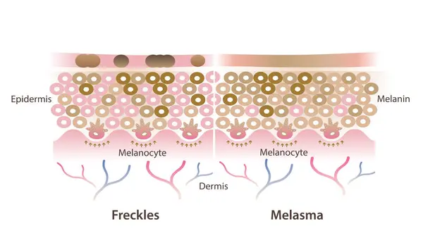
Cross section of freckles and melasma on skin layer vector illustration isolated on white background. Comparison of freckles are small flat brown spots and melasma is brown, blue gray patches on skin.
Cross Section Freckles Melasma Skin Layer Vector Illustration Isolated White Background Comparison Freckles Small Flat Brown Spots Melasma Brown Blue Gray Patches Skin
Atomic resolution of muscle contraction. Muscle Contraction. Structure of skeletal muscle. Actin filament and Myosin filament. Muscle Actin myosin interaction. Troponin or troponin complex.
Atomic Resolution Muscle Contraction Muscle Contraction Structure Skeletal Muscle Actin Filament Myosin Filament Muscle Actin Myosin Interaction Troponin Troponin Complex
Sarcomeres in different functional stages: resting and contracted. Sarcomere showing the location of the I band, A band, H band, M line, and Z lines.
Sarcomeres Different Functional Stages Resting Contracted Sarcomere Showing Location Band Band Band Line Lines
Vector Structure of kidneys. anatomy of kidneys. main organ of the urinary system. kidneys excrete metabolic waste from the body by producing urine.
Vector Structure Kidneys Anatomy Kidneys Main Organ Urinary System Kidneys Excrete Metabolic Waste Body Producing Urine
Deep sea blue background
Sea ​​horse on a blue background with actin
Skeletal muscle structure close up
Type of Muscle Cells for health education Infographic illustration
Type Muscle Cells Health Education Infographic Illustration
Anatomy of a Skeletal muscle fiber. Myofibril structure include Myosin, Z-line, M-line, Actin filaments, and Sarcomere. Isometric flat vector Illustration
Anatomy Skeletal Muscle Fiber Myofibril Structure Include Myosin Line Line Actin Filaments Sarcomere Isometric Flat Vector Illustration
Deep sea blue background with actin and corals
Muscle icons set outline vector. Fiber tissue. Muscular body
Muscle Icons Set Outline Vector Fiber Tissue Muscular Body
Muscle contraction mechanism shown through a bicep curl, nerve signal drives actin myosin sarcomere shortening, key objects, bicep, myofibril, sarcomere. Outline diagram
Muscle Contraction Mechanism Shown Bicep Curl Nerve Signal Drives Actin Myosin Sarcomere Shortening Key Objects Bicep Myofibril Sarcomere Outline Diagram
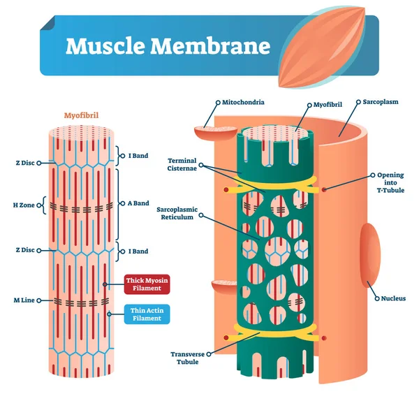
Muscle membrane vector illustration. Labeled scheme with myofibril, disc, zone, line and band. Anatomical diagram with mitochondria, sarcoplasm, reticulum and nucleus.
Cytoskeleton structure as complex protein filaments network outline diagram
Human Muscle Anatomy Infographic With Body illustration
Human Muscle Anatomy Infographic Body Illustration
Human Muscle Anatomy for health education Infographic illustration
Human Muscle Anatomy Health Education Infographic Illustration
Isolated object of muscle and cells symbol. Set of muscle and anatomy stock vector illustration.
Human Muscle Anatomy Diagram illustration
Human Muscle Anatomy Diagram Illustration
Deep sea blue background with actin and corals. Banner for your text
Healthy Lifestyle Landing Page Template. Tiny Doctor Character Presenting Skeletal Musculature on Huge Infographics
Illustration of Structure Skeletal Muscle Anatomy
Illustration Structure Skeletal Muscle Anatomy
Illustration of Structure Skeletal Muscle Anatomy
Dendritic spine. Neuron anatomy. Learning and memory. Neuronal plasticity. Neural communication. Transmission of the nerve signal between two neurons with axon and synapse. Vector illustration
Dendritic Spine Neuron Anatomy Learning Memory Neuronal Plasticity Neural Communication Transmission Nerve Signal Two Neurons Axon Synapse Vector Illustration
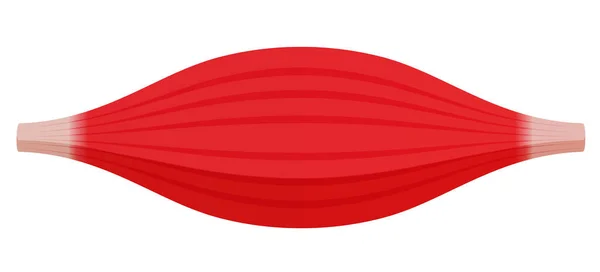
Healthy muscle vector illustration ( red muscle )
Healthy Muscle Vector Illustration Red Muscle
Skeletal muscle anatomy outline diagram. Structure with anatomical inner layers. Educational medical vector illustration.
Skeletal Muscle Anatomy Outline Diagram Structure Anatomical Inner Layers Educational Medical Vector Illustration
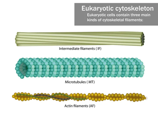
Three Eukaryotic cells cytoskeletal filaments
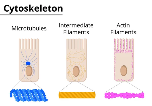
Different structures of cytoskeleton. Microtubules, intermediate filaments and actin filaments. Vector illustration. Didatic illustration.
Different Structures Cytoskeleton Microtubules Intermediate Filaments Actin Filaments Vector Illustration Didatic Illustration
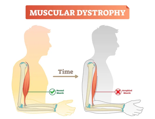
Vector illustration about muscular dystrophy. Compared normal muscle and atrophied muscle. Medical scheme how time affects health - healthy and weak human.
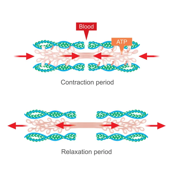
The body will use nutrients that provide energy when exercising, By based on factors from the blood system And primarily muscles, Waste such as perspiration and heat is released.
Body Use Nutrients Provide Energy Exercising Based Factors Blood System Primarily Muscles Waste Perspiration Heat Released
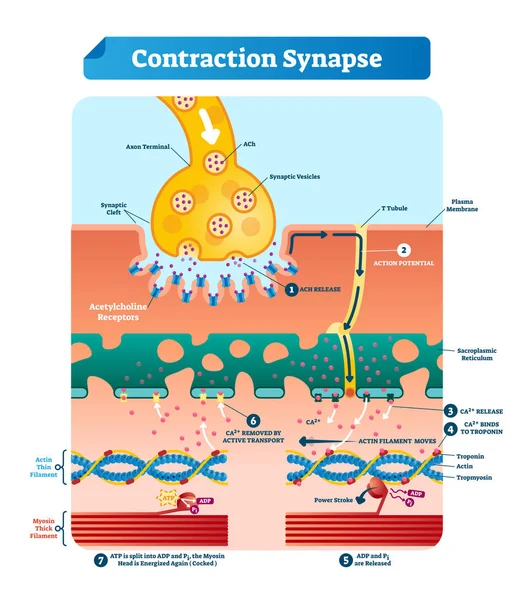
Contraction synapse vector illustration. Labeled medical structure scheme.
Smooth muscle tissue structure. Walls of internal organs and blood vessels in the human body. Spindle shaped myocyte. Intestine anatomy, digestive system diagram, medical flat vector illustration.
Smooth Muscle Tissue Structure Walls Internal Organs Blood Vessels Human Body Spindle Shaped Myocyte Intestine Anatomy Digestive System Diagram Medical Flat Vector Illustration
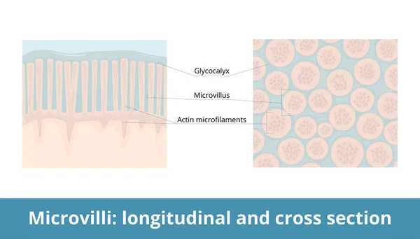
Microvilli: longitudinal and cross-section. Plasma membrane extension increases cell's surface area with its microvillus and actin microfilaments. Glycocalyx as a protective layer.
Microvilli Longitudinal Cross Section Plasma Membrane Extension Increases Cell Surface Area Its Microvillus Actin Microfilaments Glycocalyx Protective Layer