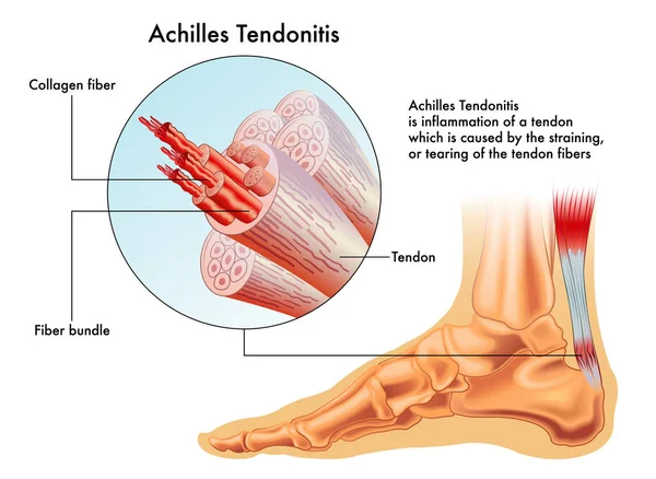
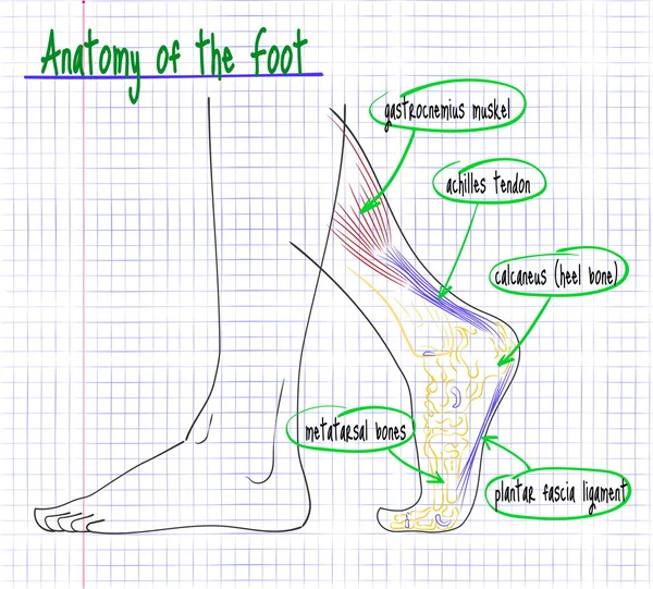
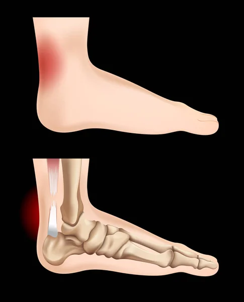
Vector illustration of achilles tendon, image of foot anatomy with all tendons and bones
Vector Illustration Achilles Tendon Image Foot Anatomy All Tendons Bones
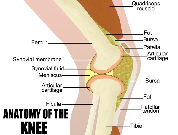
Anatomy of the knee
Anatomy of the knee
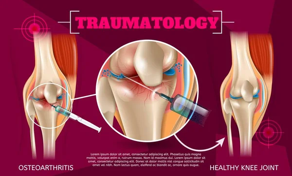
Realistic Illustration Traumatology Medicine in 3d. Vector Banner Image Anatomy Human Knee Joint Treatment Osteoarthritis Injection Drug Method Injection. Healthy Knee Joint Result Medical Treatment
Realistic Illustration Traumatology Medicine Vector Banner Image Anatomy Human Knee Joint Treatment Osteoarthritis Injection Drug Method Injection Healthy Knee Joint Result Medical Treatment
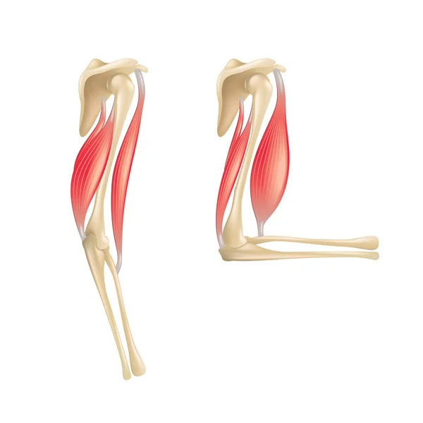
Elbow joint anatomy isolated on white vector
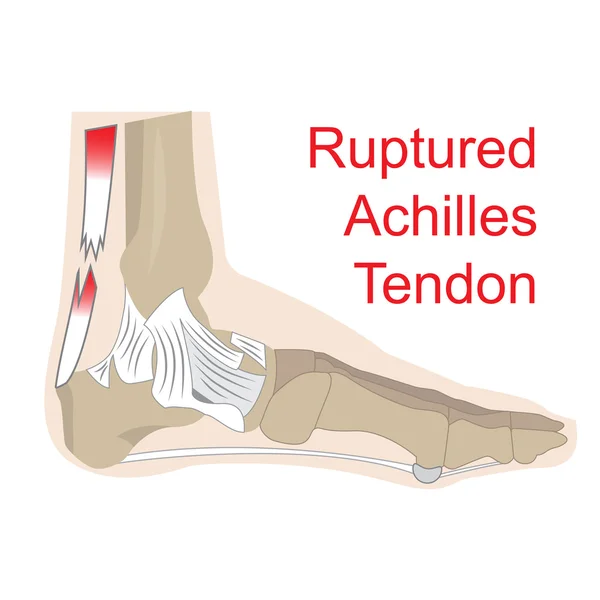
Ruptured achilles tendon
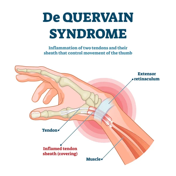
De Quervain syndrome vector illustration. Labeled thumb inflammation scheme
Anatomy of the human elbow
Shoulder bones muscles
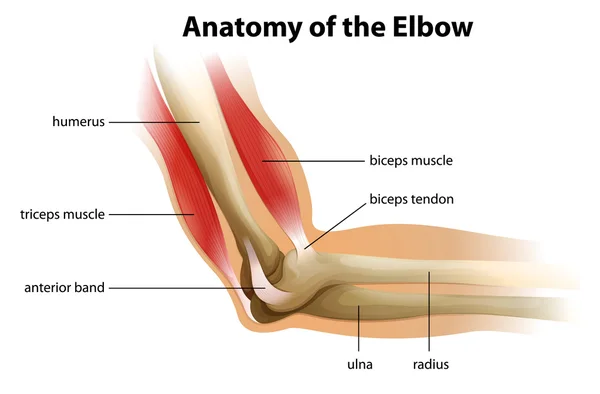
Anatomy of the Elbow
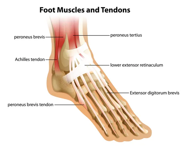
Foot Muscles and Tendons
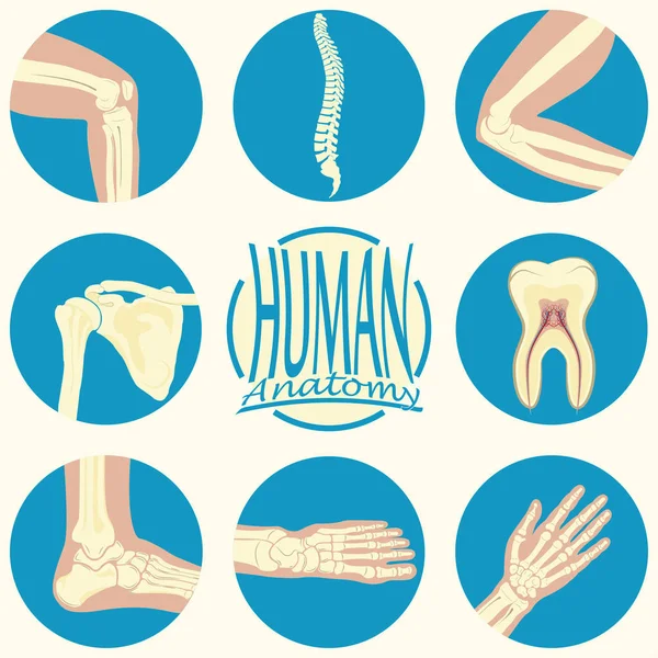
Set of humane joints
Trigger finger causes and symptoms illustration / no text
Trigger Finger Causes Symptoms Illustration Text
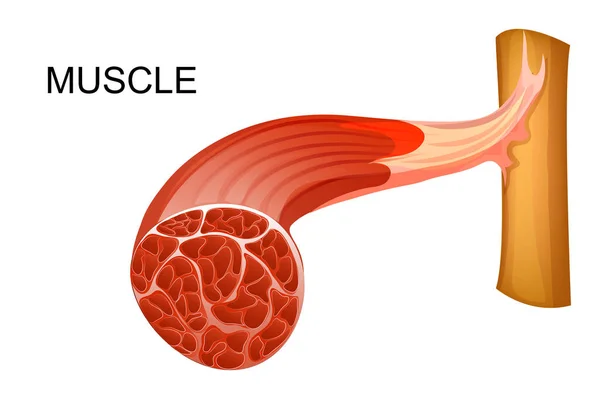
A Human Anatomy of Muscle illustration
Human Anatomy Muscle Illustration
Contractura aponeurosis palmaris
Foot swelling. edema
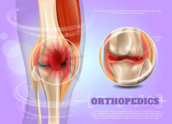
Realistic Illustration Orthopedics Medicine in 3d
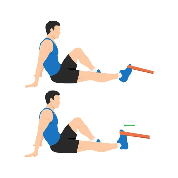
Man doing resistance band dorsiflexion exercise stretch. Ankle exercise. Flat vector illustration isolated on white background
Man Doing Resistance Band Dorsiflexion Exercise Stretch Ankle Exercise Flat Vector Illustration Isolated White Background
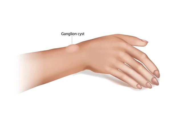
Ganglion Cyst of the Wrist and Hand. Synovial cyst or a Gideon s Disease, or a Bible Cyst, or a Bible Bump.
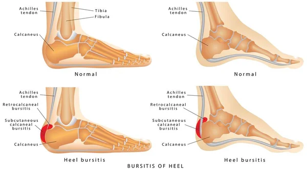
Heel Bursitis (Foot)
Detailed diagram of knee joint with inflammation
Detailed Diagram Knee Joint Inflammation
Knee joint section illustration
Knee Joint Section Illustration
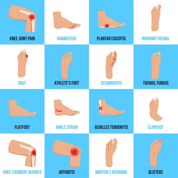
Foot Problems Flat Icons Set
Medical Illustration of posterior cruciate ligament ruptur
Medical Illustration Posterior Cruciate Ligament Ruptur
Osteochondroma knee problem as medical bone tumor overgrowth outline diagram
Finger injury types with common hand impact trauma anatomy outline diagram
Unhealthy woman sit on floor in gym touch hurt ankle. Unwell girl suffer from trauma or injury exercising or doing sports. Vector illustration.
Unhealthy Woman Sit Floor Gym Touch Hurt Ankle Unwell Girl Suffer Trauma Injury Exercising Doing Sports Vector Illustration
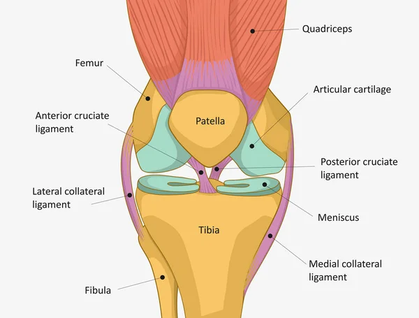
Anatomy of the knee joint front view. Knee Muscles and Ligaments Parts, knee tendons
Anatomy Knee Joint Front View Knee Muscles Ligaments Parts Knee Tendons
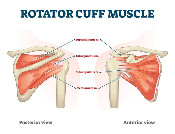
Rotator cuff muscle with anatomical posterior and anterior view expample
Anatomy of the knee joint front view, template for training a medical surgical poster, traumatology page. Vector illustration.
Anatomy Knee Joint Front View Template Training Medical Surgical Poster Traumatology Page Vector Illustration
Detailed illustration of human muscles
Anatomical illustration of muscle fibers for medical journals
Human Knee joint anatomy. Knee Tendons, anatomical diagram
Human Knee Joint Anatomy Knee Tendons Anatomical Diagram
Tensor fasciae latae stretch for muscle physical flexibility outline diagram. Labeled medical explanation with anatomical muscular stretching as part of physiotherapy activity vector illustration.
Tensor Fasciae Latae Stretch Muscle Physical Flexibility Outline Diagram Labeled Medical Explanation Anatomical Muscular Stretching Part Physiotherapy Activity Vector Illustration
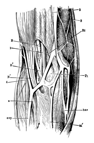
The superficial veins in front of the elbow joint, vintage line drawing or engraving illustration.
Superficial Veins Front Elbow Joint Vintage Line Drawing Engraving Illustration
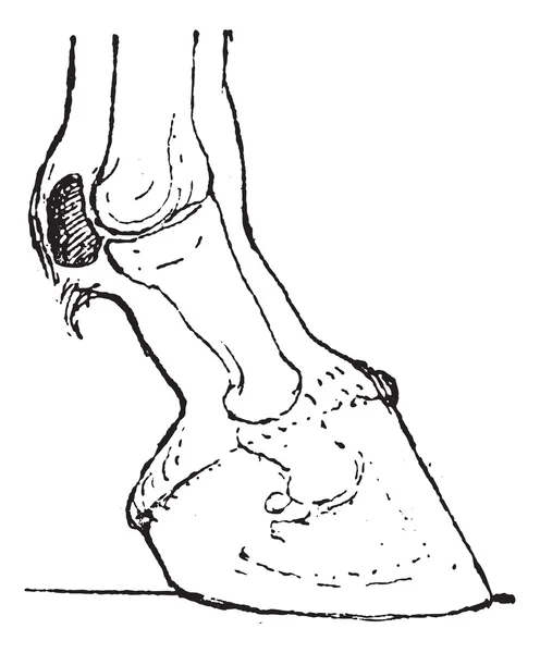
Sesamoid bones, vintage engraving.
Muscles and Tendons of the Human Arm and Hand, vintage engraved
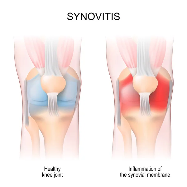
Synovitis. Human Knee joint anatomy. Healthy joint, and Inflammation of the synovial membrane. front view. anterior aspects. Vector illustration
Synovitis Human Knee Joint Anatomy Healthy Joint Inflammation Synovial Membrane Front View Anterior Aspects Vector Illustration
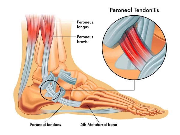
Medical illustrations of symptoms of peroneal tendonitis, with enlargement of the affected area, with annotations.
Medical Illustrations Symptoms Peroneal Tendonitis Enlargement Affected Area Annotations
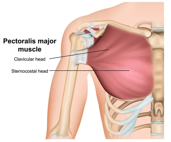
Pectoralis major breast muscle anatomy, 3d medical vector illustration on white background
Pectoralis Major Breast Muscle Anatomy Medical Vector Illustration White Background
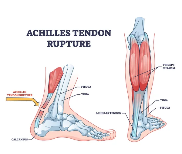
Achilles tendon rupture as painful injury and leg trauma outline diagram. Labeled educational anatomical scheme with orthopedic problem explanation vector illustration. Medical body muscle condition.
Achilles Tendon Rupture Painful Injury Leg Trauma Outline Diagram Labeled Educational Anatomical Scheme Orthopedic Problem Explanation Vector Illustration Medical Body Muscle Condition
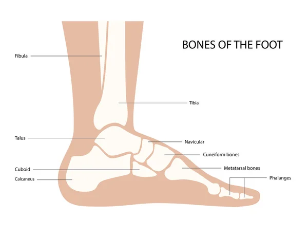
Foot bones anatomy
Foot bones anatomy
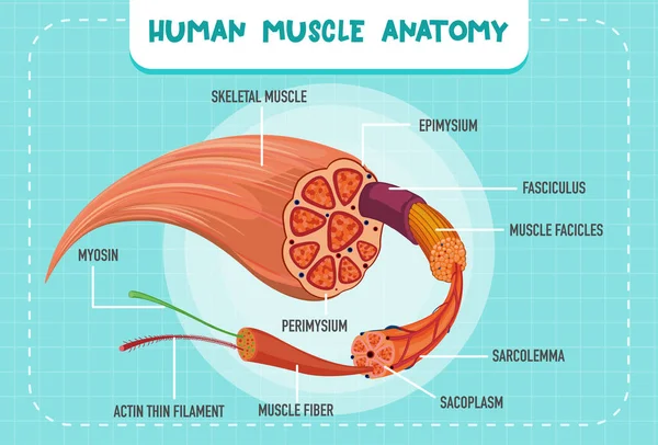
Human muscle anatomy structure illustration
Human Muscle Anatomy Structure Illustration
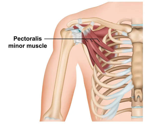
Pectoralis minor muscle anatomy, 3d medical vector illustration on white background
Pectoralis Minor Muscle Anatomy Medical Vector Illustration White Background
Carpal tunnel syndrome. Compressed median nerve. Anatomy of the carpal tunnel, showing the median nerve.
Knee damage health issue icon vector illustration
Knee Damage Health Issue Icon Vector Illustration
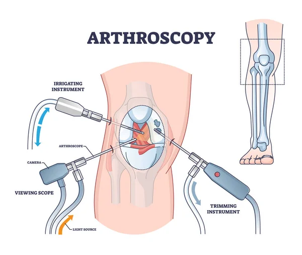
Arthroscopy procedure as knee diagnostics process explanation outline diagram
Biceps muscle with anatomical skeletal medical arm structure outline diagram
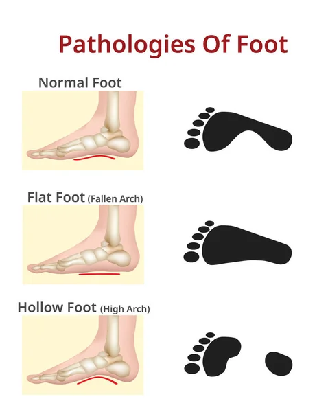
Foot pathologies, Normal, flat and hollow foot
Foot Pathologies Normal Flat Hollow Foot
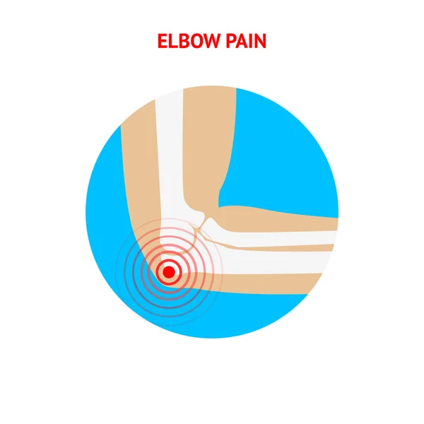
Elbow pain. Elbow pain icon isolated on white background.
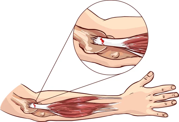
Tennis elbow - tear in the common extensor tendon of the arm
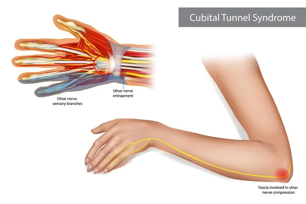
Medical illustration to explain Cubital tunnel syndrome. Ulnar nerve entrapment. Fascia involved in ulnar nerve compression.
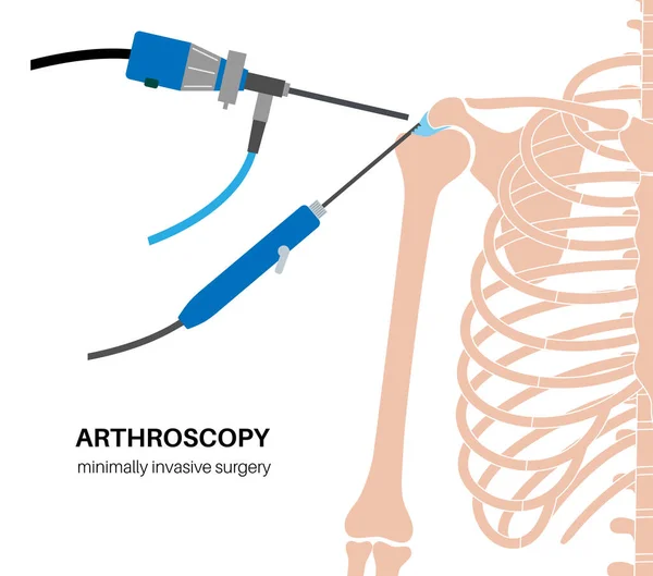
Shoulder arthroscopy procedure. Rotator cuff tears or shoulder joint replacement. Minimally invasive surgery. Ligaments treatment, tendonitis pain or arthritis inflammation medical vector illustration
Shoulder Arthroscopy Procedure Rotator Cuff Tears Shoulder Joint Replacement Minimally Invasive Surgery Ligaments Treatment Tendonitis Pain Arthritis Inflammation Medical Vector Illustration
Comparison of healthy and ruptured biceps tendon
Comparison Healthy Ruptured Biceps Tendon
Pronated foot, arch deformation, bottom and back view . Foot weight distribution.
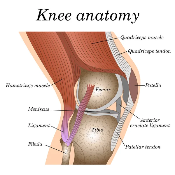
Anatomy of the knee joint side view, template for training a medical surgical poster, traumatology page. Vector illustration.
Anatomy Knee Joint Side View Template Training Medical Surgical Poster Traumatology Page Vector Illustration
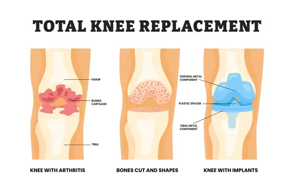
Different types of total knee replacement types, educational medical poster design healthcare scheme, labeled knee with arthritis, bones cut and shapes, knee with implant. Vector illustration.
Different Types Total Knee Replacement Types Educational Medical Poster Design Healthcare Scheme Labeled Knee Arthritis Bones Cut Shapes Knee Implant Vector Illustration
Illustration of ACL tear in the knee
Illustration Acl Tear Knee
Knee and elbow vector illustration.
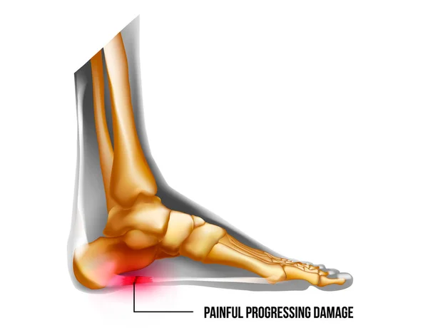
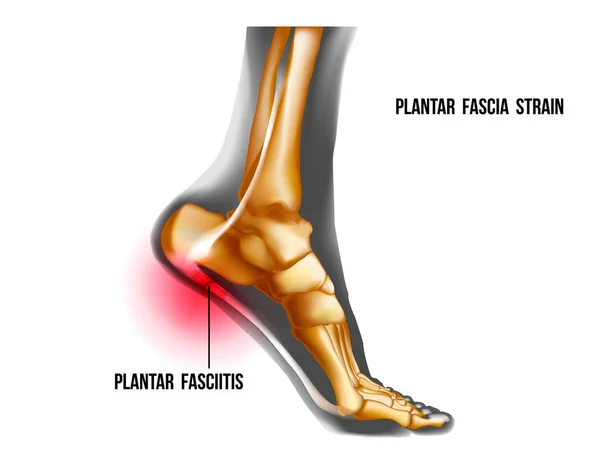
Foot pain, plantar fasciitis inflammation and ruptures strain. Bones realistic illustration. Medial view. Anatomy of joints, human leg black and yellow transparente skeleton. For medical orthopedic
Foot Pain Plantar Fasciitis Inflammation Ruptures Strain Bones Realistic Illustration Medial View Anatomy Joints Human Leg Black Yellow Transparente Skeleton Medical Orthopedic
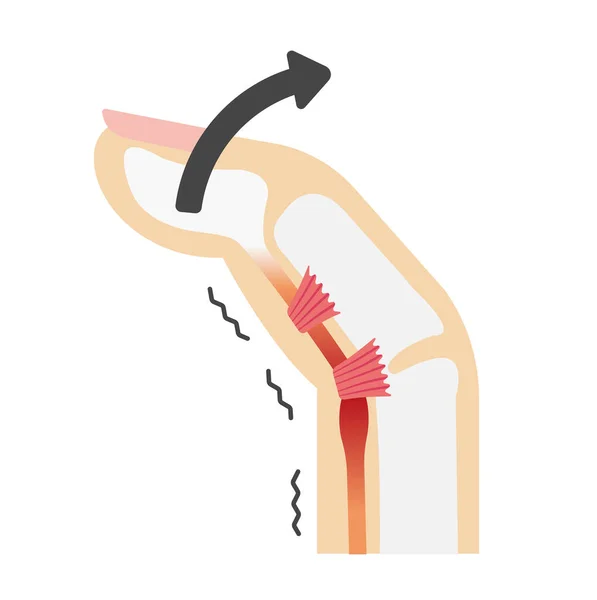
Diagram showing mallet finger injury and tendon rupture
Diagram Showing Mallet Finger Injury Tendon Rupture
Diaphragm muscle with exhalation and inhalation movement outline diagram
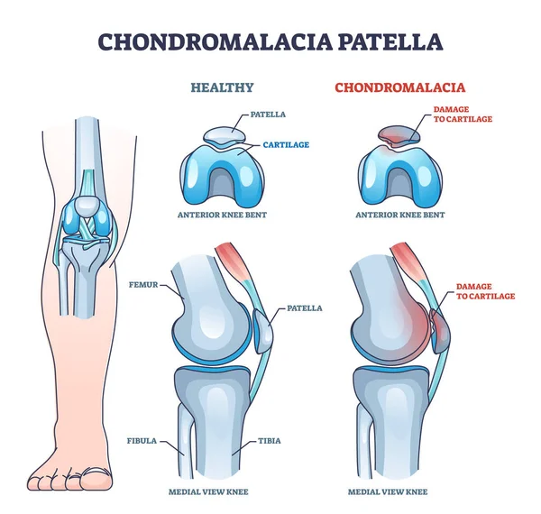
Chondromalacia patella knee breakdown compared with healthy outline diagram. Labeled educational kneecap tissue damage with cartilage problem and anatomical leg joint structure vector illustration.
Chondromalacia Patella Knee Breakdown Compared Healthy Outline Diagram Labeled Educational Kneecap Tissue Damage Cartilage Problem Anatomical Leg Joint Structure Vector Illustration
Detailed diagram of tennis elbow condition
Detailed Diagram Tennis Elbow Condition
Plantar fasciitis inflammation and ruptures strain. Foot pain, realistic anatomy illustration
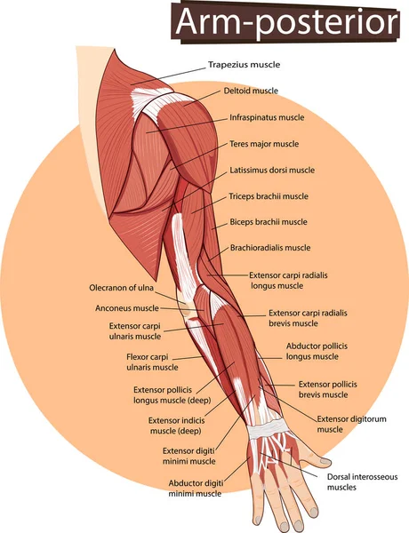
Vector of Arm anatomy
Realistic Illustration Orthopedics Medicine in 3d. Banner Vector Image Closeup Anatomy and Structure Human Knee Joint. Infographics Study Method Treatment and Elimination Foot Joint Disease
Realistic Illustration Orthopedics Medicine Banner Vector Image Closeup Anatomy Structure Human Knee Joint Infographics Study Method Treatment Elimination Foot Joint Disease
Skeletal muscle structure layers with anatomical closeups outline diagram
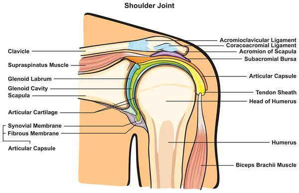
Shoulder Joint of Human Body Anatomy infographic diagram with all parts including bones ligaments muscles bursa cavity capsule cartilage membrane for medical science education and health care
Shoulder Joint Human Body Anatomy Infographic Diagram All Parts Including Bones Ligaments Muscles Bursa Cavity Capsule Cartilage Membrane Medical Science Education Health Care
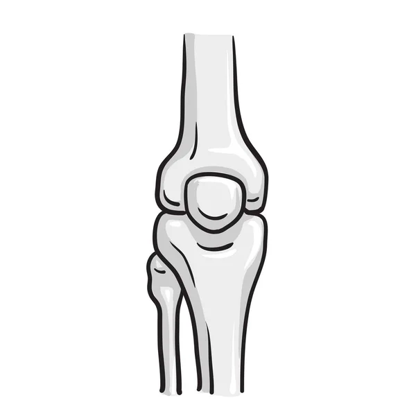
Knee bone anatomy
Knee bone anatomy
Skeletal muscle structure with anatomical inner layers outline diagram
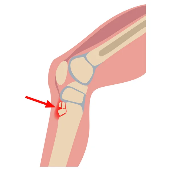
Osgood-Schlatter disease. Destruction of the tibial nucleus. Human leg on the side. Medical poster, infographics. Vector illustration
Osgood Schlatter Disease Destruction Tibial Nucleus Human Leg Side Medical Poster Infographics Vector Illustration
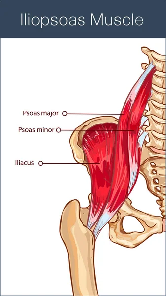
Vector illustration of a iliopsoas muscle
Vector Illustration Iliopsoas Muscle
Drawing of the anatomy of the human foot side
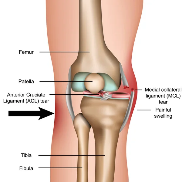
Dislocated knee, medial collateral ligament tear medical vector illustration isolated on white background
Dislocated Knee Medial Collateral Ligament Tear Medical Vector Illustration Isolated White Background
Knee pain and pain throbbing into the bone Inflammatory disease of the knee joint on black background. Realistic 3d vector file.
Knee Pain Pain Throbbing Bone Inflammatory Disease Knee Joint Black Background Realistic Vector File
Soleus muscle with anatomical leg bones skeletal structure outline diagram
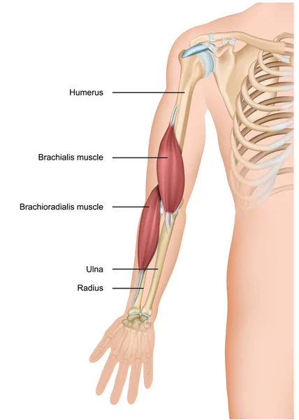
Brachialis and brachioradialis 3d medical vector illustration arm anatomy
Brachialis Brachioradialis Medical Vector Illustration Arm Anatomy
Hip labrum tear, Hip joint medical illustration flashcard
Hip Labrum Tear Hip Joint Medical Illustration Flashcard
Section passing through the middle third of the first phalanx of the middle finer, vintage line drawing or engraving illustration.
Section Passing Middle Third First Phalanx Middle Finer Vintage Line Drawing Engraving Illustration
The thigh has three sets of strong muscles. Shown here is some of the human larger muscles on the back of the thigh. Powerful tendons at the hip and on the back of the knee are well shown, vintage line drawing or engraving illustration.
Thigh Has Three Sets Strong Muscles Shown Here Some Human Larger Muscles Back Thigh Powerful Tendons Hip Back Knee Well Shown Vintage Line Drawing Engraving Illustration
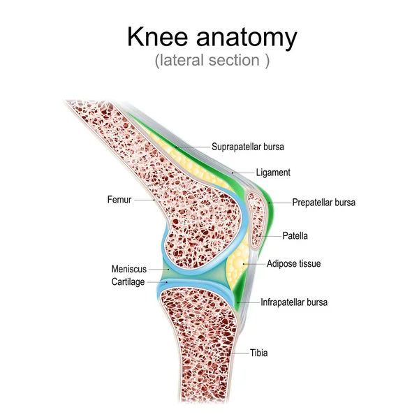
Knee anatomy. Side view. lateral section of joint with ligaments, meniscus, and bursae. knee joint cavity. Cross section of leg bones. detailed diagram. Vector poster
Knee Anatomy Side View Lateral Section Joint Ligaments Meniscus Bursae Knee Joint Cavity Cross Section Leg Bones Detailed Diagram Vector Poster
Knee joint with healthy cartilage on blue and white background with copy space for your text. side view of human knee. Vector illustration for presentation, advertising, banner, visiting card, or calling card, poster or medical education.
Knee Joint Healthy Cartilage Blue White Background Copy Space Your Text Side View Human Knee Vector Illustration Presentation Advertising Banner Visiting Card Calling Card Poster Medical Education
Knee Tendons medical vector illustration scheme, anatomical diagram.
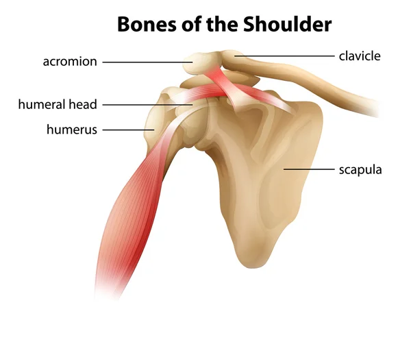
Bones of the Shoulder
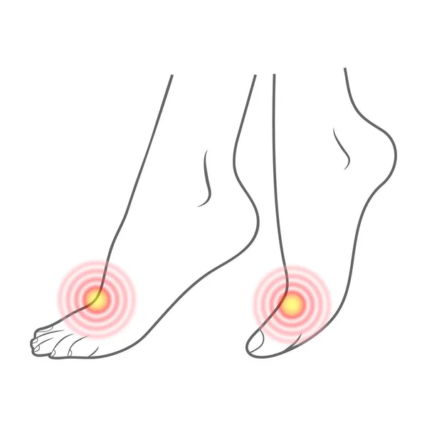
Vector illustration of foot pain with highlighted painful area. Isolated flat style medical concept for health, podiatry, arthritis, injury or anatomy design
Vector Illustration Foot Pain Highlighted Painful Area Isolated Flat Style Medical Concept Health Podiatry Arthritis Injury Anatomy Design
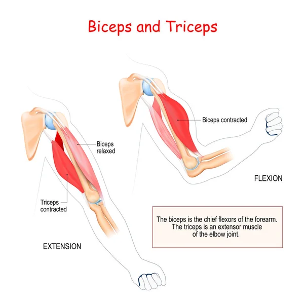
Biceps and triceps. Antagonist muscles. The biceps is the chief flexors of the forearm. The triceps is an extensor muscle of the elbow joint.
Biceps Triceps Antagonist Muscles Biceps Chief Flexors Forearm Triceps Extensor Muscle Elbow Joint
Trigger finger diagram. Joints stuck in bent position. Anatomy of tendons and bones of hand index finger. Medical trauma flexor tendon from inside educational scheme. Cartoon flat vector illustration
Trigger Finger Diagram Joints Stuck Bent Position Anatomy Tendons Bones Hand Index Finger Medical Trauma Flexor Tendon Educational Scheme Cartoon Flat Vector Illustration
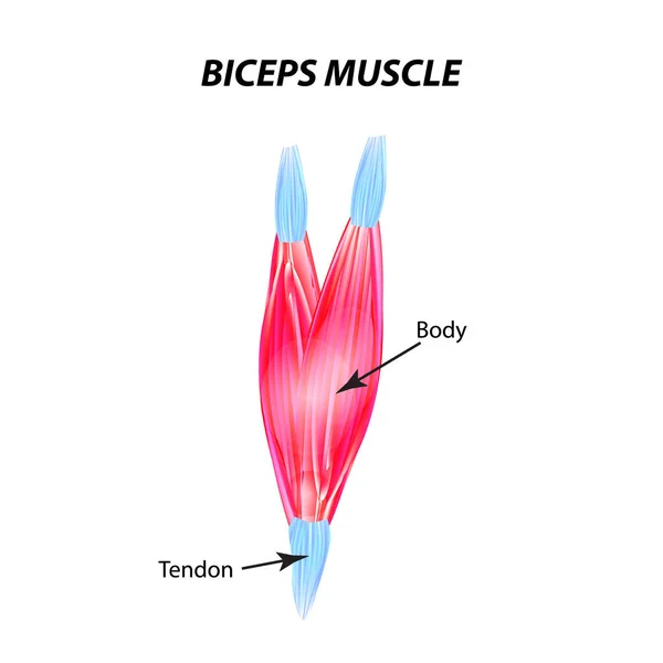
The structure of skeletal muscle. Biceps muscle. Tendon. Infographics. Vector illustration on isolated background.
Anatomy of human muscles in the front, a template for medical tutorial, banner. Vector illustration.
Anatomy Human Muscles Front Template Medical Tutorial Banner Vector Illustration
Cartoon structure of healthy knee joint. Concept of human anatomy skeleton. Design element for medical book or flyer. Detailed flat vector icon
Cartoon Structure Healthy Knee Joint Concept Human Anatomy Skeleton Design Element Medical Book Flyer Detailed Flat Vector Icon
Sever's disease, calcaneal apophysitis. Ankle injury broken bone
Sever Disease Calcaneal Apophysitis Ankle Injury Broken Bone
Human Foot Contour And Red Spots Of Pain. Ready Element For Medicine And Orthopedics Isolated On A White Background. Vector Illustration.
Pain in feet - Arthritis - Icon as EPS 10 File
Pain Feet Arthritis Icon Eps File
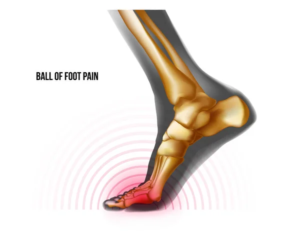
Ball of foot pain bones skeleton realistic anatomy illustration
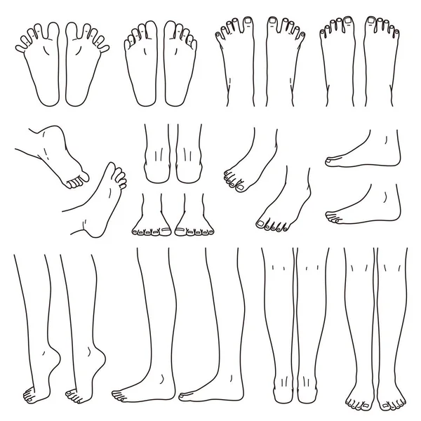
Foot and leg, knee and toe, vector file set, monochrome illustration
Foot Leg Knee Toe Vector File Set Monochrome Illustration
Comparison of normal shoulder and shoulder impingement
Comparison Normal Shoulder Shoulder Impingement
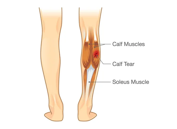
Calf muscle tear.
Calf muscle tear.
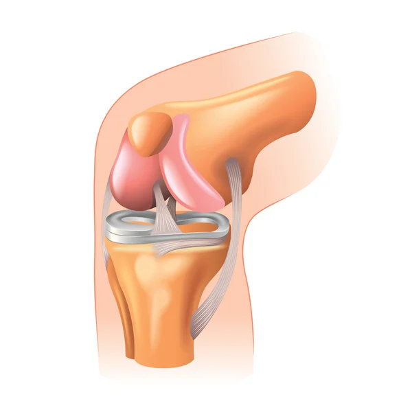
Meniscus structure. Knee joint anatomy. Cartilage and Ligaments. Vector illustration
Meniscus Structure Knee Joint Anatomy Cartilage Ligaments Vector Illustration
Diagram showing tendon injury
Knee joint anatomy isolated on white vector
Page