Arthroscopy Stock Vector Images

42 Arthroscopy vector art & graphics are available under a royalty-free license
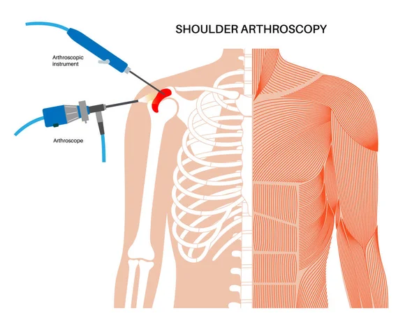
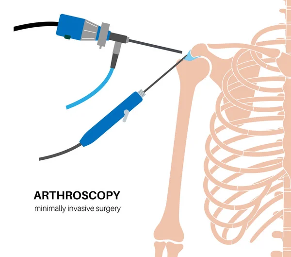
Shoulder arthroscopy procedure. Rotator cuff tears, shoulder muscles problem. Minimally invasive surgery. Arthroscopic joint and ligaments treatment, pain or inflammation in the human body flat vector
Shoulder Arthroscopy Procedure Rotator Cuff Tears Shoulder Muscles Problem Minimally Invasive Surgery Arthroscopic Joint Ligaments Treatment Pain Inflammation Human Body Flat Vector
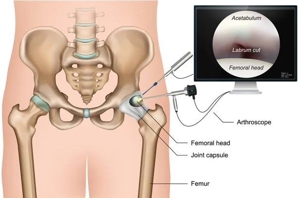
Hip arthroscopy 3d medical vector illustration on white background
Hip Arthroscopy Medical Vector Illustration White Background
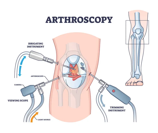
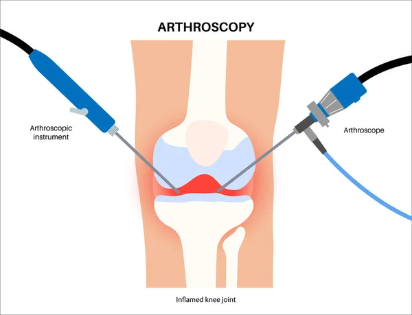
Arthroscopy procedure as knee diagnostics process explanation outline diagram
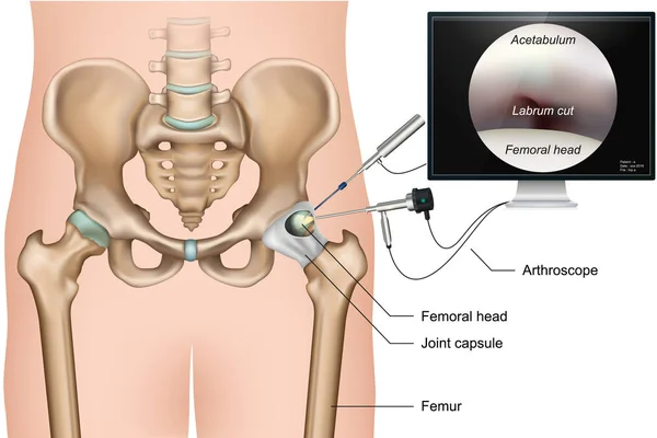
Hip arthroscopy 3d medical vector illustration on white background
Hip Arthroscopy Medical Vector Illustration White Background
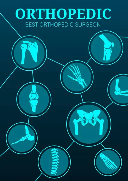
Orthopedic surgery and hematology clinic, vector poster. Orthopedics surgeon medicine, joint and bones treatment technology, medical center X-ray for osteoporosis and arthritis
Orthopedic Surgery Hematology Clinic Vector Poster Orthopedics Surgeon Medicine Joint Bones Treatment Technology Medical Center Ray Osteoporosis Arthritis
Realistic Illustration Orthopedics Medicine in 3d
Shoulder arthroscopy procedure. Rotator cuff tears or shoulder joint replacement. Minimally invasive surgery. Ligaments treatment, tendonitis pain or arthritis inflammation medical vector illustration
Shoulder Arthroscopy Procedure Rotator Cuff Tears Shoulder Joint Replacement Minimally Invasive Surgery Ligaments Treatment Tendonitis Pain Arthritis Inflammation Medical Vector Illustration
Elbow joint minimally invasive surgery. Arthroscopy medical procedure. Anatomy of humerus, ulna and radius bones. Arm pain, fracture, osteoarthritis or rheumatoid arthritis x ray vector illustration
Elbow Joint Minimally Invasive Surgery Arthroscopy Medical Procedure Anatomy Humerus Ulna Radius Bones Arm Pain Fracture Osteoarthritis Rheumatoid Arthritis Ray Vector Illustration
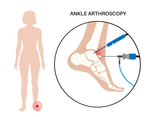
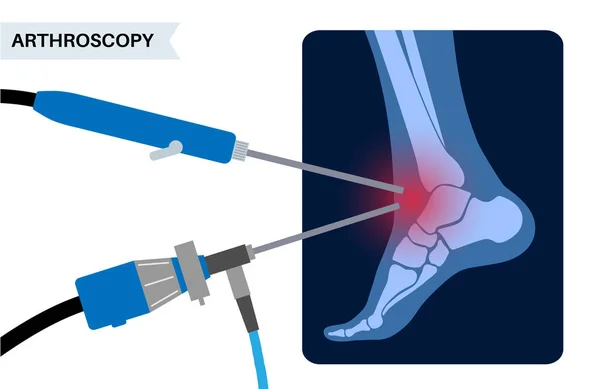
Ankle arthroscopy procedure. Feet joint minimally invasive surgery concept. Arthroscope and arthroscopic instrument. Foot treatment, leg pain and inflammation. Anatomical x ray vector illustration
Ankle Arthroscopy Procedure Feet Joint Minimally Invasive Surgery Concept Arthroscope Arthroscopic Instrument Foot Treatment Leg Pain Inflammation Anatomical Ray Vector Illustration
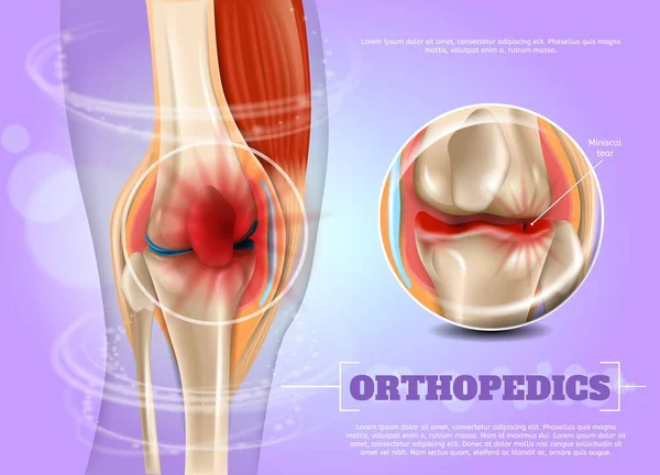
Realistic 3d Illustration Knee with Torn Miniscus
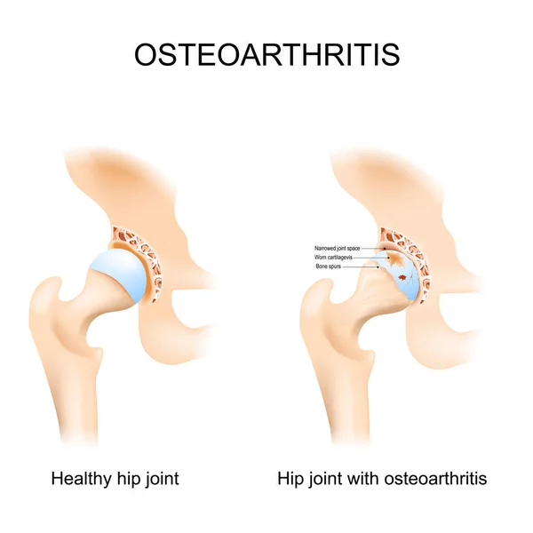
Hip joint with osteoarthritis with femoral head and the acetabulum of the pelvis. A comparison between a healthy hip joint and one with osteoarthritis, highlighting the key differences in structure. This can help patients understand diagnosis. Vector
Hip Joint Osteoarthritis Femoral Head Acetabulum Pelvis Comparison Healthy Hip Joint One Osteoarthritis Highlighting Key Differences Structure Can Help Patients Understand Diagnosis Vector
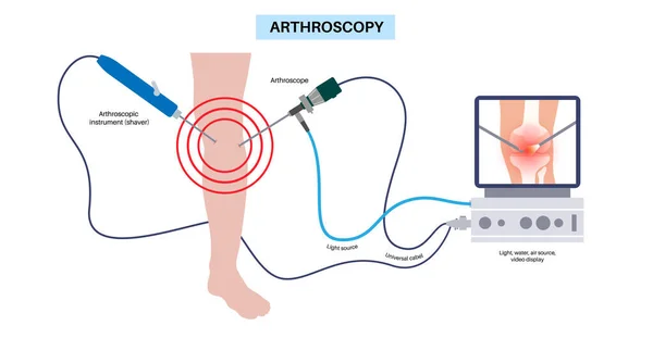
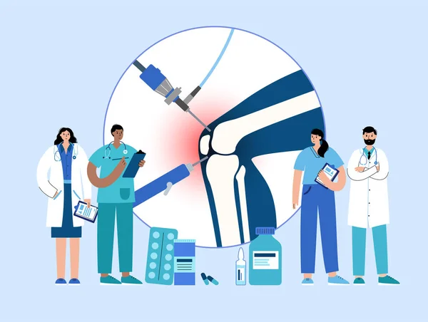
Arthroscopy medical procedure. Knee joint minimally invasive surgery. Arthroscope and arthroscopic instrument. Patella replacement, leg injury, kneecap reconstruction. Ligament and meniscus vector.
Arthroscopy Medical Procedure Knee Joint Minimally Invasive Surgery Arthroscope Arthroscopic Instrument Patella Replacement Leg Injury Kneecap Reconstruction Ligament Meniscus Vector
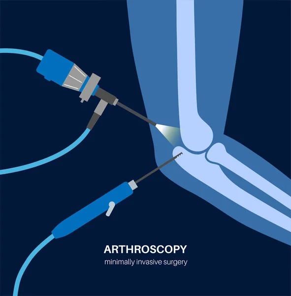
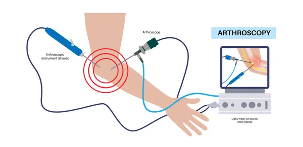
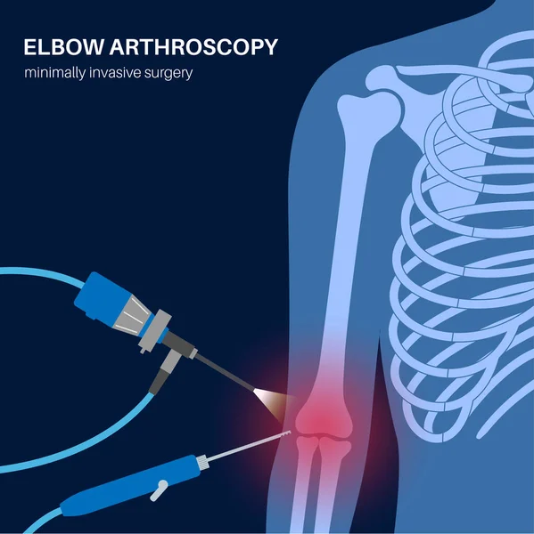
Elbow joint minimally invasive surgery. Arthroscopy medical procedure. Arthroscope and arthroscopic instrument. Arm pain, fracture, osteoarthritis or rheumatoid arthritis treatment, x ray flat vector
Elbow Joint Minimally Invasive Surgery Arthroscopy Medical Procedure Arthroscope Arthroscopic Instrument Arm Pain Fracture Osteoarthritis Rheumatoid Arthritis Treatment Ray Flat Vector
Meniscal tear. Knee anatomy. Close-up of Torn meniscus and cartilage damage. Sports knee injury. Vector illustration
Meniscal Tear Knee Anatomy Close Torn Meniscus Cartilage Damage Sports Knee Injury Vector Illustration
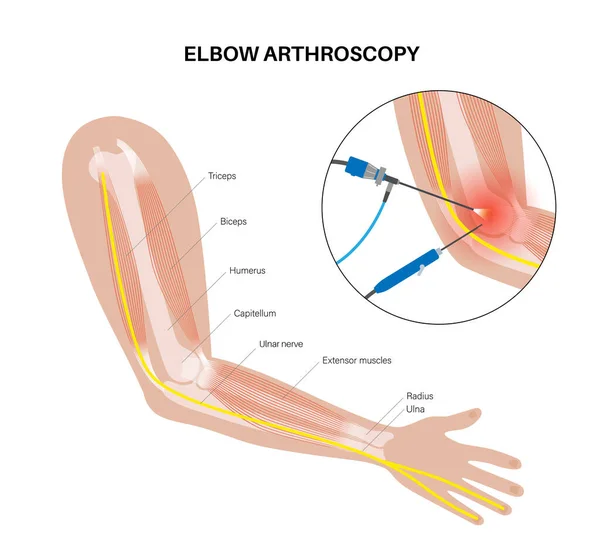
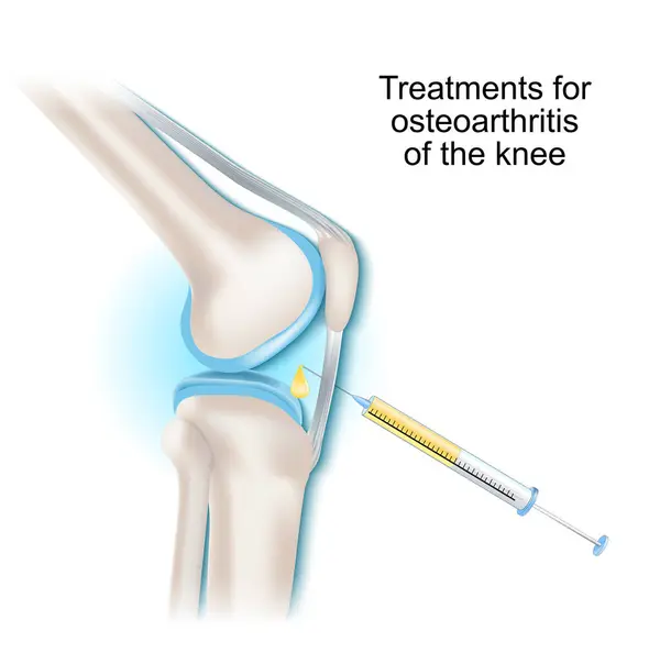
Treatments for osteoarthritis of the knee joint. Syringe and Intra-articular injection. Vector illustration.
Treatments Osteoarthritis Knee Joint Syringe Intra Articular Injection Vector Illustration
Meniscus injury. Close-up of knee joint. Meniscal tear before and after surgery. Suture repair. Meniscal healing. Top view of a human knee. Vector poster
Meniscus Injury Close Knee Joint Meniscal Tear Surgery Suture Repair Meniscal Healing Top View Human Knee Vector Poster
Arthroscopy medical procedure. Knee joint minimally invasive surgery. Arthroscope and arthroscopic instrument. Patella replacement, leg injury, kneecap reconstruction. Ligament and meniscus vector.
Arthroscopy Medical Procedure Knee Joint Minimally Invasive Surgery Arthroscope Arthroscopic Instrument Patella Replacement Leg Injury Kneecap Reconstruction Ligament Meniscus Vector
Ankle arthroscopy procedure. Feet joint minimally invasive surgery concept. Arthroscope and arthroscopic instrument. Foot treatment, leg pain and inflammation. Anatomical x ray vector illustration
Ankle Arthroscopy Procedure Feet Joint Minimally Invasive Surgery Concept Arthroscope Arthroscopic Instrument Foot Treatment Leg Pain Inflammation Anatomical Ray Vector Illustration
Arthroscopy medical procedure. Knee joint minimally invasive surgery. Arthroscope and arthroscopic instrument. Patella replacement, leg injury, kneecap reconstruction. Ligament and meniscus vector.
Arthroscopy Medical Procedure Knee Joint Minimally Invasive Surgery Arthroscope Arthroscopic Instrument Patella Replacement Leg Injury Kneecap Reconstruction Ligament Meniscus Vector
Arthroscopy medical procedure. Knee joint minimally invasive surgery. Arthroscope and arthroscopic instrument. Patella replacement, leg injury, kneecap reconstruction. Ligament and meniscus vector.
Arthroscopy Medical Procedure Knee Joint Minimally Invasive Surgery Arthroscope Arthroscopic Instrument Patella Replacement Leg Injury Kneecap Reconstruction Ligament Meniscus Vector
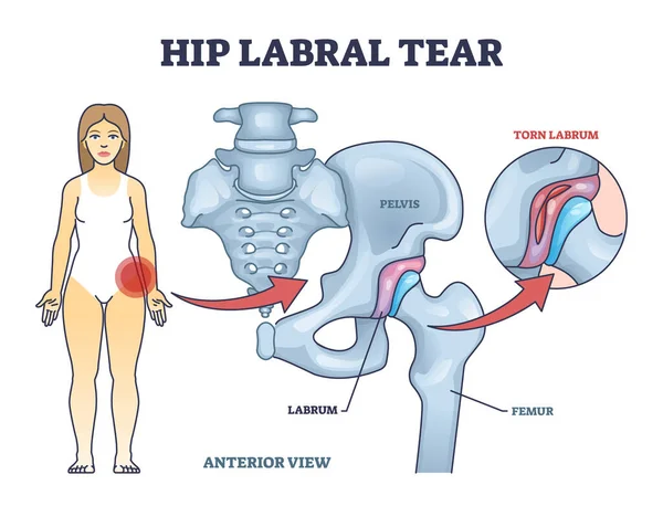
Hip labral tear anatomical explanation with medical labrum bone damage outline diagram. Labeled educational skeletal anatomy with pelvis, femur and torn labrum condition example vector illustration.
Hip Labral Tear Anatomical Explanation Medical Labrum Bone Damage Outline Diagram Labeled Educational Skeletal Anatomy Pelvis Femur Torn Labrum Condition Example Vector Illustration
Wrist arthroscopy surgery. Minimal invasive procedure. Pain, injury and inflammation in the human arm. Arthroscope and arthroscopic instrument. Treatment of hand bones flat vector medical illustration
Wrist Arthroscopy Surgery Minimal Invasive Procedure Pain Injury Inflammation Human Arm Arthroscope Arthroscopic Instrument Treatment Hand Bones Flat Vector Medical Illustration
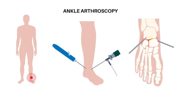
Ankle arthroscopy procedure. Feet joint minimally invasive surgery concept. Arthroscope and arthroscopic instrument. Foot treatment, leg pain and inflammation. Anatomical x ray vector illustration
Ankle Arthroscopy Procedure Feet Joint Minimally Invasive Surgery Concept Arthroscope Arthroscopic Instrument Foot Treatment Leg Pain Inflammation Anatomical Ray Vector Illustration
Meniscal tear. Torn meniscus. Knee injury. Cartilage damage. Meniscal repair. Sports injury. Head of tibia seen from above. Vector illustration
Meniscal Tear Torn Meniscus Knee Injury Cartilage Damage Meniscal Repair Sports Injury Head Tibia Seen Vector Illustration
Joint replacement surgery thin line icon . Editable stroke . Vector .
Joint Replacement Surgery Thin Line Icon Editable Stroke Vector
Osteophyte. Anatomy of human knee joint Osteoarthritis . Bone spurs. Close-up of a Synovial Joint with cartilage degeneration. Osteophyte formation. Vector illustration
Osteophyte Anatomy Human Knee Joint Osteoarthritis Bone Spurs Close Synovial Joint Cartilage Degeneration Osteophyte Formation Vector Illustration
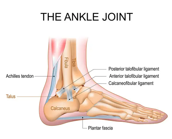
The ankle joint anatomy. Talocrural region or the jumping bone. Part of human body where the foot and the leg meet. Plantar fascia and Achilles tendon. Lateral collateral ligaments of ankle joint Anterior talofibular, Posterior talofibular, and Calca
Ankle Joint Anatomy Talocrural Region Jumping Bone Part Human Body Foot Leg Meet Plantar Fascia Achilles Tendon Lateral Collateral Ligaments Ankle Joint Anterior Talofibular Posterior Talofibular Calca
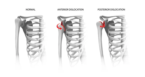
Shoulder dislocation options. Vector illustration.
Shoulder Dislocation Options Vector Illustration
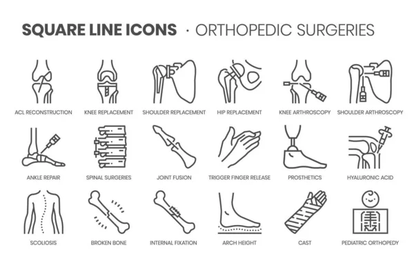
Orthopedic surgery related, pixel perfect, editable stroke, up scalable square line vector icon set.
Orthopedic Surgery Related Pixel Perfect Editable Stroke Scalable Square Line Vector Icon Set
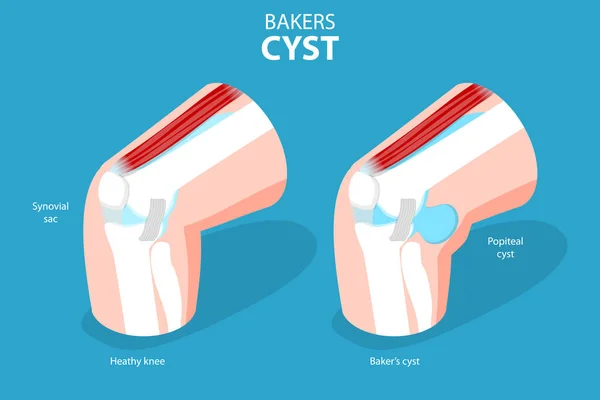
3D Isometric Flat Vector Conceptual Illustration of Bakers CYST, Traumatology and Prthopedics
Isometric Flat Vector Conceptual Illustration Bakers Cyst Traumatology Prthopedics
Arthroscopy medical procedure. Knee joint minimally invasive surgery. Arthroscope and arthroscopic instrument. Patella replacement, leg injury, kneecap reconstruction. Ligament and meniscus vector.
Arthroscopy Medical Procedure Knee Joint Minimally Invasive Surgery Arthroscope Arthroscopic Instrument Patella Replacement Leg Injury Kneecap Reconstruction Ligament Meniscus Vector
Arthroscopy medical procedure. Knee joint minimally invasive surgery. Arthroscope and arthroscopic instrument. Patella replacement, leg injury, kneecap reconstruction. Ligament and meniscus vector.
Arthroscopy Medical Procedure Knee Joint Minimally Invasive Surgery Arthroscope Arthroscopic Instrument Patella Replacement Leg Injury Kneecap Reconstruction Ligament Meniscus Vector
Multiracial orthopedics doctors examine foot
Elbow joint minimally invasive surgery. Arthroscopy medical procedure. Anatomy of humerus, ulna and radius bones. Arm pain, fracture, osteoarthritis or rheumatoid arthritis x ray vector illustration
Elbow Joint Minimally Invasive Surgery Arthroscopy Medical Procedure Anatomy Humerus Ulna Radius Bones Arm Pain Fracture Osteoarthritis Rheumatoid Arthritis Ray Vector Illustration
Arthroscopy medical procedure. Knee joint minimally invasive surgery. Arthroscope and arthroscopic instrument. Patella treatment, leg pain, kneecap inflammation. Ligament and meniscus anatomy vector.
Arthroscopy Medical Procedure Knee Joint Minimally Invasive Surgery Arthroscope Arthroscopic Instrument Patella Treatment Leg Pain Kneecap Inflammation Ligament Meniscus Anatomy Vector
3d rheumatoid anatomical knee joint illustration with realistic bone structure, vector banner with injured pain leg. Medical graphic for orthopedic ads, health protection and arthritis care concepts
Rheumatoid Anatomical Knee Joint Illustration Realistic Bone Structure Vector Banner Injured Pain Leg Medical Graphic Orthopedic Ads Health Protection Arthritis Care Concepts
Glenohumeral joint coronal anatomy highlights humeral head, glenoid cavity, and supraspinatus tendon, conveying shoulder articulation and stabilizing soft tissues. Outline diagram
Glenohumeral Joint Coronal Anatomy Highlights Humeral Head Glenoid Cavity Supraspinatus Tendon Conveying Shoulder Articulation Stabilizing Soft Tissues Outline Diagram
Arthroscopy check icon vector outline illustration
Elbow joint minimally invasive surgery. Arthroscopy medical procedure. Anatomy of humerus, ulna and radius bones. Arm pain, fracture, osteoarthritis or rheumatoid arthritis x ray vector illustration
Elbow Joint Minimally Invasive Surgery Arthroscopy Medical Procedure Anatomy Humerus Ulna Radius Bones Arm Pain Fracture Osteoarthritis Rheumatoid Arthritis Ray Vector Illustration
Urinary System icon pack with silhouette and stroke web icons.
Urinary System Icon Pack Silhouette Stroke Web Icons
Rheumatoid arthritis. A comparison between a healthy knee and joint with Bone erosion, Cartilage wears, Reduced joint space and Swollen inflamed synovial membrane. Vector illustration.
Rheumatoid Arthritis Comparison Healthy Knee Joint Bone Erosion Cartilage Wears Reduced Joint Space Swollen Inflamed Synovial Membrane Vector Illustration
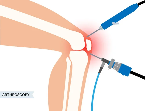
Illustration of a knee joint with a surgical instrument performing a procedure.
Illustration Knee Joint Surgical Instrument Performing Procedure