Chromatyna Stock Vector Images

173 Chromatyna vector art & graphics are available under a royalty-free license
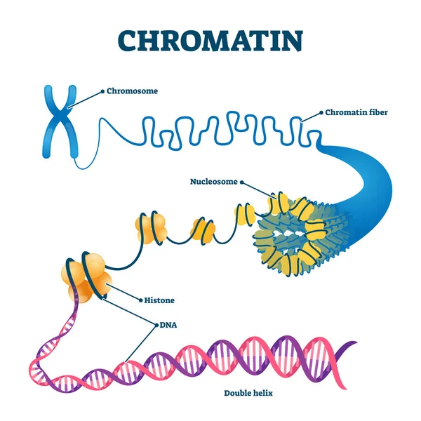
Chromation biological diagram vector illustration
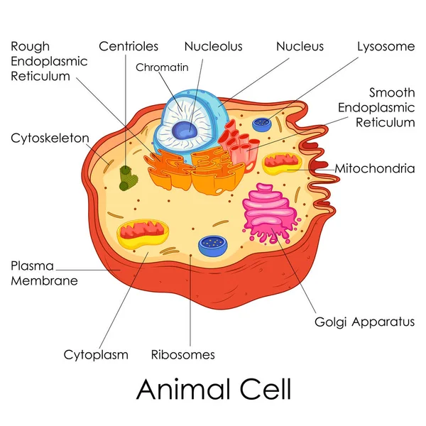
Education Chart of Biology for Animal Cell Diagram
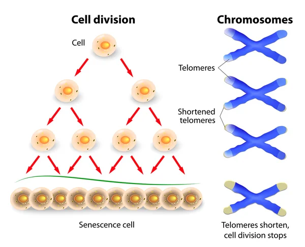
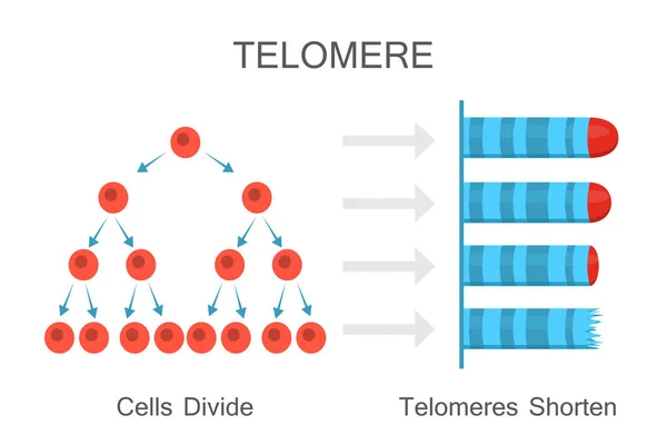
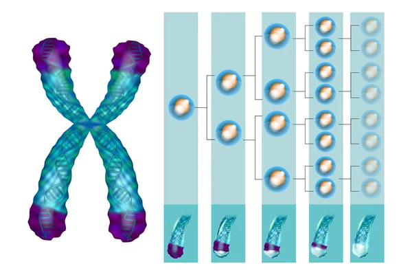
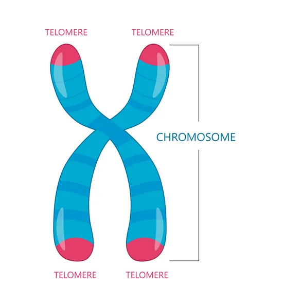
Telomere, cell division and human chromosomes
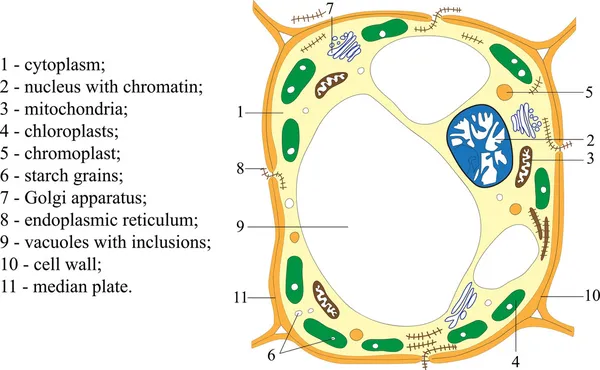
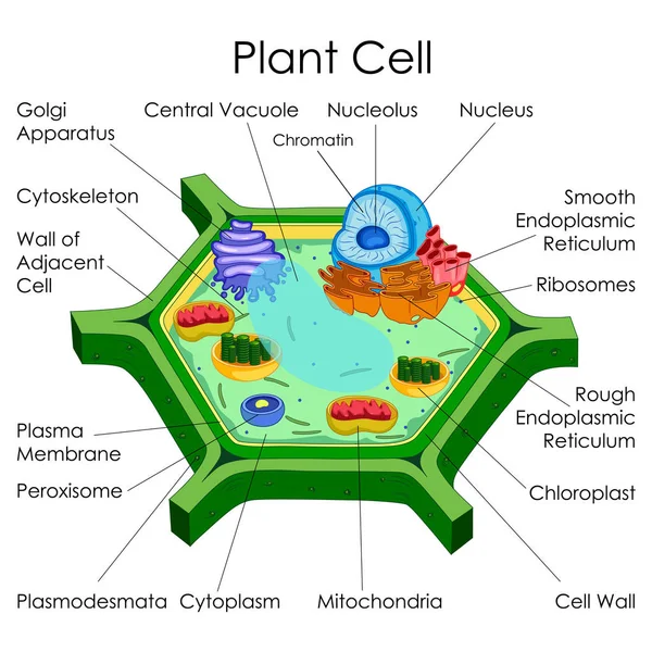
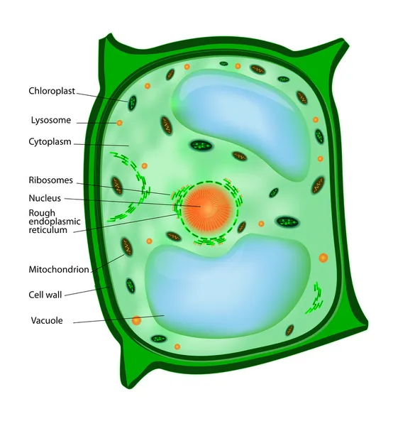
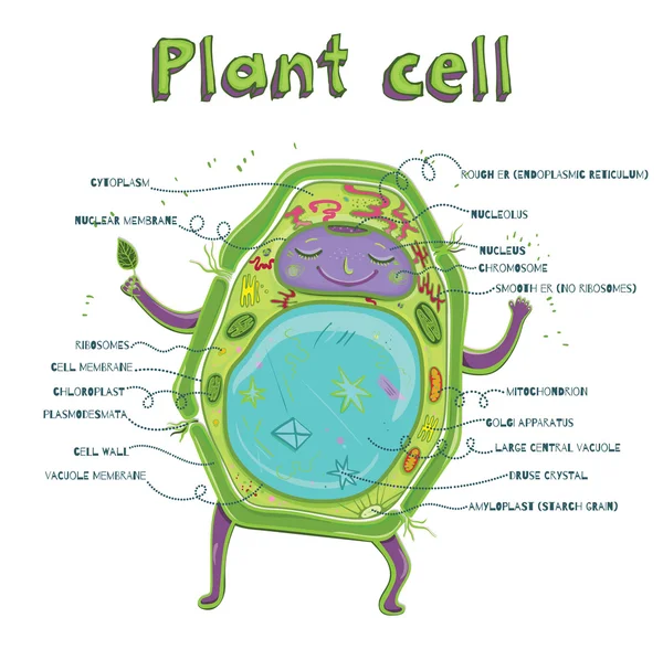
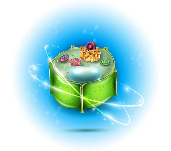
Plant cell
Plant cell
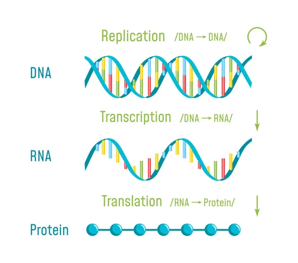
DNA Replication, Transcription and Translation
DNA chromosome
DNA chromosome
Education Chart of Biology for Plant Cell Diagram
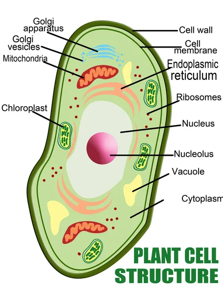
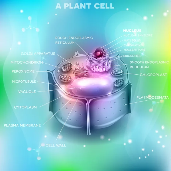
Plant cell structure
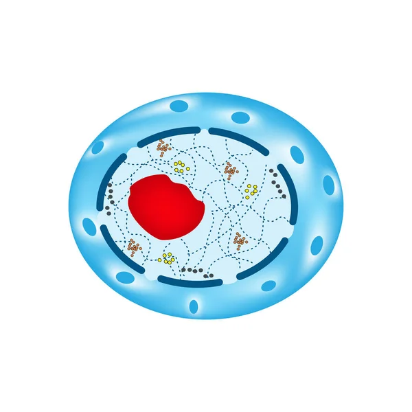
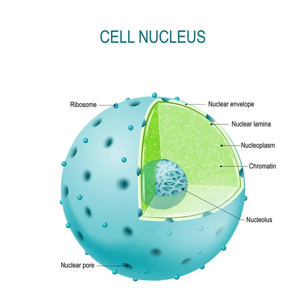
The structure of the human cell nucleus. Infographics. Vector illustration on isolated background
Telomeres Shorten with Age Diagram
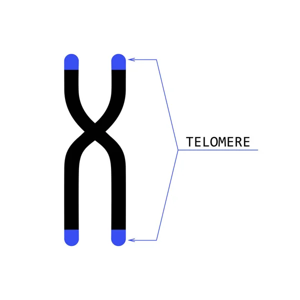
Telomere end of chromosome
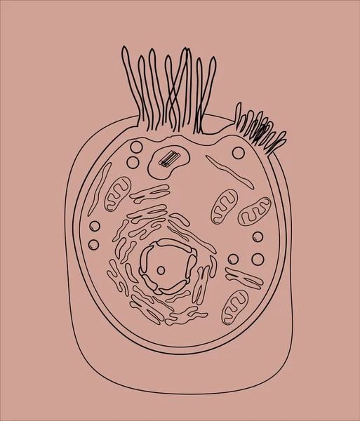
Handdrawn Cell Pattern
Chromatin is the complex of DNA and proteins that makes up chromosomes within the nucleus of eukaryotic cells. The structure of chromatin is dynamic and undergoes changes that regulate gene expression and DNA replication.
Chromatin Complex Dna Proteins Makes Chromosomes Nucleus Eukaryotic Cells Structure Chromatin Dynamic Undergoes Changes Regulate Gene Expression Dna Replication
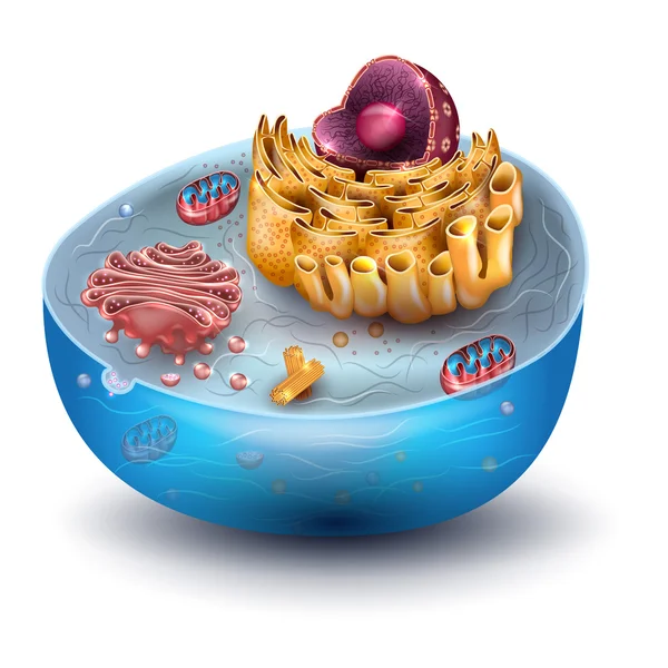
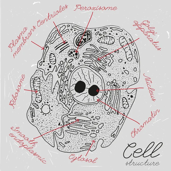
Cell structure
Cell structure
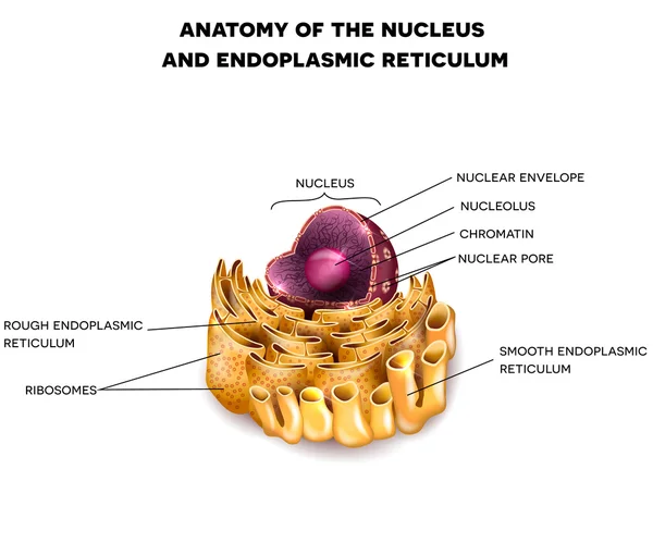
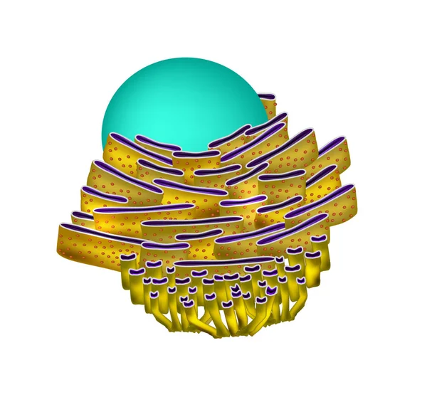
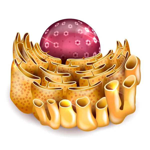
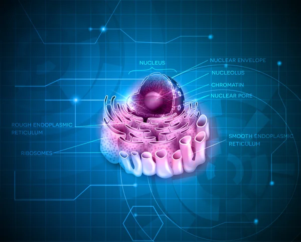
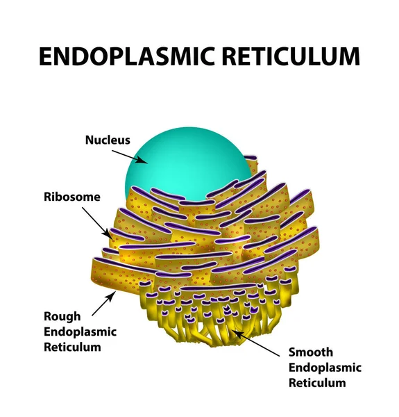
Cell Nucleus and Endoplasmic reticulum
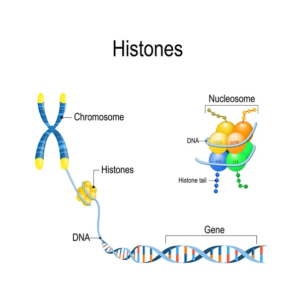
Histones close up. Organization and packaging of genetic materia
Cell structure cross section
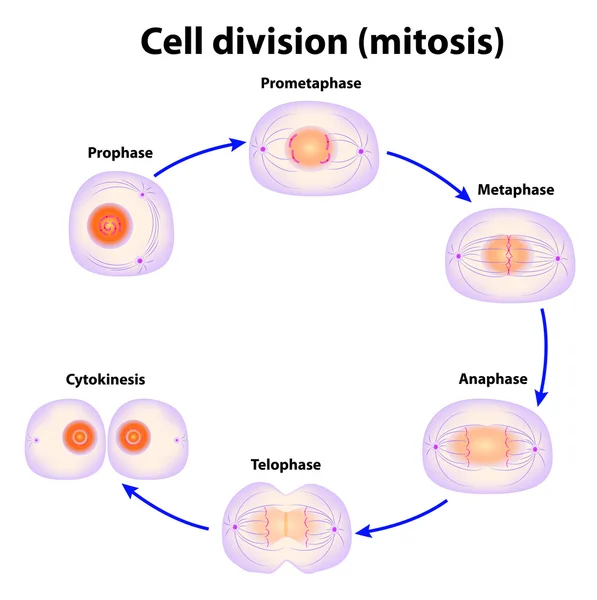
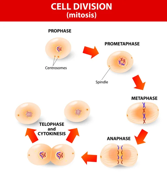
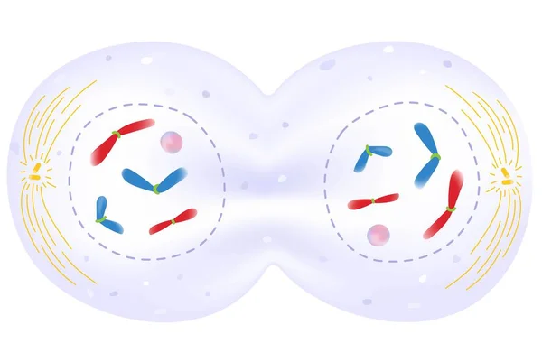
Cell division. Mitosis
Plant cell
Plant cell
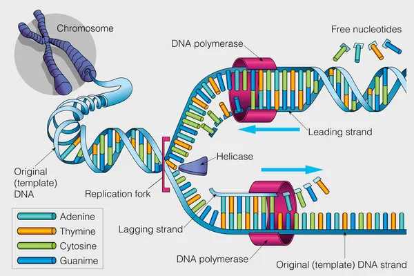
The graphic shows the duplication of a DNA chain on a gray background. Vector image
Graphic Shows Duplication Dna Chain Gray Background Vector Image
Hand Drawn Cell
Hand Drawn Cell
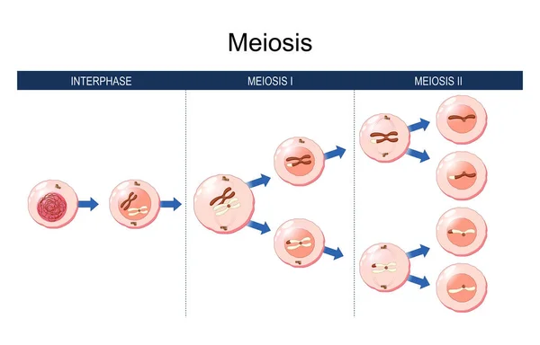
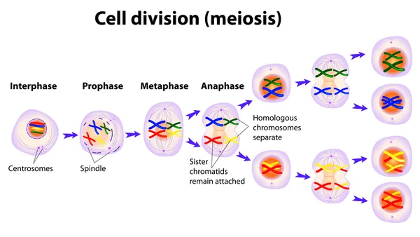
Meiosis. cell division. homologous chromosomes exchange genetic information. during the first meiosis. The daughter cells divide for to form haploid gametes. Two gametes fuse during fertilization, forming a diploid cell. Vector illustration
Meiosis Cell Division Homologous Chromosomes Exchange Genetic Information First Meiosis Daughter Cells Divide Form Haploid Gametes Two Gametes Fuse Fertilization Forming Diploid Cell Vector Illustration
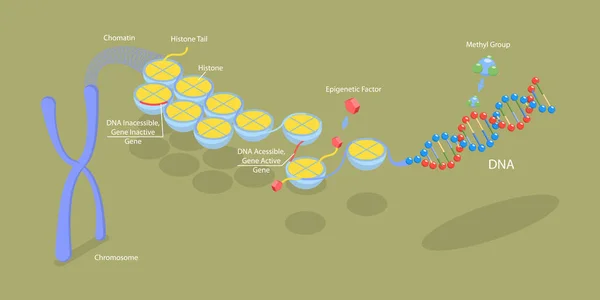
Structure of a chromosome. Epigenetic mechanism. Epigenetic factor, Methyl group, Gene, DNA, Chromosome, Chromatin. Genome sequence. Molecular biology. Vector illustration
Structure Chromosome Epigenetic Mechanism Epigenetic Factor Methyl Group Gene Dna Chromosome Chromatin Genome Sequence Molecular Biology Vector Illustration
Simple set of icons such as cell division, laboratory, cell, chemical reaction, chromosome, molecular, hazardous, scientific, microbiology, genetic. related chemistry icons collection. editable
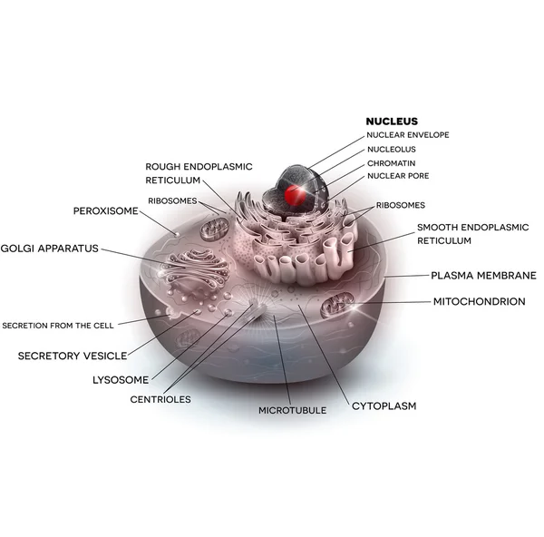
The structure of the human cells in a graphic image, cell components on a colored background
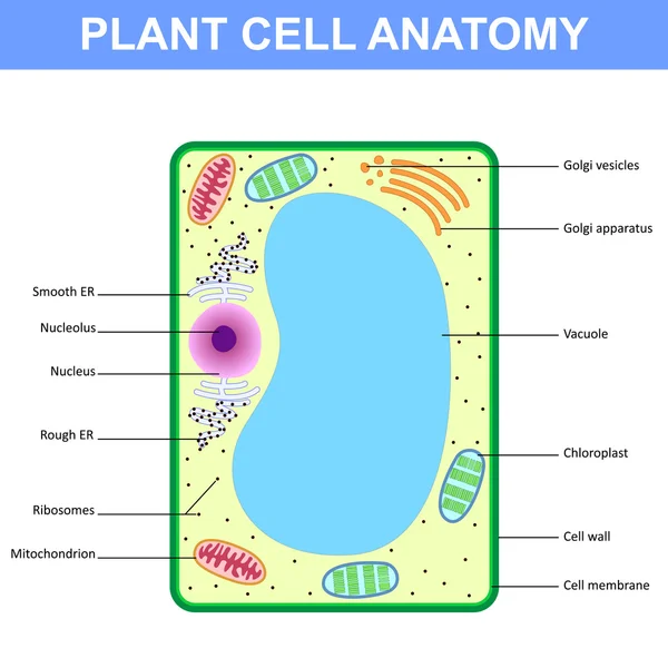
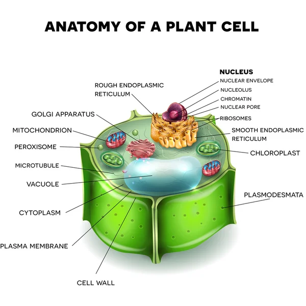
Plant cell anatomy
Plant cell anatomy
Endoplasmic reticulum structure. Infographics. Vector illustration on isolated background
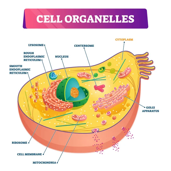
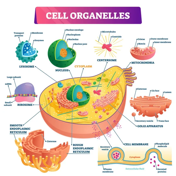
Cell organelles biological anatomy vector illustration diagram
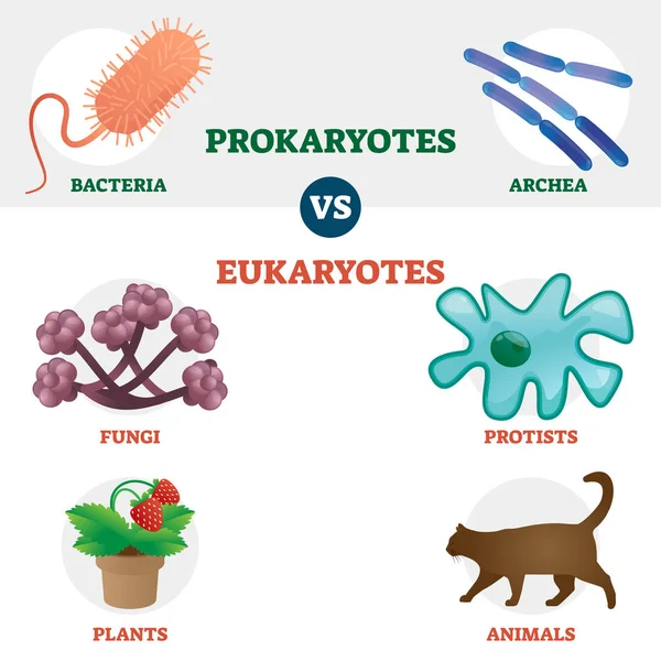
Eukaryote vs Prokaryote cell type organisms educational set
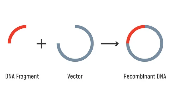
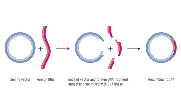
Gene Cloning. Plasmids and Recombinant DNA.
Cell Nucleus and Endoplasmic reticulum
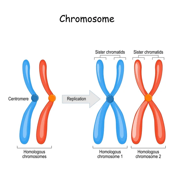
Difference between homologous chromosomes, a pair of homologous
Chromosome parts. Structure of a chromosome. Centromere, telomere, chromatids. Diagram showing elements of a threadlike structures made of protein and a single molecule of DNA. Vector illustration.
Chromosome Parts Structure Chromosome Centromere Telomere Chromatids Diagram Showing Elements Threadlike Structures Made Protein Single Molecule Dna Vector Illustration
Cell structure beautiful color illustration
3D Isometric Flat Vector Conceptual Illustration of Molecular Biology, Cell Structure
Isometric Flat Vector Conceptual Illustration Molecular Biology Cell Structure
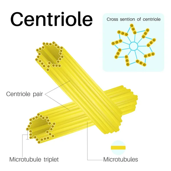
Centrioles are cylindrical organelles.Found in most eukaryotic cells.
Centrioles Cylindrical Organelles Found Most Eukaryotic Cells
Eukaryotic vs Prokaryotic cells, educational biology vector illustration diagram
Types of cells in the human body.
Types Cells Human Body
Cell nucleus. vector diagram
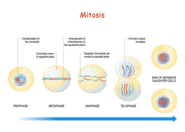
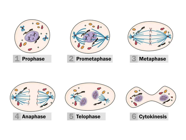
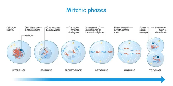
Mitosis. cell division stages from Interphase, Prophase, and Prometaphase to Metaphase, Anaphase, and Telophase. Process of multiplication. Vector illustration
Mitosis Cell Division Stages Interphase Prophase Prometaphase Metaphase Anaphase Telophase Process Multiplication Vector Illustration
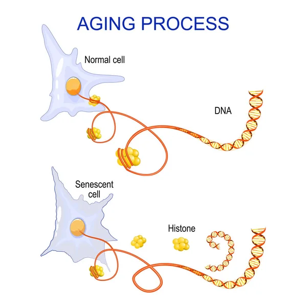
Aging process into cells. chromatin, DNA and histones change in ageing and senescent cells. Vector
Aging Process Cells Chromatin Dna Histones Change Ageing Senescent Cells Vector
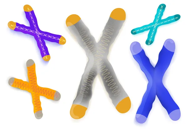
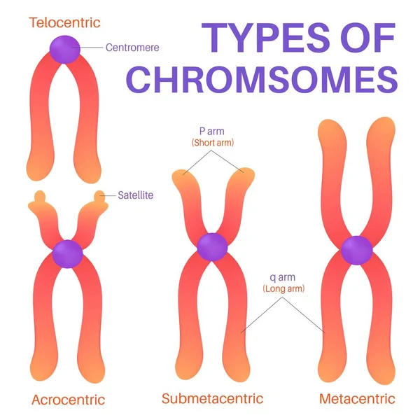
Types of chromosomes. Structure of chromosome with centromere, long and short arms. Metacentric, Submetacentric, Acrocentric, Telocentric. transparent Chromosomes with glowing effect on white background. vector poster
Types Chromosomes Structure Chromosome Centromere Long Short Arms Metacentric Submetacentric Acrocentric Telocentric Transparent Chromosomes Glowing Effect White Background Vector Poster
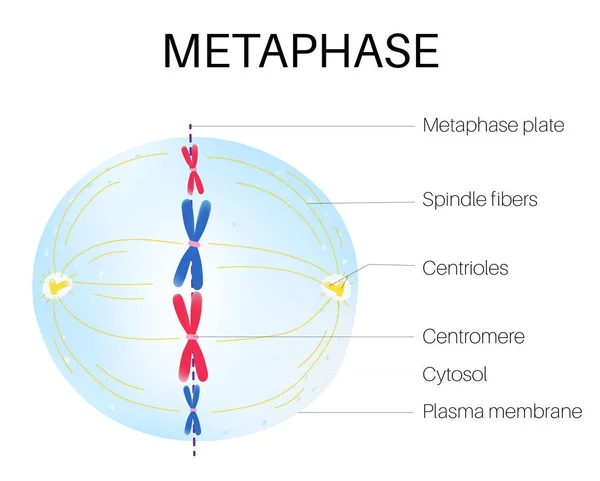
Metaphase is a stage of mitosis in the eukaryotic cell cycle.
Metaphase Stage Mitosis Eukaryotic Cell Cycle
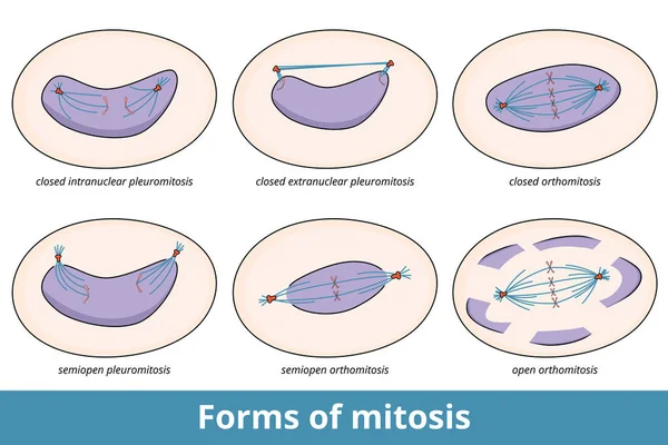
Forms of mitosis. 6 common mitosis forms, based on state of nuclear envelope and symmetry and location of central spindle, including closed orthomitosis and semiopen pleuromitosis.
Forms Mitosis Common Mitosis Forms Based State Nuclear Envelope Symmetry Location Central Spindle Including Closed Orthomitosis Semiopen Pleuromitosis
Cell structure chromosome histone and DNA illustration
Cell Structure Chromosome Histone Dna Illustration
Genetic engineering, DNA cells vector isolated illustration
Meiosis cell division
Cell anatomy grey background
Illustration showing the position of telomeres at the end of our chromosomes. Telomere shortening - with every cell division and during different pathological processes.
Illustration Showing Position Telomeres End Our Chromosomes Telomere Shortening Every Cell Division Different Pathological Processes
Mitosis. process cell division
Chromosome on a white background
Animal cell anatomy
Animal cell anatomy
Plant cell anatomy
Plant cell anatomy
3D Isometric Flat Vector Conceptual Illustration of Epigenetic Mechanisms, Educational Labeled Scheme
Isometric Flat Vector Conceptual Illustration Epigenetic Mechanisms Educational Labeled Scheme
DNA Research Project
The process of karyokinesis (or mitosis) is divided into five stages: prophase, prometaphase, metaphase, anaphase, and telophase.
Process Karyokinesis Mitosis Divided Five Stages Prophase Prometaphase Metaphase Anaphase Telophase
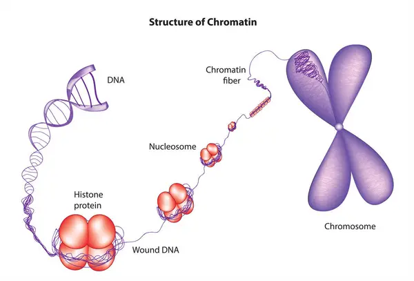
Nucleosome model - Wound DNA complex around histones
Nucleosome Model Wound Dna Complex Histones
Cell organelles biological vector illustration diagram
Hand Drawn Cell
Hand Drawn Cell
Telomere end of chromosome
Plant Cell structure, cross section of the cell detailed colorfu
Plant Cell structure
Human Genome Project
Illustration of biology, Crossing over and recombination, Genetic recombination happens as a result of the separation of genes that occurs during gamete formation in meiosis, Chromosome Structure
Illustration Biology Crossing Recombination Genetic Recombination Happens Result Separation Genes Occurs Gamete Formation Meiosis Chromosome Structure
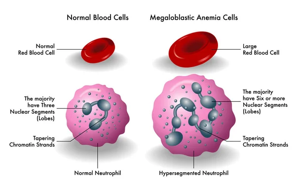
Medical illustration showing megaloblastic anemia cells
Medical Illustration Showing Megaloblastic Anemia Cells
Gene Cloning. DNA Recombinant
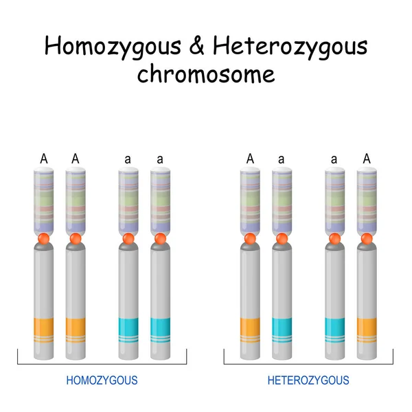
Homozygous Heterozygous chromosome. Vector illustration. Poster
Homozygous Heterozygous Chromosome Vector Illustration Poster
Gene regulation pathway visualizes epigenetic, transcriptional, and translational control with DNA, nucleosomes, and ribosome, highlighting on-off switches and mRNA edits shaping protein output.
Gene Regulation Pathway Visualizes Epigenetic Transcriptional Translational Control Dna Nucleosomes Ribosome Highlighting Switches Mrna Edits Shaping Protein Output
Cell Nucleus and Endoplasmic reticulum
Cell division. differences between mitosis and meiosis. Cell cycle. Genetic variation. Vector illustration
Cell Division Differences Mitosis Meiosis Cell Cycle Genetic Variation Vector Illustration
Two chromosome illustrations crossing in vibrant colors, detailed vector design with gradient shading and striped patterns.
Two Chromosome Illustrations Crossing Vibrant Colors Detailed Vector Design Gradient Shading Striped Patterns
Endoplasmic reticulum structure. Infographics. Vector illustration on isolated background
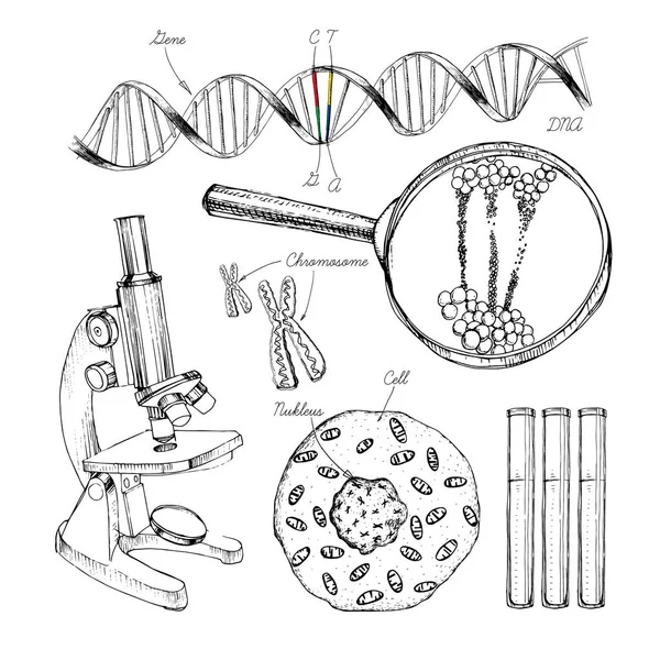
Hand drawn set structure of DNA. genome sequence. Health and biochemistry laboratory of nanotechnology. Molecule helix of dna, genome or gene evolution. Doodle elements. Gene editing tool.
Telophase is the phase of the cell cycle.
Telophase Phase Cell Cycle
Structure of a plant cell
Organelle color line icon. Organisation in organism.
Organelle Color Line Icon Organisation Organism
Plant Cell anatomy scientific background
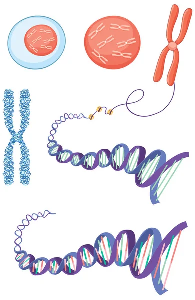
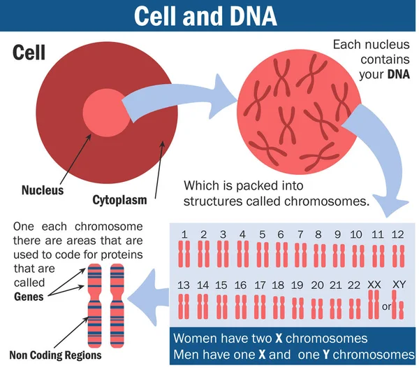
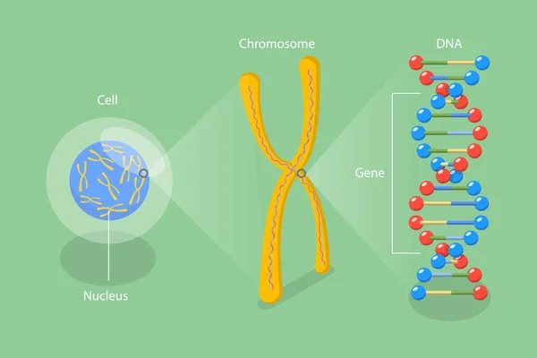
Cell, chromosome and DNA vector illustration. illustration of Biology. Chromosome structure. Genome study. Vector illustration for medical, science, and educational use
Cell Chromosome Dna Vector Illustration Illustration Biology Chromosome Structure Genome Study Vector Illustration Medical Science Educational Use
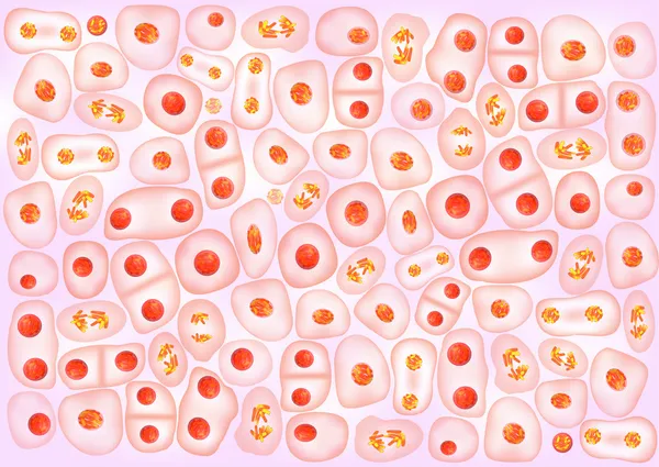
Cell division background
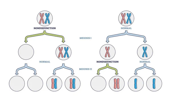
Nondisjunction as abnormal meiosis chromosome number failure outline diagram
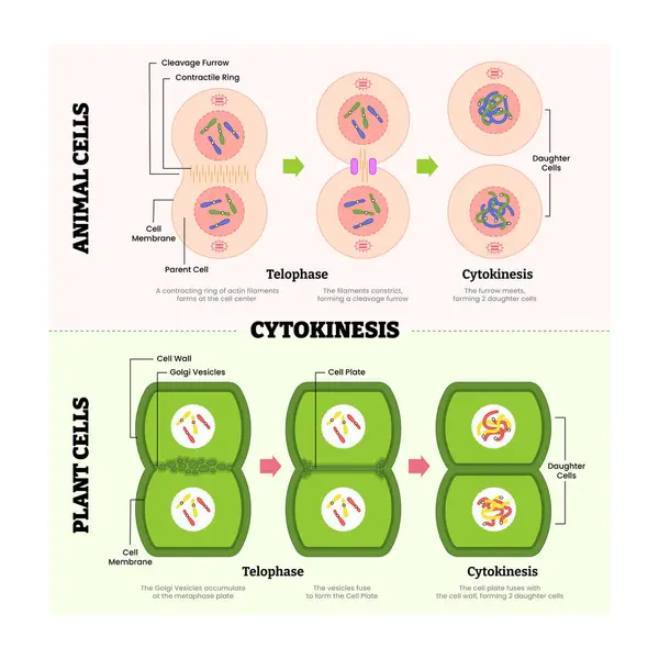
Cytokines in animal cell and plant cell diagram process, produce protein for immune system, labeled telophase, cytokinesis, cleavage furrow, parent and daughter cell division. Vector illustration
Cytokines Animal Cell Plant Cell Diagram Process Produce Protein Immune System Labeled Telophase Cytokinesis Cleavage Furrow Parent Daughter Cell Division Vector Illustration
Chromosomes on Black Background
Chromosomes  on Black Background
Mitosis stages from Interphase, Prophase, and Prometaphase to Metaphase, Anaphase, and Telophase. cell division. Vector illustration
Mitosis Stages Interphase Prophase Prometaphase Metaphase Anaphase Telophase Cell Division Vector Illustration
DNA methylation diagram showing how CpG sites gain CH3 by DNMT to condense chromatin and repress transcription, highlights DNA strand, methyl groups, and promoter states. Outline diagram
Dna Methylation Diagram Showing How Cpg Sites Gain Ch3 Dnmt Condense Chromatin Repress Transcription Highlights Dna Strand Methyl Groups Promoter States Outline Diagram
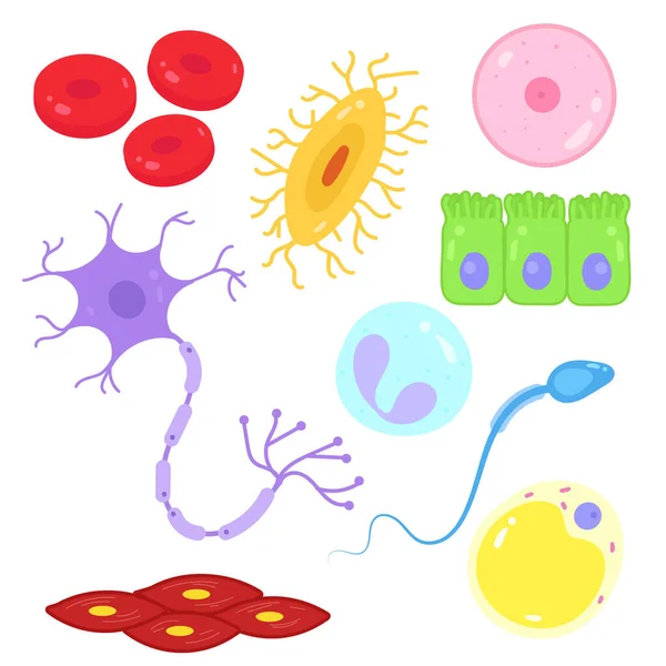
Cell anatomy beautiful abstract design
Meiosis cell division
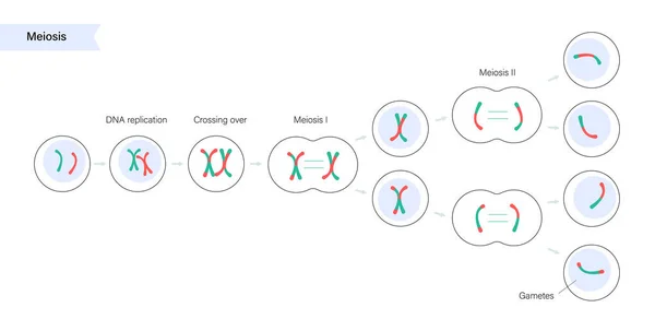
Cell division. meiosis. Vector scheme
Meiosis cell division illustration
Vector diagram of the meiosis phases
DNA Strands Icon
DNA Strands Icon
Histones structure. Histone proteins. Organization and packaging of genetic material in Chromosome. Vector poster. Isometric Flat illustration. Schematic diagram
Histones Structure Histone Proteins Organization Packaging Genetic Material Chromosome Vector Poster Isometric Flat Illustration Schematic Diagram
Four Types Of Human Chromosome.
Four Types Human Chromosome
DNA Research Project
Plant Cell structure, cross section of the cell detailed colorfu
Cell division background
Telomere is the End of a Chromosome
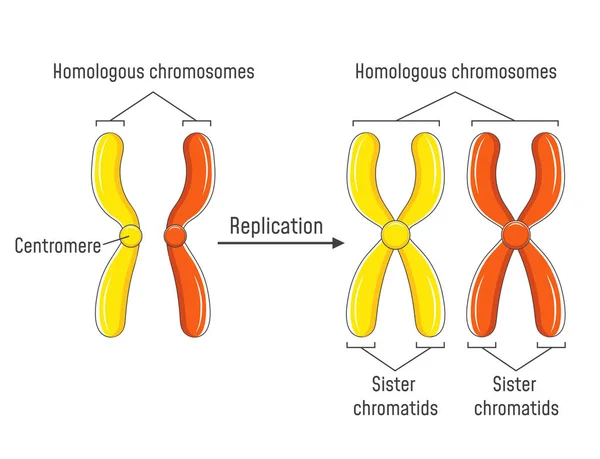
Homologous Chromosomes and Chromatids
Mitosis cell division.
Malignant neoplasm. Cancer and Normal cells. comparison and difference between healthy tissue and tumor. details about chromatin, nucleus and cytoplasm
Malignant Neoplasm Cancer Normal Cells Comparison Difference Healthy Tissue Tumor Details Chromatin Nucleus Cytoplasm
Page