Ciliar Stock Vector Images

117 Ciliar vector art & graphics are available under a royalty-free license
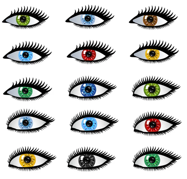
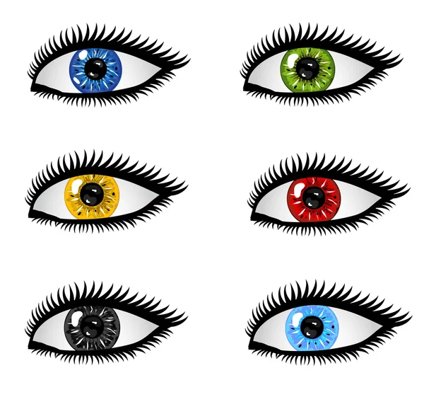
Set of human eyes
Set of human eyes
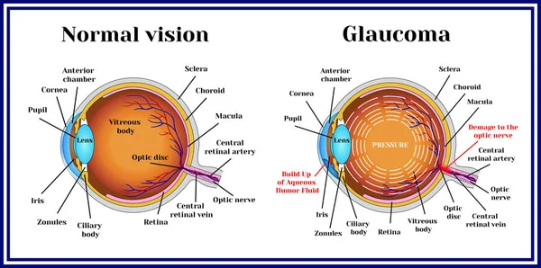
Normal vision. Glaucoma.
Medical illustration of the symptoms of blepharitis
Medical Illustration Symptoms Blepharitis
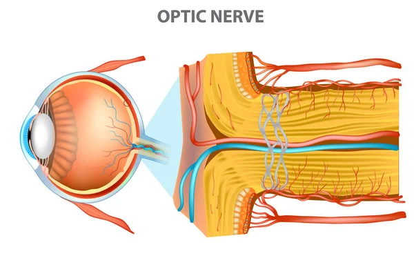
The Optic Nerve. Anatomy of the Eye
Optic Nerve Anatomy Eye
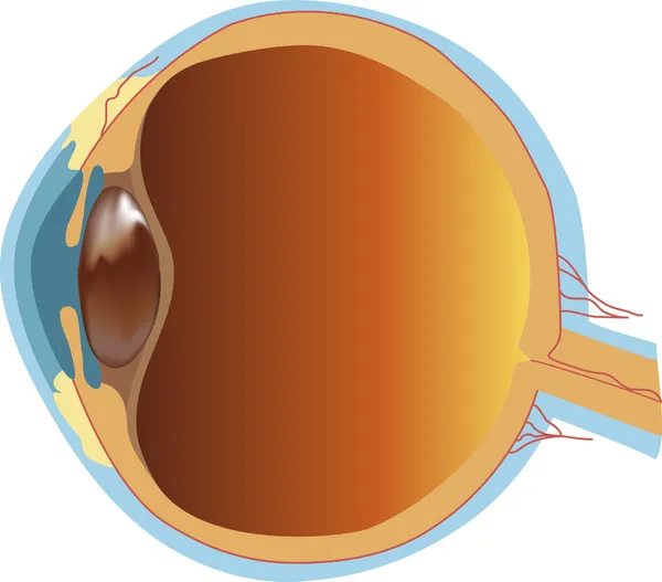
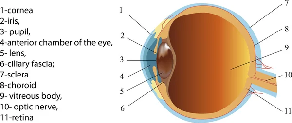
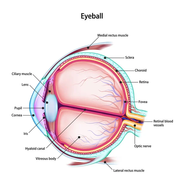
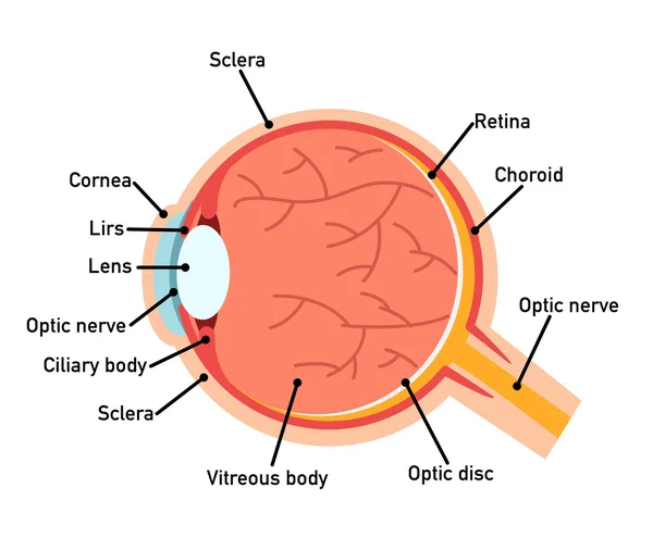
Structure of human eye (section)
Vector image of eyeball anatomy
Human eye anatomy. Retina structure. Cross-section of the eye. Cells in section of the retina. Detailed vector poster
Human Eye Anatomy Retina Structure Cross Section Eye Cells Section Retina Detailed Vector Poster
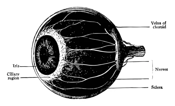
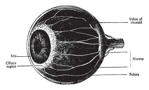
This illustration represents The Choroid Ciliary Body and Iris, vintage line drawing or engraving illustration.
Illustration Represents Choroid Ciliary Body Iris Vintage Line Drawing Engraving Illustration
Human eye anatomy labeled diagram, educational medical poster design healthcare scheme, tarsal plate, ocular structure. Labeled cornea, iris, lens, retina, choroid, optic nerve, sclera, eyelid
Human Eye Anatomy Labeled Diagram Educational Medical Poster Design Healthcare Scheme Tarsal Plate Ocular Structure Labeled Cornea Iris Lens Retina Choroid Optic Nerve Sclera Eyelid
Motor nerves of the eye anatomy poster. Abducens, trochlear and oculomotor nerves in the human brain. Ciliary gland and muscle, coordinate eye position. Sensory and motor functions vector illustration
Motor Nerves Eye Anatomy Poster Abducens Trochlear Oculomotor Nerves Human Brain Ciliary Gland Muscle Coordinate Eye Position Sensory Motor Functions Vector Illustration
Oculomotor nerve in human brain, eye anatomy. Cranial nerves poster, motor and parasympathetic fibers. Movement and coordinate eye position. Vestibulo ocular and optokinetic reflex vector illustration
Oculomotor Nerve Human Brain Eye Anatomy Cranial Nerves Poster Motor Parasympathetic Fibers Movement Coordinate Eye Position Vestibulo Ocular Optokinetic Reflex Vector Illustration
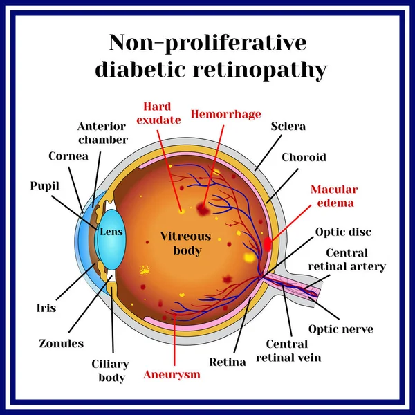
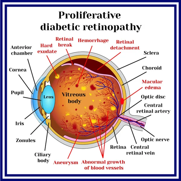
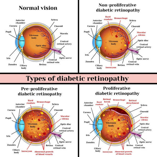
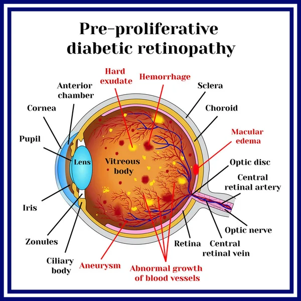
Types of diabetic retinopathy
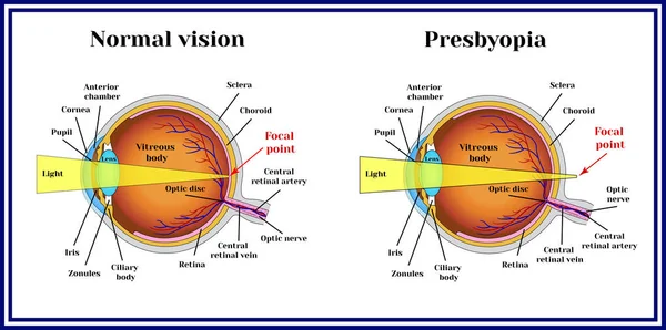
Refractive errors eyeball. Presbyopia.
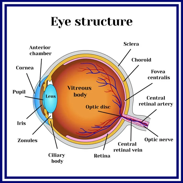
Eyeball structure. Medicine.
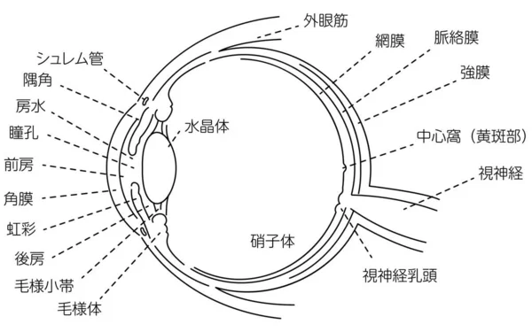
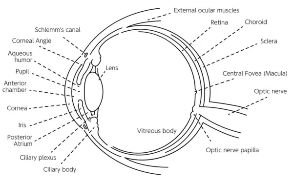
Illustration of the eye - Translation: Schlemm's canal, corner angle, aqueous humor, pupil, anterior chamber, cornea, iris, posterior chamber, ciliary body, ciliary body, lens, vitreous body, external eye muscle, retina, choroid, sclera, central foss
Illustration Eye Translation Schlemm Canal Corner Angle Aqueous Humor Pupil Anterior Chamber Cornea Iris Posterior Chamber Ciliary Body Ciliary Body Lens Vitreous Body External Eye Muscle Retina Choroid Sclera Central Foss
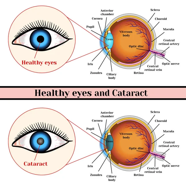
Healthy eyes and cataract (opacity of the eye lens).
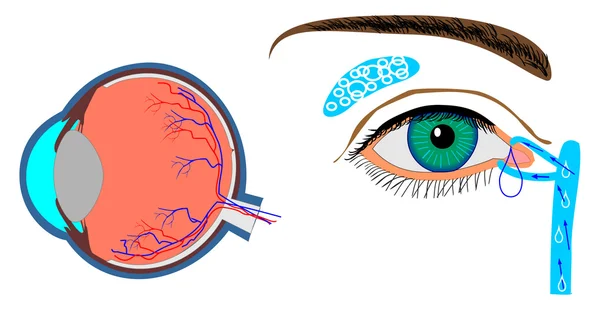
Structure of eyeball and localization lachrymal gland
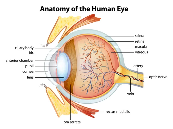
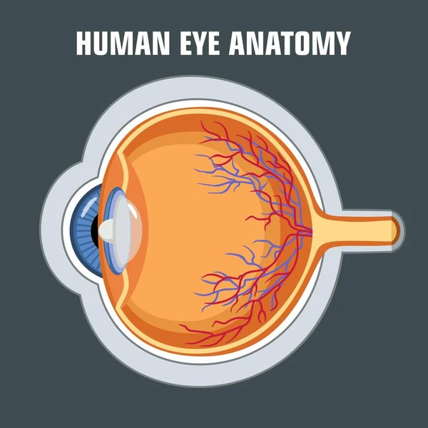
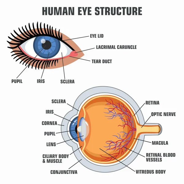
Human eye anatomy
Human eye anatomy
Linear hand drawn illustration of human eye for logotype or banner / design template
Linear Hand Drawn Illustration Human Eye Logotype Banner Design Template
Macular degeneration. Age-related dry macular degeneration. AMD. Cross section of human eye. Central vision loss. Close-up of a macula, optic disc, choroid, retina, sclera, and fovea. Vector poster
Macular Degeneration Age Related Dry Macular Degeneration Amd Cross Section Human Eye Central Vision Loss Close Macula Optic Disc Choroid Retina Sclera Fovea Vector Poster
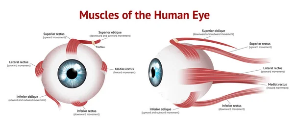
Refractive errors eyeball. Astigmatism.
Types of diabetic retinopathy
Medical illustration of the cross-sectional structure of the human eye
Medical Illustration Cross Sectional Structure Human Eye
Eye system. Vector design.
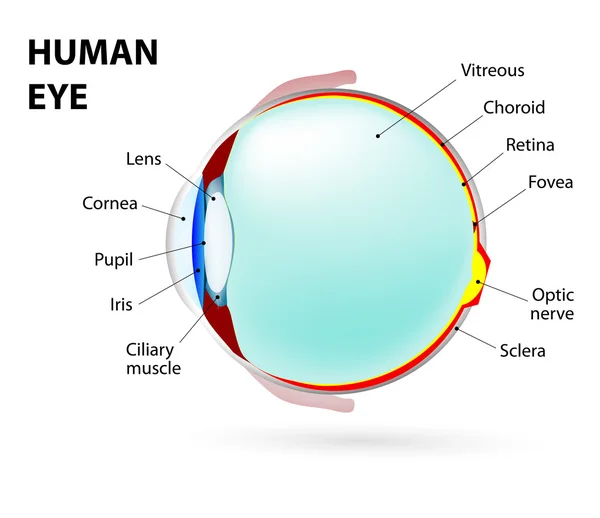
Human eye
Human eye
Oculomotor nerve in human brain, eye anatomy. Cranial nerves poster, motor and parasympathetic fibers. Movement and coordinate eye position. Vestibulo ocular and optokinetic reflex vector illustration
Oculomotor Nerve Human Brain Eye Anatomy Cranial Nerves Poster Motor Parasympathetic Fibers Movement Coordinate Eye Position Vestibulo Ocular Optokinetic Reflex Vector Illustration
This illustration represents The Choroid Ciliary Body and Iris, vintage line drawing or engraving illustration.
Illustration Represents Choroid Ciliary Body Iris Vintage Line Drawing Engraving Illustration
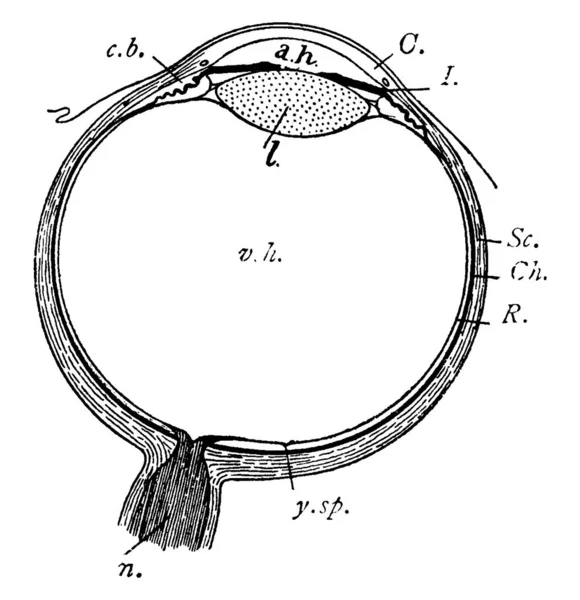
Represents, C., Cornea; a.h., aqueous humour; c.b., ciliary body; l., lens; I., iris; Sc., sclerotic; Ch., choroid; R., retina; and other, vintage line drawing or engraving illustration.
Represents Cornea Aqueous Humour Ciliary Body Lens Iris Sclerotic Choroid Retina Other Vintage Line Drawing Engraving Illustration
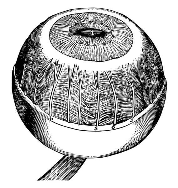
This illustration represents Iris Ciliary Muscle and Choroid, vintage line drawing or engraving illustration.
Illustration Represents Iris Ciliary Muscle Choroid Vintage Line Drawing Engraving Illustration
Diagram of human eyeball anatomy illustration
Diagram Human Eyeball Anatomy Illustration
Human eye anatomy labeled diagram, educational medical poster design healthcare scheme, cross-sectional ocular structure. Labeled cornea, iris, lens, retina, choroid, optic nerve, sclera, fovea optic
Human Eye Anatomy Labeled Diagram Educational Medical Poster Design Healthcare Scheme Cross Sectional Ocular Structure Labeled Cornea Iris Lens Retina Choroid Optic Nerve Sclera Fovea Optic
Structure of the human eyeball with the name and description of all sites. Medical didactic anatomy poster. Vector illustration isolated on a white background.
Eye accommodation infographic. Retina and sclera, ciliary muscle are contracted and relaxed, rounded and flattened lens. Objects at different distances and maintain clear images vector illustration
Eye Accommodation Infographic Retina Sclera Ciliary Muscle Contracted Relaxed Rounded Flattened Lens Objects Different Distances Maintain Clear Images Vector Illustration
Eye. human anatomy
Eye. human anatomy
Human eye anatomy showcasing retinal detachment, including cornea, lens, and optic nerve structure diagram hand drawn schematic vector illustration. Medical science educational illustration
Human Eye Anatomy Showcasing Retinal Detachment Including Cornea Lens Optic Nerve Structure Diagram Hand Drawn Schematic Vector Illustration Medical Science Educational Illustration
Motor nerves of the eye anatomy poster. Abducens, trochlear and oculomotor nerves in the human brain. Ciliary gland and muscle, coordinate eye position. Sensory and motor functions vector illustration
Motor Nerves Eye Anatomy Poster Abducens Trochlear Oculomotor Nerves Human Brain Ciliary Gland Muscle Coordinate Eye Position Sensory Motor Functions Vector Illustration
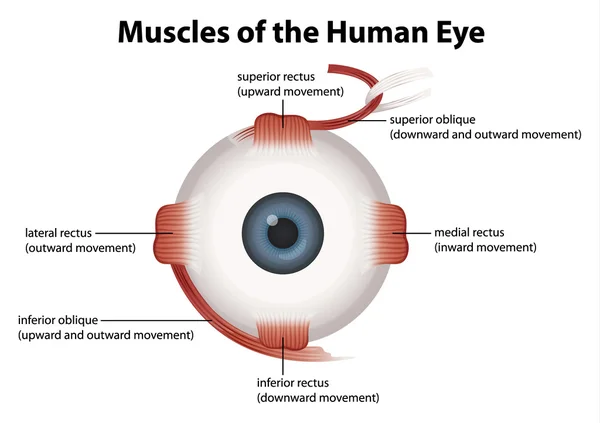
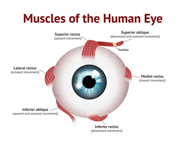
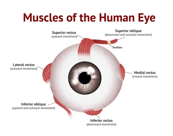
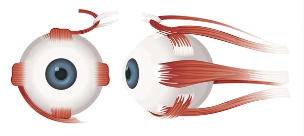
Muscle-eye-v3Muscles of Human eye, Eye muscle anatomy, , Blue eye, Vector Illustration on white background.
Eye anatomy diagram,illustration.
Eye accommodation infographic. Retina and sclera, ciliary muscle are contracted and relaxed, rounded and flattened lens. Objects at different distances and maintain clear images vector illustration
Eye Accommodation Infographic Retina Sclera Ciliary Muscle Contracted Relaxed Rounded Flattened Lens Objects Different Distances Maintain Clear Images Vector Illustration
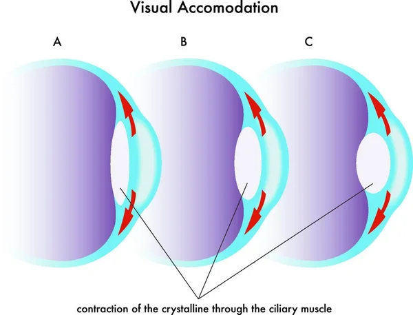
Medical illustration of the visual accomodation
Medical Illustration Visual Accomodation
How eye work medical illustration, eye - brain diagram
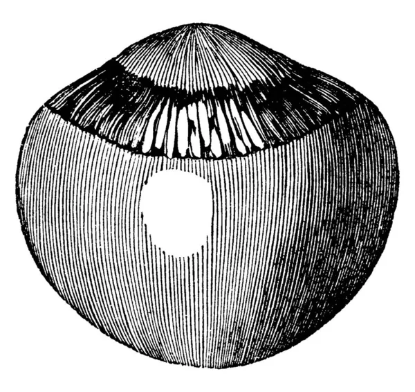
He vitreous humor and crystalline lens of the eyeball magnified, with the stains of the pigmentum nigrum left by the ciliary processes. Showing three different size of images in it, vintage line drawing or engraving
Vitreous Humor Crystalline Lens Eyeball Magnified Stains Pigmentum Nigrum Left Ciliary Processes Showing Three Different Size Images Vintage Line Drawing Engraving
Refractive errors eyeball. Astigmatism.
Herpes simplex keratitis HSV1 infection infographic showing human eye macro layout and detailed cornea cross section anatomy with dendritic lesion on corneal surface vector illustration.
Herpes Simplex Keratitis Hsv1 Infection Infographic Showing Human Eye Macro Layout Detailed Cornea Cross Section Anatomy Dendritic Lesion Corneal Surface Vector Illustration
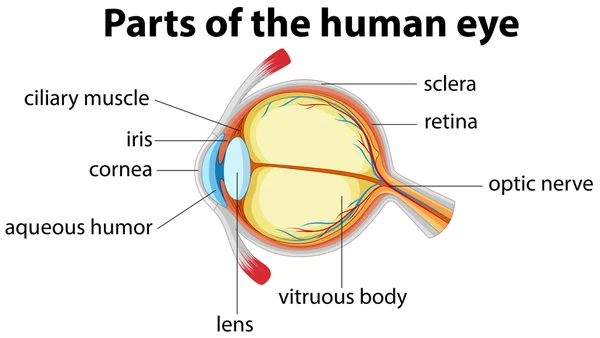
Parts of human eye with name
Human eye
Human eye
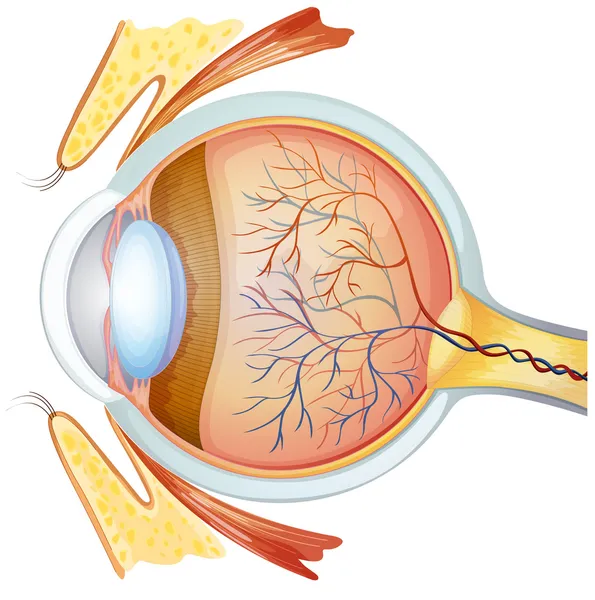
Human eye cross section
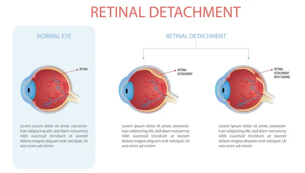
Infographic of a normal eye and an eye with retinal detachment, two types with and without tear.
Infographic Normal Eye Eye Retinal Detachment Two Types Tear
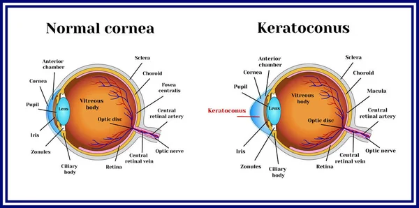
Keratoconus. Dystrophic disease of the cornea.
Set of human eyes
Set of human eyes
Human eye
Human eye
Herpes simplex keratitis HSV1 infection detailed infographic showing human eyeball cross section anatomy with lines pointing to internal structures and focus zone vector illustration.
Herpes Simplex Keratitis Hsv1 Infection Detailed Infographic Showing Human Eyeball Cross Section Anatomy Lines Pointing Internal Structures Focus Zone Vector Illustration
Iridocyclitis Eye Inflammation Comparison with Healthy Eye Medical Anatomy Illustration. Iridocyclitis Ocular Inflammation Educational Ophthalmology. Conjunctival hyperemia, and inflammation of the iris and ciliary body.
Iridocyclitis Eye Inflammation Comparison Healthy Eye Medical Anatomy Illustration Iridocyclitis Ocular Inflammation Educational Ophthalmology Conjunctival Hyperemia Inflammation Iris Ciliary Body
The posterior chamber, trabecular meshwork, and eye drainage. Crucial role in removing intraocular fluid. Intraocular pressure. Eye health concept, glaucoma treatment research vector illustration
Posterior Chamber Trabecular Meshwork Eye Drainage Crucial Role Removing Intraocular Fluid Intraocular Pressure Eye Health Concept Glaucoma Treatment Research Vector Illustration
Muscles of Human eye, Eye muscle anatomy, , Blue eye, Vector Illustration on white background.
Eye accommodation infographic. Retina and sclera, ciliary muscle are contracted and relaxed, rounded and flattened lens. Objects at different distances and maintain clear images vector illustration
Eye Accommodation Infographic Retina Sclera Ciliary Muscle Contracted Relaxed Rounded Flattened Lens Objects Different Distances Maintain Clear Images Vector Illustration
Keratoconus. Dystrophic disease of the cornea.
Comprehensive Vector Infographic Showing Eye Anatomy With Labeled Parts And Common Vision Defects. Visual Guide Includes Details Of Eye Components And Conditions Like Myopia, Hyperopia And Astigmatism
Comprehensive Vector Infographic Showing Eye Anatomy Labeled Parts Common Vision Defects Visual Guide Includes Details Eye Components Conditions Myopia Hyperopia Astigmatism
Human eye anatomy
Human eye anatomy
Human eye anatomy diagram. Flat vector poster on white background.
Human Eye Anatomy Diagram Flat Vector Poster White Background
Structural design of the human eye
Structural Design Human Eye
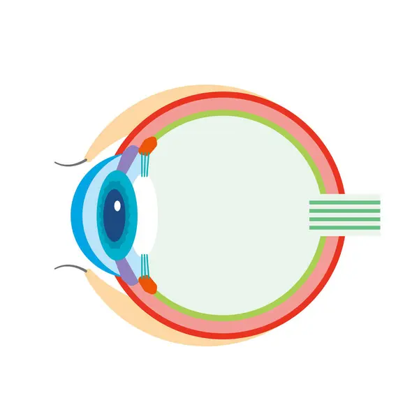
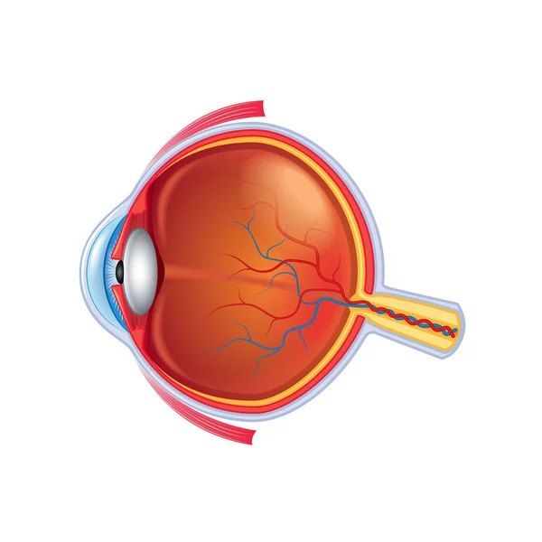
Cross section illustration of an eyeball
Cross Section Illustration Eyeball
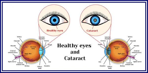
Healthy eyes and cataract (opacity of the eye lens).
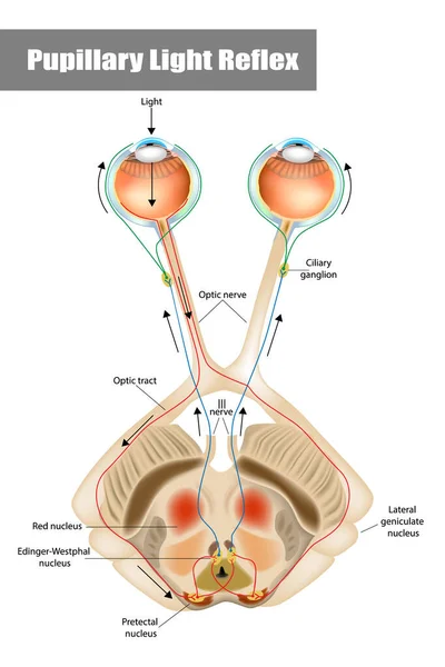
Pupillary light reflex PLR or photopupillary reflex.
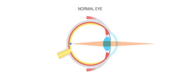
Normal vision. Glaucoma.
Eye anatomy isolated on white vector
Eyes are the organs of vision. They detect light and convert it into electro-chemical impulses in neurons. Illustration anatomy body.
Eyes Organs Vision Detect Light Convert Electro Chemical Impulses Neurons Illustration Anatomy Body
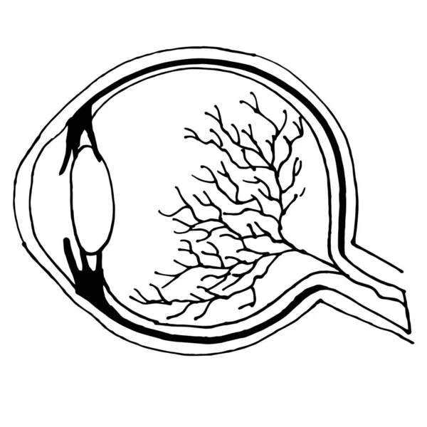
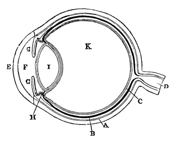
Diagrammatic illustration of the eye (line drawing)
Diagrammatic Illustration Eye Line Drawing
Plan of the eye seen in section showing the Sclerotic Coat, The Choroid Coat, The Retina, The Optic Nerve; The Vitreous Humor. showing three different size of images in it, vintage line drawing or engraving
Plan Eye Seen Section Showing Sclerotic Coat Choroid Coat Retina Optic Nerve Vitreous Humor Showing Three Different Size Images Vintage Line Drawing Engraving
Herpes simplex keratitis HSV1 corneal infection detailed diagram showing eye cross section anatomy with highlighted red target focus over dendritic lesion vector illustration.
Herpes Simplex Keratitis Hsv1 Corneal Infection Detailed Diagram Showing Eye Cross Section Anatomy Highlighted Red Target Focus Dendritic Lesion Vector Illustration
Anterior Uveitis Iridocyclitis Clinical Signs Medical Eye Anatomy. Keratic Precipitates and Ciliary Injection Medical Illustration. Ophthalmology Educational
Anterior Uveitis Iridocyclitis Clinical Signs Medical Eye Anatomy Keratic Precipitates Ciliary Injection Medical Illustration Ophthalmology Educational
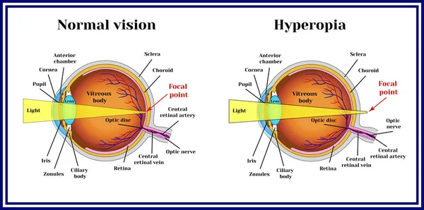
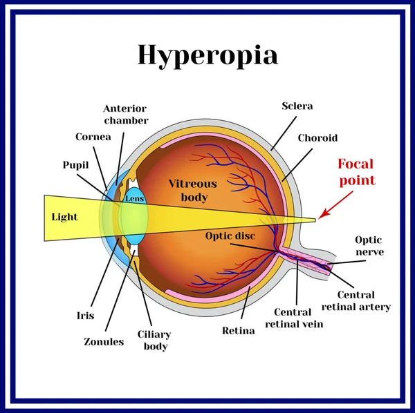
Refractive errors eyeball. Hyperopia.
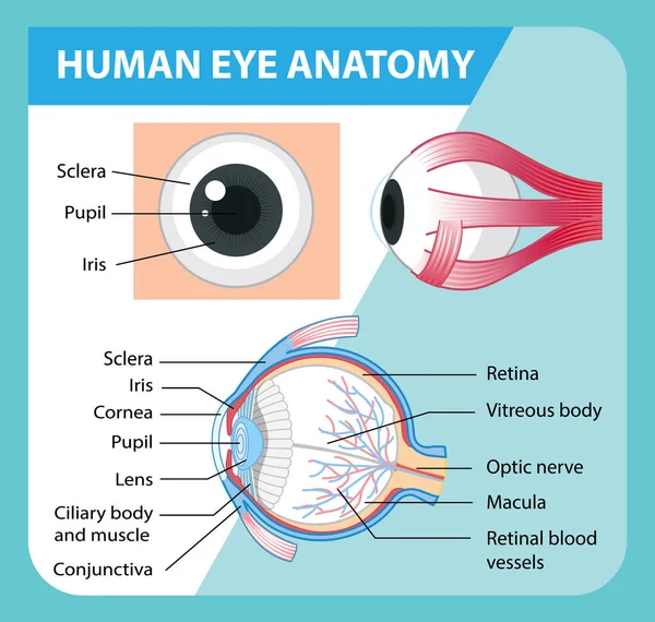
Diagram of human eye anatomy with label illustration
Diagram Human Eye Anatomy Label Illustration
Vector Education poster eye structure. Image human eye anatomy poster
Vector Education Poster Eye Structure Image Human Eye Anatomy Poster
Diagram of human eye anatomy with label illustration
Diagram Human Eye Anatomy Label Illustration
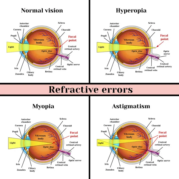
Refractive errors eyeball: hyperopia, myopia, astigmatism.
Eye accommodation infographic. Retina and sclera, ciliary muscle are contracted and relaxed, rounded and flattened lens. Objects at different distances and maintain clear images vector illustration
Eye Accommodation Infographic Retina Sclera Ciliary Muscle Contracted Relaxed Rounded Flattened Lens Objects Different Distances Maintain Clear Images Vector Illustration
Human retina anatomy. Cross-section of the eye. Close-up of Photoreceptors Rod cell and Cone cells. Structure of retina. infographic diagram for ophthalmology. Medical illustration. Detailed vector poster
Human Retina Anatomy Cross Section Eye Close Photoreceptors Rod Cell Cone Cells Structure Retina Infographic Diagram Ophthalmology Medical Illustration Detailed Vector Poster
The posterior chamber, trabecular meshwork, and eye drainage. Crucial role in removing intraocular fluid. Intraocular pressure. Eye health concept, glaucoma treatment research vector illustration
Posterior Chamber Trabecular Meshwork Eye Drainage Crucial Role Removing Intraocular Fluid Intraocular Pressure Eye Health Concept Glaucoma Treatment Research Vector Illustration
Causes and mechanism of cataract vector illustration
Causes Mechanism Cataract Vector Illustration
Refractive errors eyeball: hyperopia, myopia, astigmatism.
Types of diabetic retinopathy
Diagrammatic illustration of the eye (line drawing)
Diagrammatic Illustration Eye Line Drawing
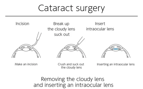
Illustrations, Cataract surgery, Medical Illustrations.
Illustrations Cataract Surgery Medical Illustrations
Human eye
Human eye
Types of diabetic retinopathy
Motor nerves of the eye anatomy poster. Abducens, trochlear and oculomotor nerves in the human brain. Ciliary gland and muscle, coordinate eye position. Sensory and motor functions vector illustration
Motor Nerves Eye Anatomy Poster Abducens Trochlear Oculomotor Nerves Human Brain Ciliary Gland Muscle Coordinate Eye Position Sensory Motor Functions Vector Illustration
Irrigation of nasal passages. Relief and treatment from allergies, reducing inflammation, cleaning the nasal passages, hydration, improvement of nasal function
Irrigation Nasal Passages Relief Treatment Allergies Reducing Inflammation Cleaning Nasal Passages Hydration Improvement Nasal Function
Muscles of Human eye, Eye muscle anatomy, Vector Illustration on white background
Refractive errors eyeball. Hyperopia.
Vector Education poster eye structure. Image human eye anatomy poster
Vector Education Poster Eye Structure Image Human Eye Anatomy Poster
Human eye profiles
Human eye profiles
Human eye
Human eye
Flow of cataract surgery, illustrated illustration (line drawing)
Flow Cataract Surgery Illustrated Illustration Line Drawing
Medical diagram of eye work. Anatomical infographics with structure of visual system, connection with brain and function of eyes. Cartoon flat vector illustration isolated on white background
Medical Diagram Eye Work Anatomical Infographics Structure Visual System Connection Brain Function Eyes Cartoon Flat Vector Illustration Isolated White Background
Human eye
Human eye
Herpes simplex keratitis HSV1 infection infographic showing eyeball cross section transition to detailed cornea anatomy diagram with highlighted red dendritic lesion focus vector illustration.
Herpes Simplex Keratitis Hsv1 Infection Infographic Showing Eyeball Cross Section Transition Detailed Cornea Anatomy Diagram Highlighted Red Dendritic Lesion Focus Vector Illustration
The posterior chamber, trabecular meshwork, and eye drainage. Crucial role in removing intraocular fluid. Intraocular pressure. Eye health concept, glaucoma treatment research vector illustration
Posterior Chamber Trabecular Meshwork Eye Drainage Crucial Role Removing Intraocular Fluid Intraocular Pressure Eye Health Concept Glaucoma Treatment Research Vector Illustration
Page