Current page requires JavaScript, this web browser either does not support JavaScript, or scripts are being blocked.
Please turn on JavaScript or use different browser.
Photos
Vectors
Illustrations
Free Pictures
Videos
Music & SFX
AI Assistant
AI Image Generator
Free Background Remover
Free Image Upscaler
Reverse Image Search
AI training data
Pricing
Vectors
Log In
Sign Up
Epithel Stock Vector Images
324 Epithel vector art & graphics are available under a royalty-free license
Create with AI
Landscape
Happiness
Nature
Lifestyle
Science
Animals
Business
Food
Architecture
Sport
Best Match
Fresh
Popular
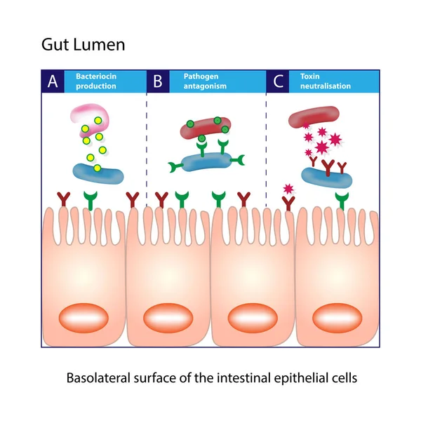
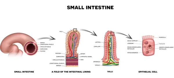
Gut lumen. Enterocytes, or intestinal absorptive cells. Small intestine. Columnar epithelial cells
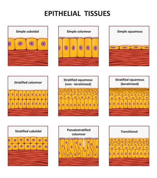
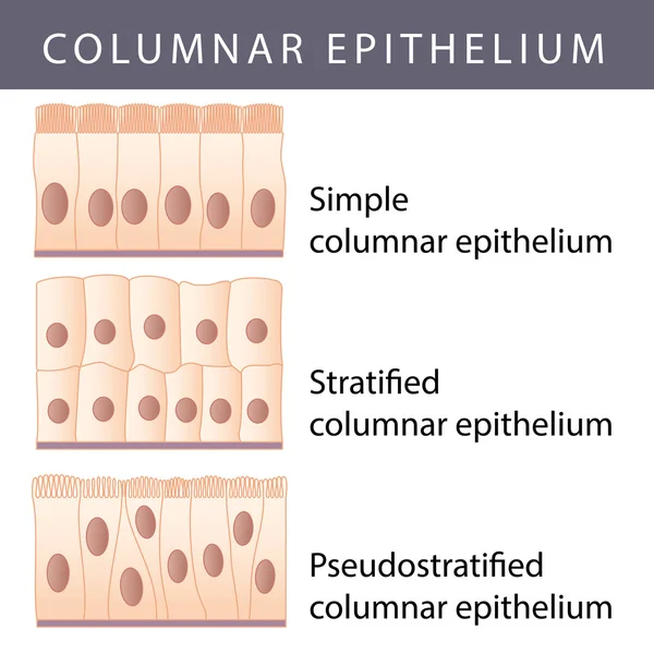
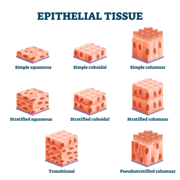
Different types of epithelial tissue collection.
Ciliated columnar epithelium. epithelial cells forms the lining of the stomach and intestines, duodenum, fallopian tubes, uterus, central canal of the spinal cord, nose, ears and the taste buds.
Structure of Villi and microvilli
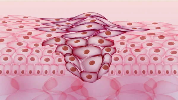
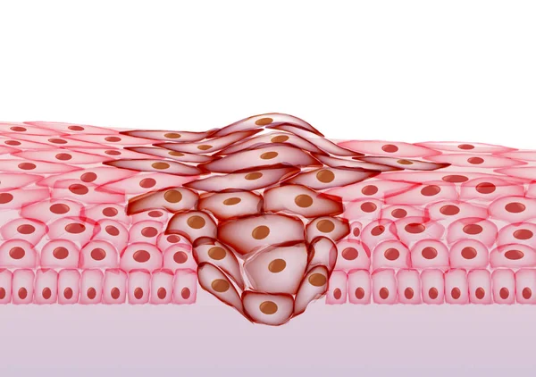
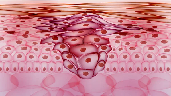
Growing Tumor, Tissue Section - Vector Illustration
Epithelium
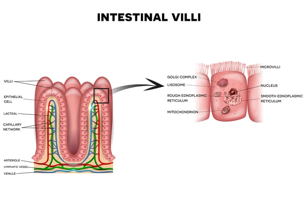
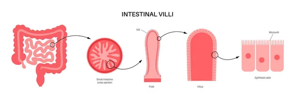
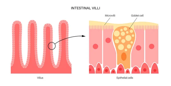
Intestinal villi and microvilli
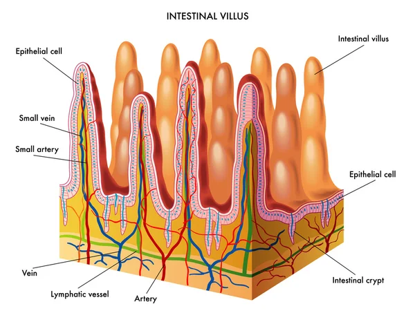
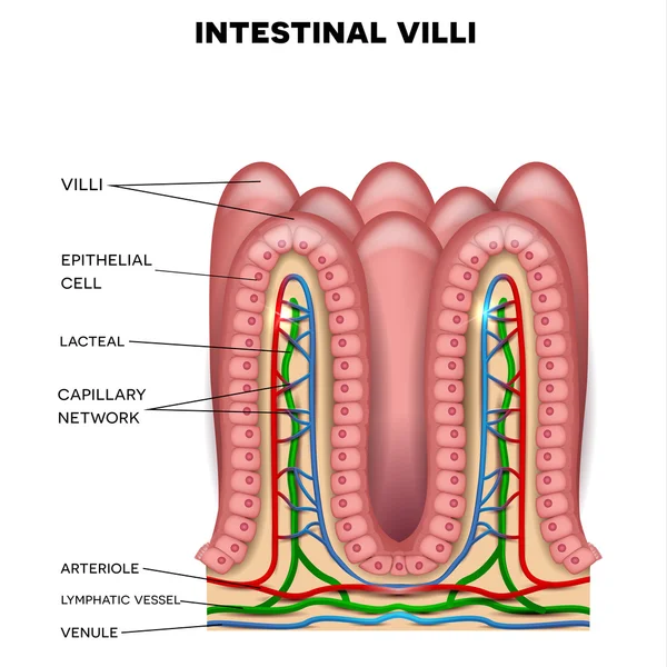
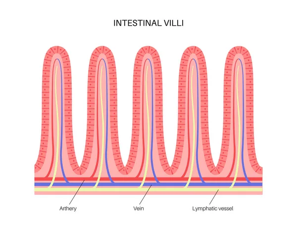
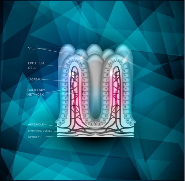
Intestinal villi anatomy
Growing Tumor, Tissue Section - Vector Illustration
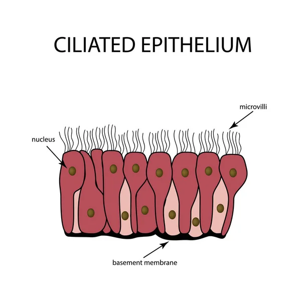
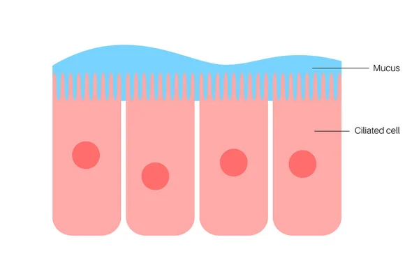
The structure of the ciliated epithelium. Infographics. Vector illustration on isolated background
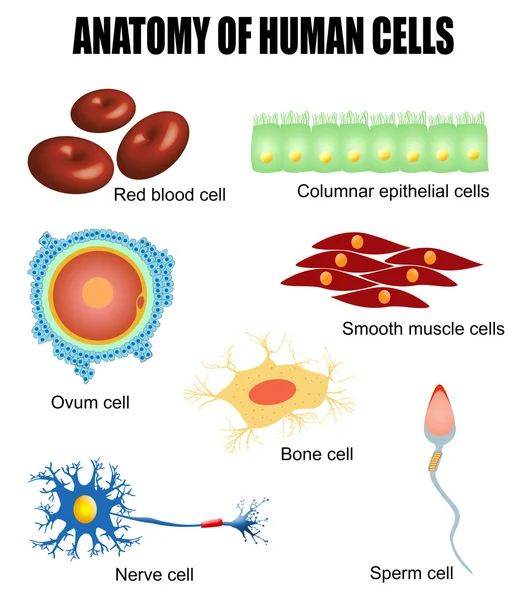
Anatomy of human cells
Epithelium
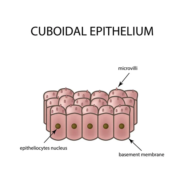
The structure of cubic epithelium. Infographics. Vector illustration on isolated background
The structure of the glandular epithelium. Infographics. Vector illustration on isolated background
Olfactory Bulb Anatomy. Epithelium receptor cells. Nasal mucosa
Celiac disease inflammation
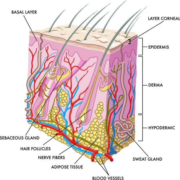
Medical illustration of the section of skin
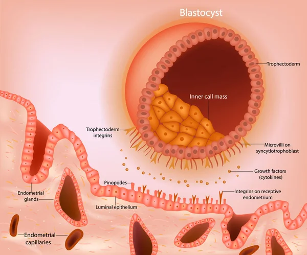
Blastocyst implantation. A schematic representation of a blastocyst approaching the receptive endometrium. Early signaling between the blastocyst. Embryonic Development
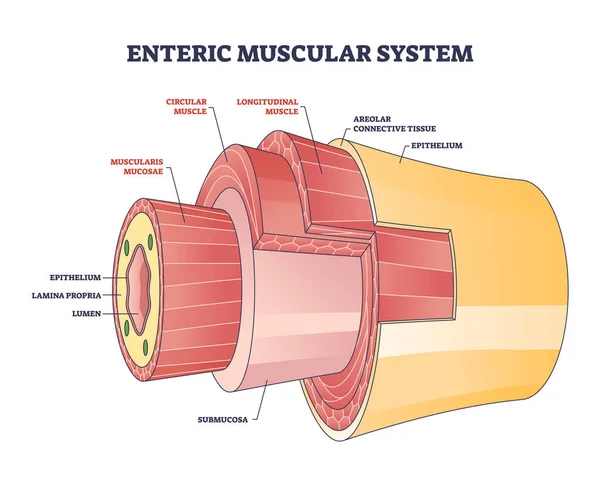
Enteric muscular system in gut wall of the small intestine outline diagram. Labeled educational scheme with layers and structure of digestive tract muscle vector illustration. Lamina propria location.
Olfactory1
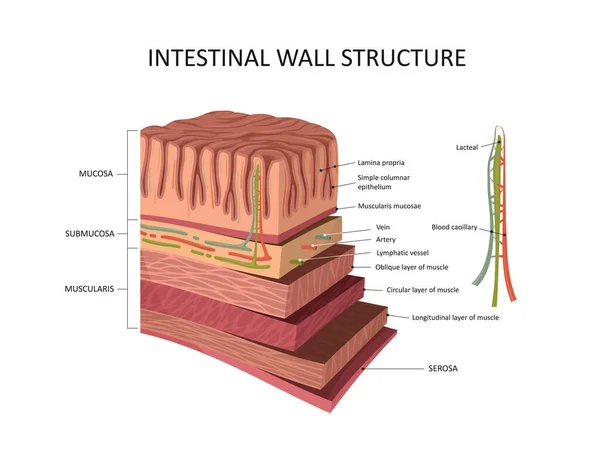
Intestinal wall structure. Stomach wall layers detailed anatomy
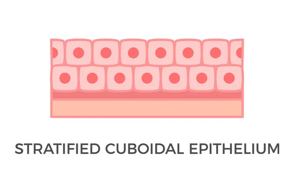
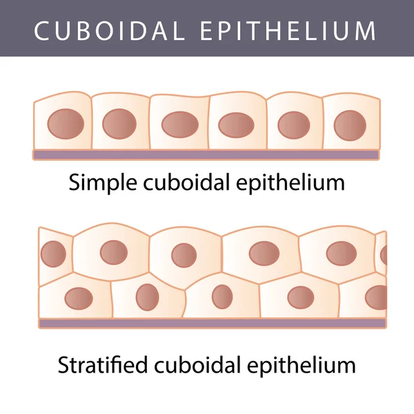
Stratified cuboidal epithelium. Epithelial tissue types. Multiple layers of cube-like cells. Occurs in the excretory ducts of sweat glands and salivary glands. Medical illustration. Vector.
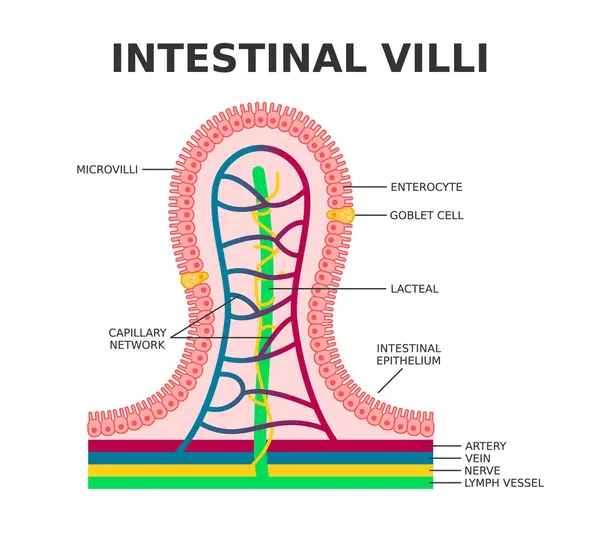
Intestinal villi. Microvilli. Intestinal epithelium. Villi absorb nutrients from the food. Intestinal epithelial cells with capillary network. Enterocyte and Goblet cell. Vector illustration.
Nasal epithelium concept
Cervical cancer cells elongated Dysplasia stages infographic Female reproductive system squamous cell carcinoma in uterus pap smear test. Human anatomy internal organs flat style icon
Intestinal villi anatomy
Melanocytes are specialized cells that produce melanin pigments.
Epithelium
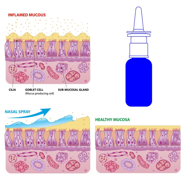
Nasal mucosa cells and micro cilia vector scheme
Human eye anatomy. Retina structure. Cross-section of the eye. Cells in section of the retina. Detailed vector poster
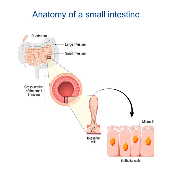
Small intestine Anatomy. Cross section of a ileum with Internal villi. Close-up of Epithelial cells with Microvilli. Vector illustration
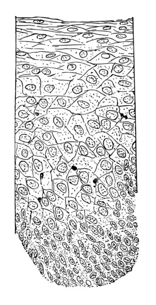
This illustration represents Cross Section of the Epithelium, vintage line drawing or engraving illustration.
Epidermis structure. Skin anatomy. Cell, and layers of a human skin. Cross section of the epidermis. Skin care. vector illustration.
Types of muscle tissue
Celiac disease info poster
Intestinal villi anatomy
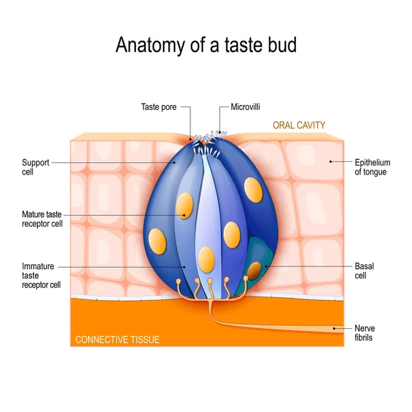
Taste bud. Mature and Immature taste Receptor, Support and Basal Cells, Epithelium Of tongue. Human Anatomy. Vector diagram for educational, biological, science and medical use
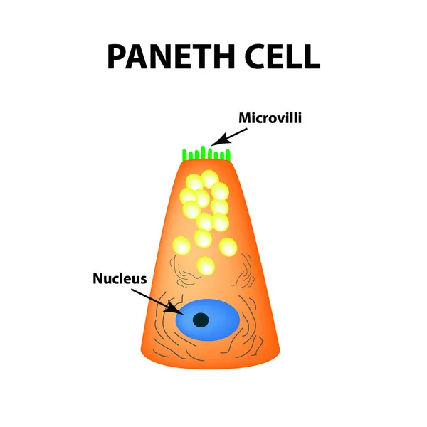
The structure of Paneth cells. Davidoff's cell. fographics. Vector illustration on isolated background.
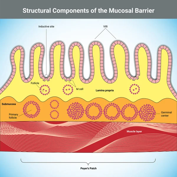
Structural Components of the Mucosal Barrier medical illustration
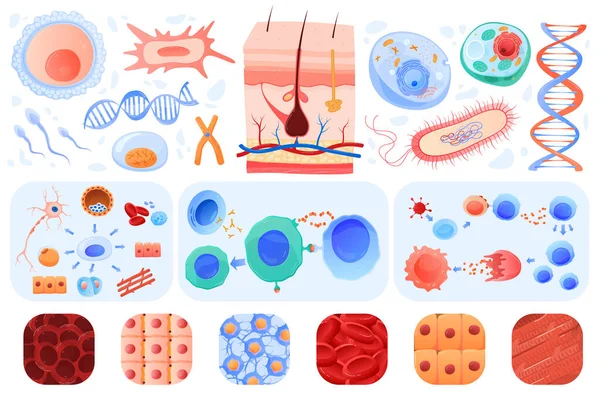
Anatomy of human cells, skin, blood cells bacillus, set of vector illustrations. Biology or anatomy education in schools and medical clinics.
Anatomy of human cells
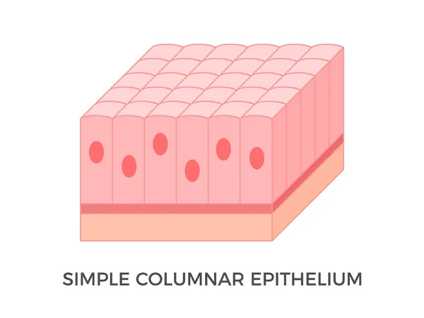
Simple columnar epithelium. Epithelial tissue types. Tall and slender cells with oval-shaped nuclei. Lines most organs of the digestive tract like stomach, intestines. Medical illustration. Vector.
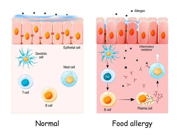
Food allergy is an abnormal immune response to food. Cell of normal Epithelium and Food allergy. development of an allergic reaction from allergens penetration to Inflammatory mediators synthesis. vector
Gastric chief cell. Gastric gland cell that releases pepsinogen and gastric lipase. Cell responsible for secretion of chymosin in ruminant animals.
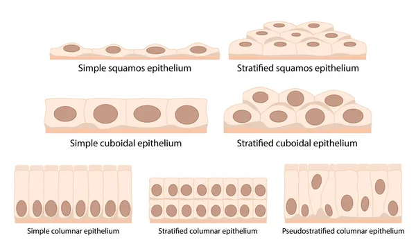
Cells of epithelial tissue: squamous (flattened and thin), cuboidal (boxy, as wide as it is tall), columnar (rectangular, taller than it is wide), pseudostratified.
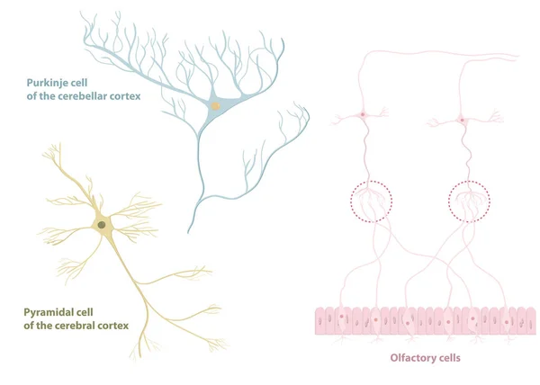
Types of neurons: pyramidal cell from the cerebral cortex, Purkinje cell from the cerebral cortex, olfactory cells from the olfactory epithelium and olfactory bulb
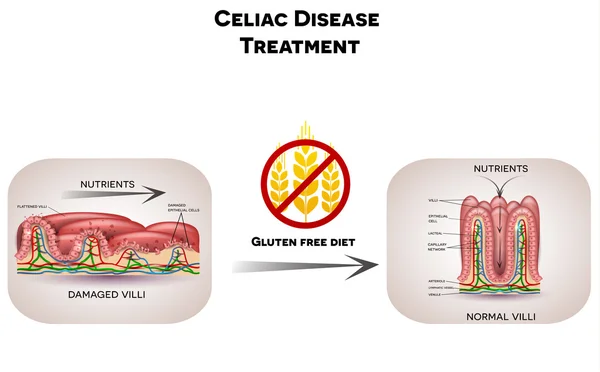
Celiac disease treatment gluten free diet
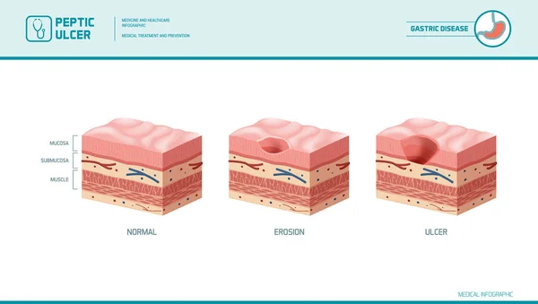
Stomach erosion and peptic ulcer
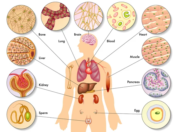
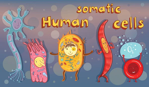
Vector illustration of human somatic cells
3D Isometric Flat Vector Conceptual Illustration of Nasal Mucosa Cells, Medical Educational Diagram
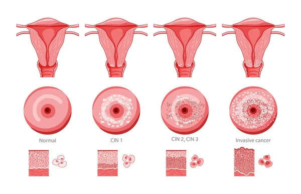
Set of Cervical invasive cancer intraepithelial neoplasia Dysplasia 1 2,3 and normal stage cell morphology on pap smear test infographic Female reproductive system Carcinoma view. medical flat style
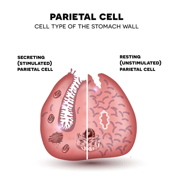
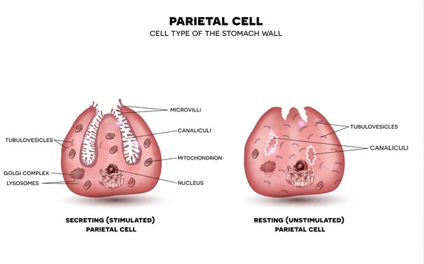
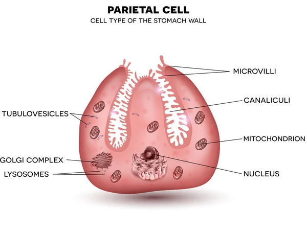
Parietal cell of stomach wall, located in the gastric glands se
Epithelial cells vector illustration. Medical location and meaning diagram.
Intestinal villus. Different between Villi and microvilli. Cross section of a duodenum with submucosa, mucosa and muscularis layers. Vertical section of a villus with Lymph capillary or Lacteal and Blood vessels. Close-up of a Columnar epithelium, an
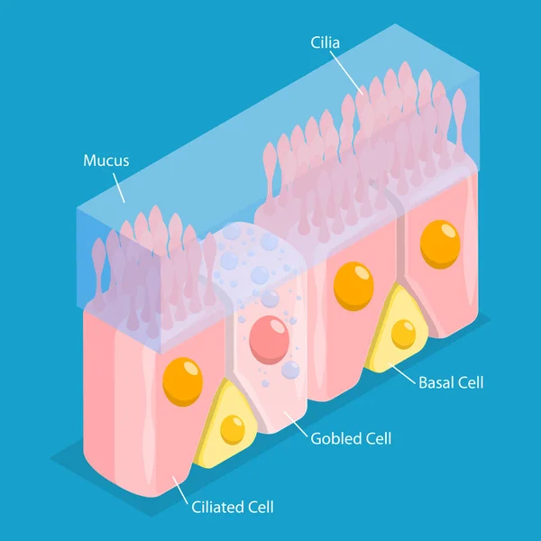
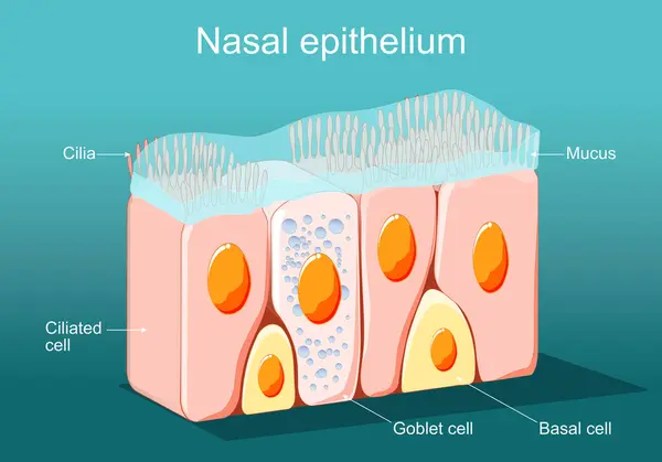
Nasal Epithelium structure anatomy, labeled with cilia, mucus, ciliated cell, goblet cell, basal cell. Human tissue nasal cavity, immune response to nasal diseases, diagram biology education.
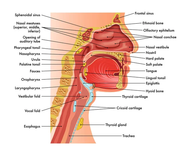
Medical diagram of anatomy of nose, mouth, larynx, and pharynx, with annotations.
Leaky gut. Close-up of cells of a intestinal epithelium. Difference between Healthy cells, and inflamed intestinal cells. Comparison normal tissue of the gastrointestinal tract, and Intestinal permeability. Vector poster. Isometric Flat illustration.
Parietal cell of stomach wall, located in the gastric glands sec
A rare skin disease manifested by the appearance of multiple red hyperkeratic papules. Hand-drawn versions of the appearance of lichenoid keratosis chronicus on the skin.
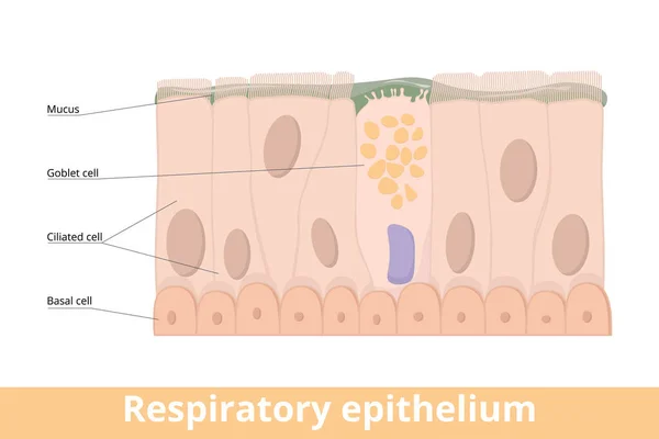
Respiratory epithelium. A type of ciliated columnar epithelium found lining most of the respiratory tract as respiratory mucosa including goblet cell, basal and ciliated cells, mucus.
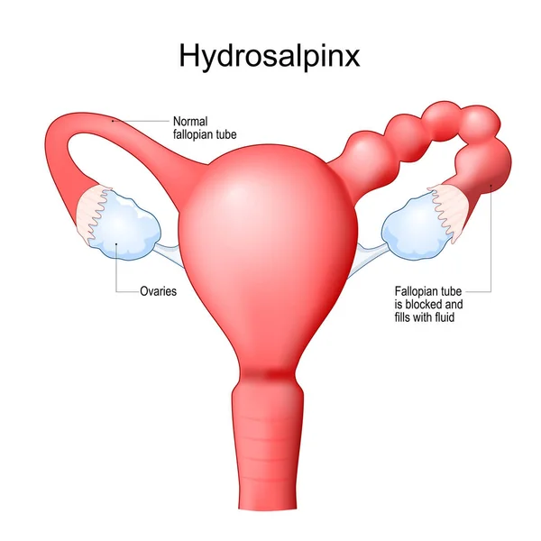
Human uterus with blocked of Fallopian tube. Hydrosalpinx cause infertility. Female reproductive system. Frontal view. Human anatomy. Realistic medical illustration. Vector poster
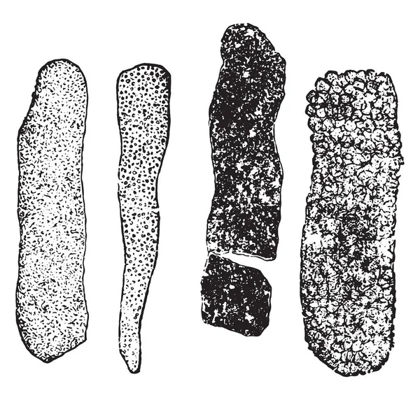
Granular casts, vintage engraved illustration
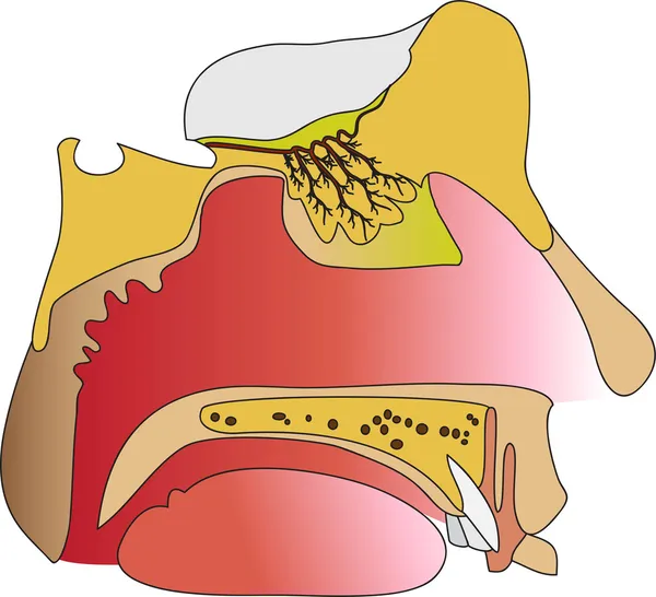
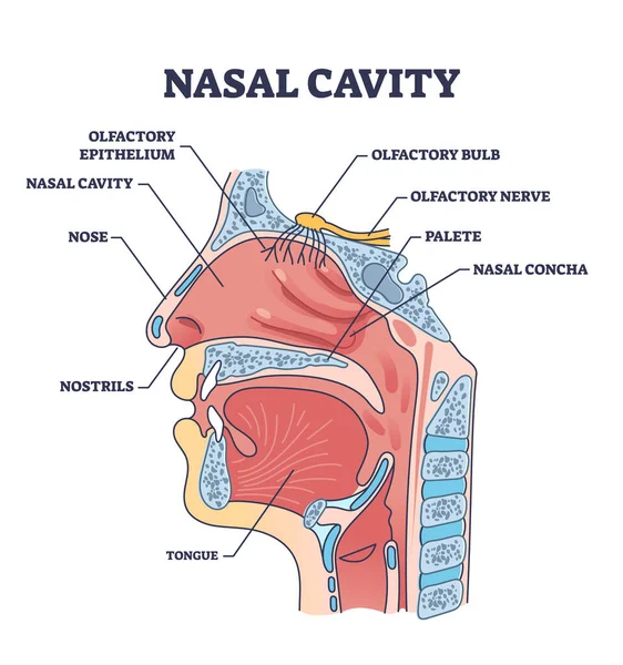
Nasal cavity anatomy with medical nose parts description outline diagram. Labeled educational cross section scheme with mouth and oral structure vector illustration. Nostrils and olfactory location.
Olfactory system anatomy
Intestinal villi background
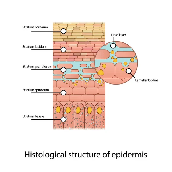
Histological structure of epidermis - skin layers shcematic vector illustration showing stratum basale, spinosum, granulosum, lucidum and corneum and lamellar bodies
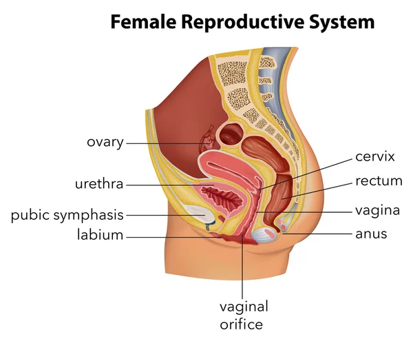
Female Reproductive System
Anatomy of the retina. Human eye. Photoreceptor cells. Cone cell and rod cell. Vector illustration
Small intestine anatomy
Nasal epithelium concept
Ion channels with different membrane gated types examples outline diagram. Labeled educational anatomical scheme with mechanically, ligand and voltage gated channel differences vector illustration.
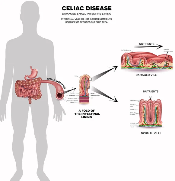
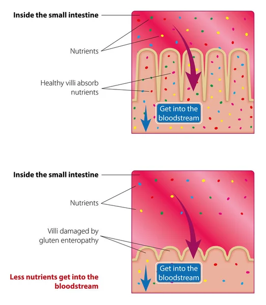
Absorption of nutrients in the small intestine. Healthy and damaged villi. Medical vector illustration
Intestinal Epithelial Cells background vector
Cervical intraepithelial neoplasia. Cervical dysplasia. CIN. Pre-cancerous lesion. Close-up of a cervix. Cancer development from normal cell to Precancerous transformation of cells of the cervix. Vector. Schematic diagram. Detailed poster.
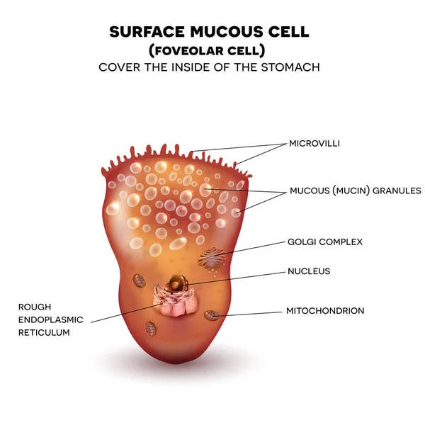
Foveolar cell or surface mucous cell of the stomach wall
Intestinal villi concept
Epithelium of nasal mucosa
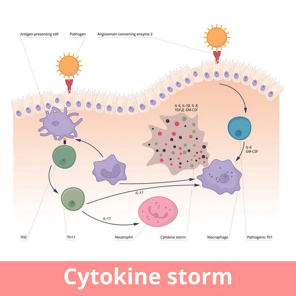
Cytokine storm. Hypercytokinemia during which the immune system causes a release of cytokines with help of macrophages, t helper cells, neutrophils.
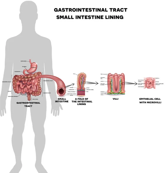
Small intestine lining
Intestinal villi anatomy
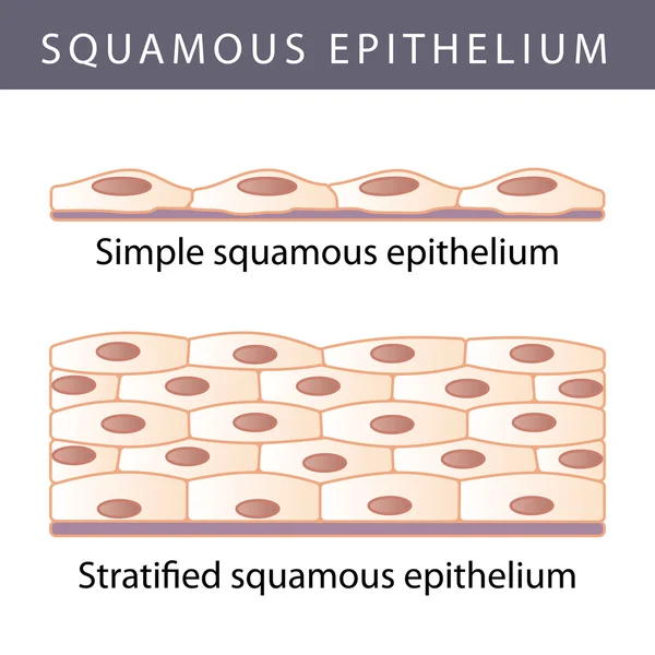
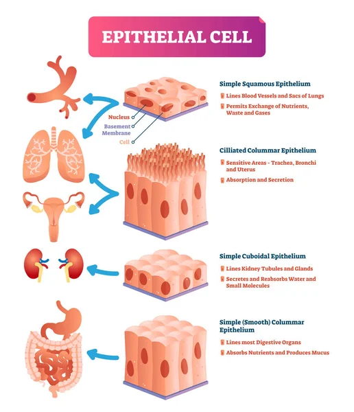
Epithelial tissue with labeled squamous, cuboidal and columnar examples.
Growing Tumor, Tissue Section Inflamed Skin - Vector Illustratio
Retina structure
Parietal cell of stomach wall, located in the gastric glands se
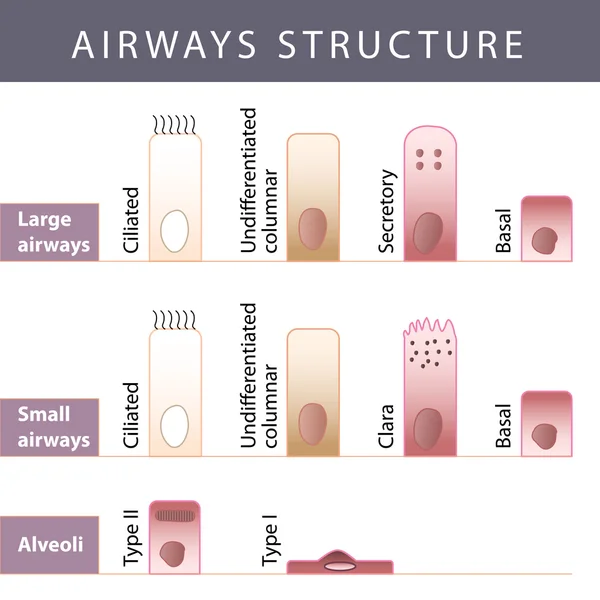
Airways structure
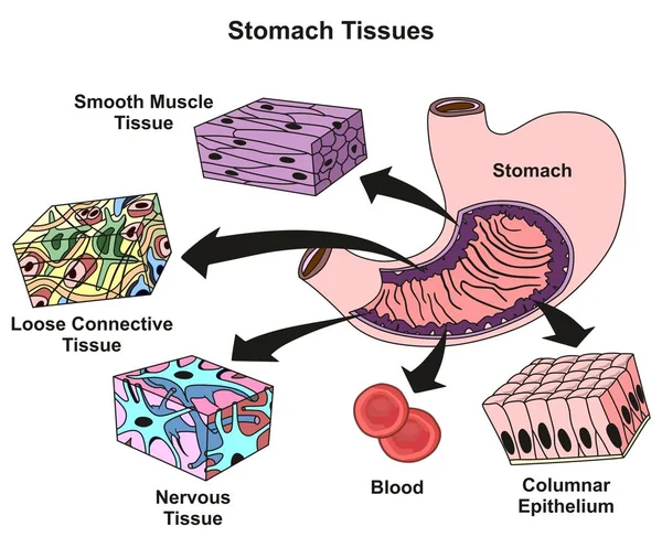
Stomach Tissues Types and Structure infographic diagram including smooth muscle loose connective nervous blood, columnar epithelium for medical science education and health care
Small intestine lining
Respiratory Epithelium Cell Structure Illustration. Ciliated, Basal, and Goblet Cells Diagram. Mucociliary Epithelium Cross Section Illustration
Set of Types of epithelial tissue: cilliated columnar, simple columnar, simple cuboidal, and simple squamous cells, labeled educational anatomical structure with microbiology elements.
Nasal epithelium. Ciliated columnar epithelium. epithelial cells forms the lining of the stomach and intestines, duodenum, fallopian tubes, uterus, central canal of the spinal cord, nose, ears and the taste buds. Ciliated cells. Respiratory defense
Cervical dysplasia. difference in covering epithelium affected of Cervical Intraepithelial Neoplasia. Cross section of epithelium. Close-up of a normal Squamous cell and Abnormal cells. Cervical cancer evolution. Vector illustration
Bronchiectasis and Normal Bronchus Cross Section Medical Illustration. Airway Damage Comparison in Bronchiectasis Anatomy Diagram
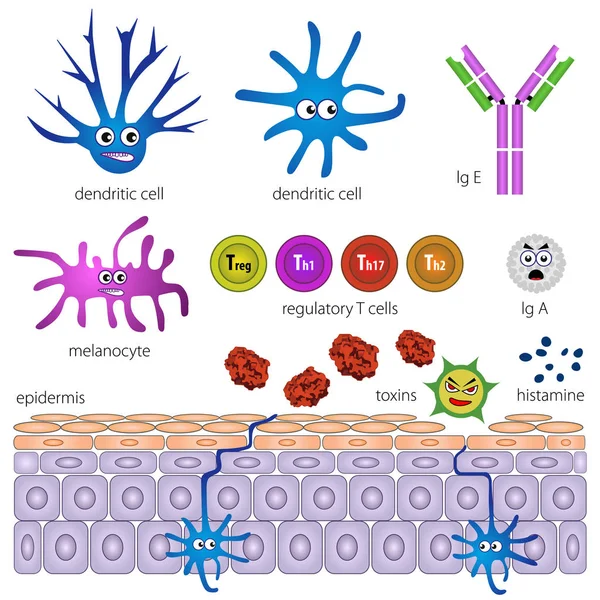
The cells of the protective layer of the skin. Protection against inflammation
Intestinal villi anatomy
Simple cuboidal epithelium. Epithelial tissue types. A single layer of cube-like cells that provide protection and may be active or passive depending on the location. Medical illustration. Vector.
IgA immunity. Peyers patch. Lymphoid follicles of the small intestine generate IgA immune response with B cells, T cells, dendritic cells, and plasma cells secreting IgA.
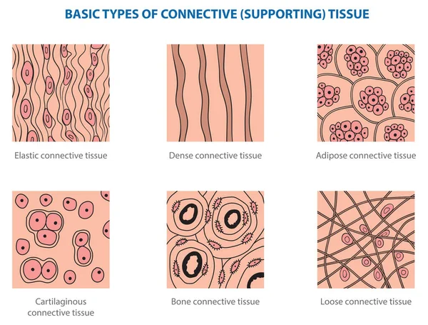
Types of animal tissues by structure - connective tissue.
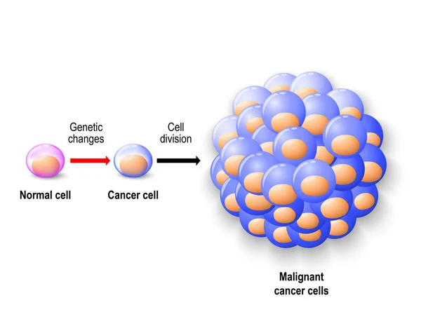
Normal human cell, cancer cell and malignant cancer
Folliculitis. Healthy skin before and after Infection and inflammation of the hair follicle. Acne, Pus under epithelium. Hair follicle infection. Hot tub infection. Vector poster. Isometric Flat illustration. Schematic diagram
Exocrine Glands Secrete Hormones. Illustration showing cross section for learning exocrine glands human
Next Page
1
2
3
4
Page
Go
Home
Vectors
epithel vector images
Related vector searches
cell
,
histology
,
basal cell carcinoma
,
nerve cell
,
fallopian tube
,
wedding icons pink
,
cherry blossom clipart
,
pink flowers clip art
,
intestine anatomy
,
proteinuria