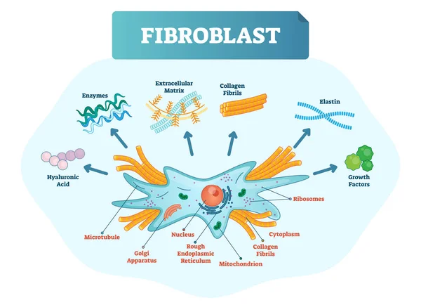
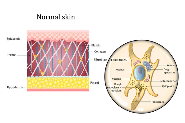
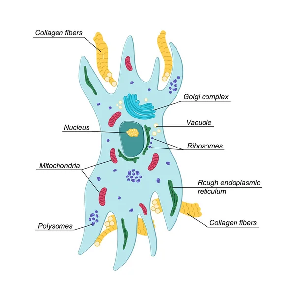
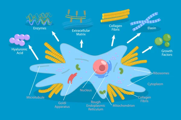
Fibroblast vector illustration. Scheme with extracellular, collagen fibrils, elastin, hyaluronic acid, microtubule, golgi apparatus, nucleus and ribosomes.
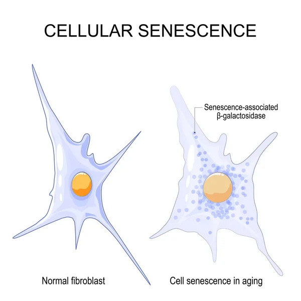
Cellular senescence. changes senescent cells During ageing. Comparison and difference between Normal fibroblast and Cell senescence in aging. Vector illustration
Cellular Senescence Changes Senescent Cells Ageing Comparison Difference Normal Fibroblast Cell Senescence Aging Vector Illustration
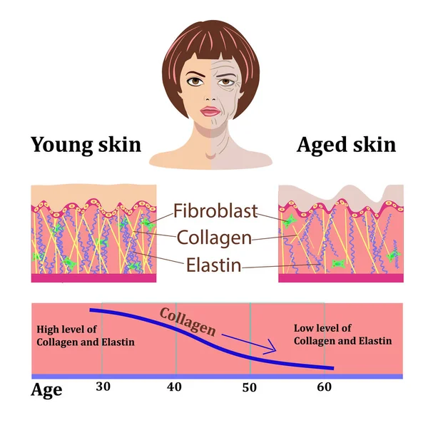
Vector faces and two types of skin - aged and young for medical and cosmetological illustrations isolated
Skin aging
Skin aging
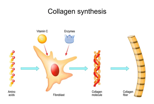
Process of Collagen synthesis. From Amino acid to Collagen fiber and molecule. Role of fibroblast of aging process. vector illustration
Process Collagen Synthesis Amino Acid Collagen Fiber Molecule Role Fibroblast Aging Process Vector Illustration
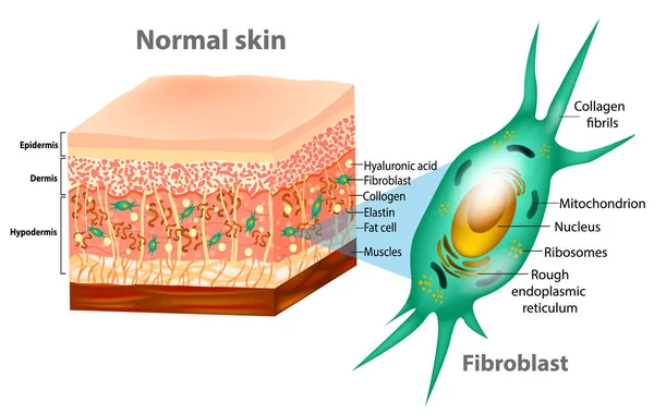
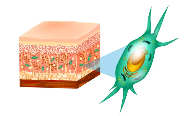
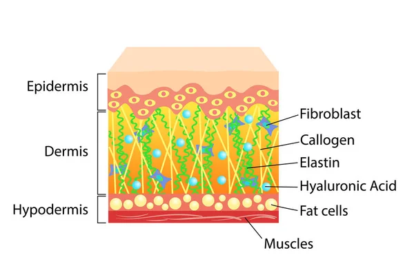
Fibroblast and Human skin structure (Muscles, Fat cell, Hyaluronic acid, Elastin, Collagen, Fibroblast).
Fibroblast Human Skin Structure Muscles Fat Cell Hyaluronic Acid Elastin Collagen Fibroblast
Fibroblast and Human skin structure (Muscles, Fat cell, Hyaluronic acid, Elastin, Collagen, Fibroblast).
Fibroblast Human Skin Structure Muscles Fat Cell Hyaluronic Acid Elastin Collagen Fibroblast
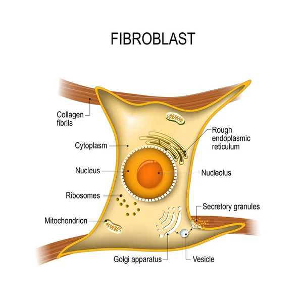
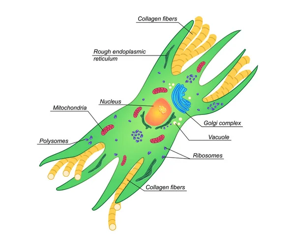
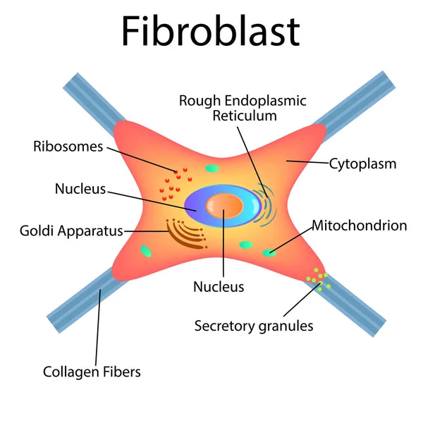
Fibroblast anatomy. structure of cell. Diagram with golgi apparatus, nucleus, mitochondrion and ribosomes. vector illustration. Poster
Fibroblast Anatomy Structure Cell Diagram Golgi Apparatus Nucleus Mitochondrion Ribosomes Vector Illustration Poster
Collagen synthesis with Vitamin C and Enzymes. From Fibroblast and Amino acids to Collagen fiber that comprises molecules of protein. Vector
Collagen Synthesis Vitamin Enzymes Fibroblast Amino Acids Collagen Fiber Comprises Molecules Protein Vector
Cytokine. Vector concept
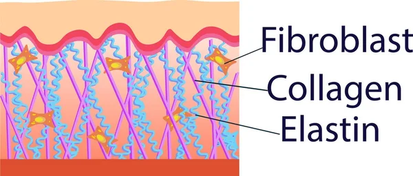
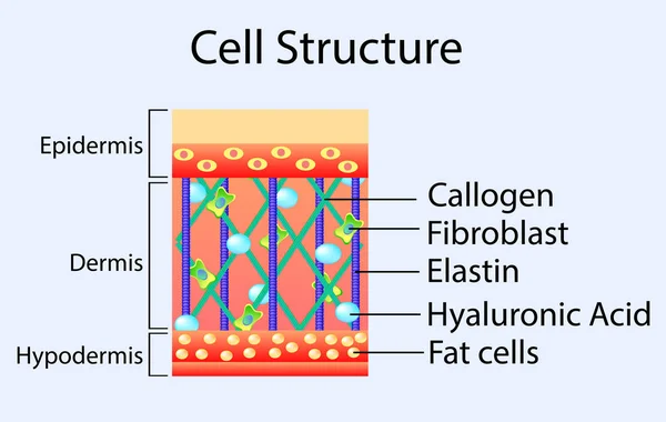
Human skin structure and fibroblast structure. Fat cell, Elastin, Collagen, Fibroblast. Detailed diagram. Vector illustration
Structure human skin with collagen and elastin fibers, fibroblasts.Layers of the human skin. skin and health care concept Vector diagram
Hands skin aging, vector illustration with a chart
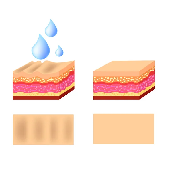
Vector diagram with schemes of two types of skin, for healthcare illustrations
Vector illustration. Fibroblast is a dermis cell. Structure of Fibroblast cell.
Fibroblast is vital to the skin's strength and elasticity. Struc
Vector illustration of structure cells with collagen, elastin and fibroblast
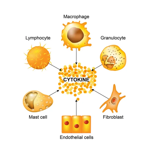
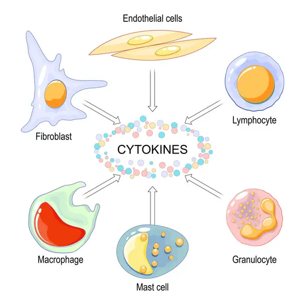
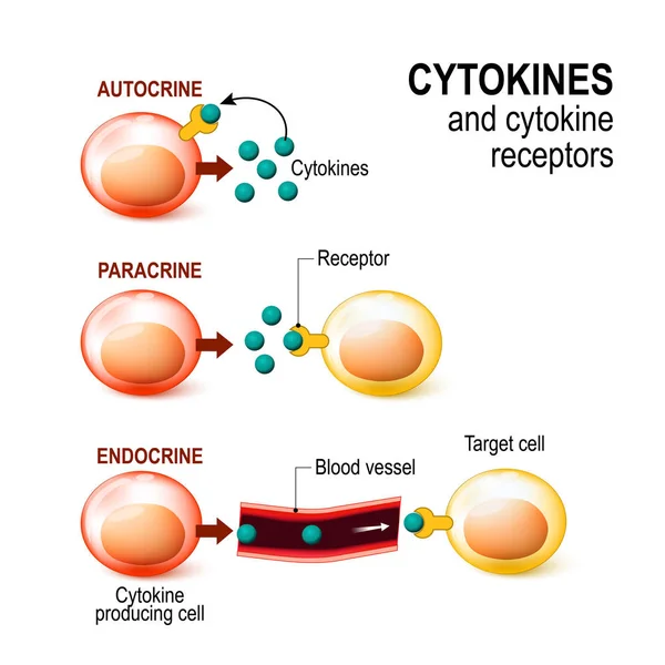
Cells that produce cytokines
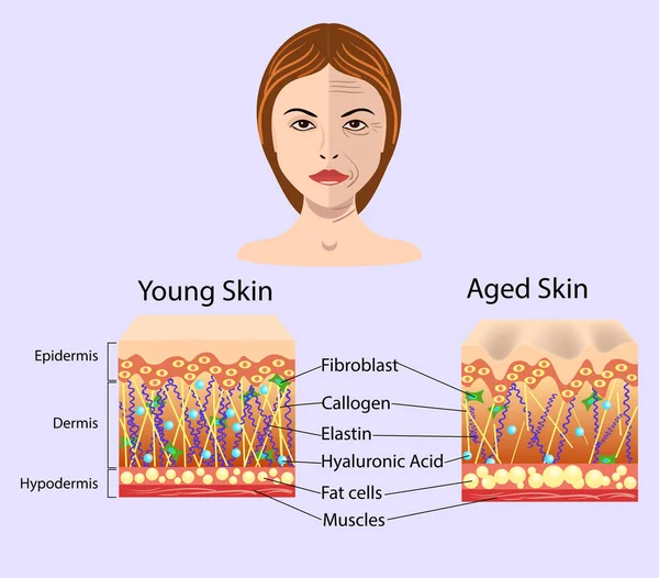
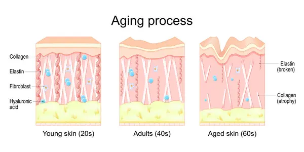
Aging process, educational medical poster design healthcare scheme, labeled comparison of Young, Adults and aged skin, Collagen, Elastin, Hyaluronic acid and fibroblasts. Face skin structure changes.
Aging Process Educational Medical Poster Design Healthcare Scheme Labeled Comparison Young Adults Aged Skin Collagen Elastin Hyaluronic Acid Fibroblasts Face Skin Structure Changes
Structure of the Chondrocytes. Chondrocytes cells in healthy cartilage.
Structure Chondrocytes Chondrocytes Cells Healthy Cartilage
Ovarian cyst, follicle and Corpus luteum.
Human anatomy disease. medulloblastoma vector illustration on white background. eps 10
Human Anatomy Disease Medulloblastoma Vector Illustration White Background Eps
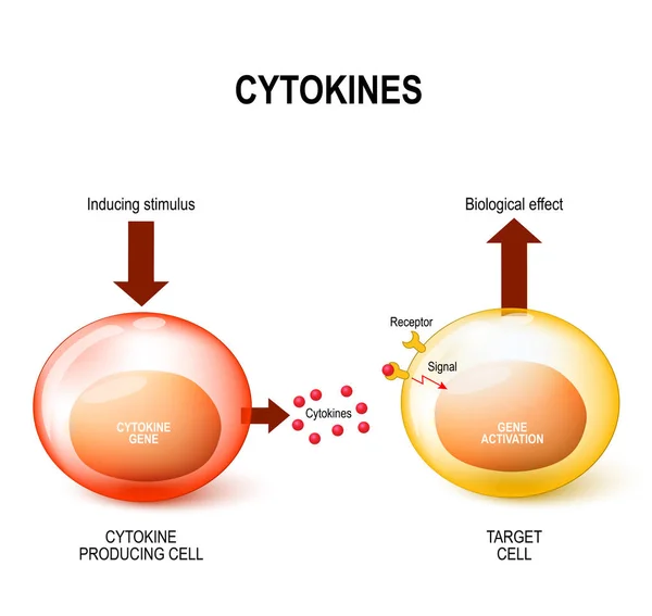
Cytokine Education diagram. It shows that the cytokine cell is induced by the stimulus and the cytokine is released into the target cell, which then responds to biological effects. Interferons, interleukins, lymphokines.
Cytokine Education Diagram Shows Cytokine Cell Induced Stimulus Cytokine Released Target Cell Which Responds Biological Effects Interferons Interleukins Lymphokines
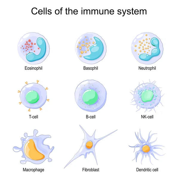
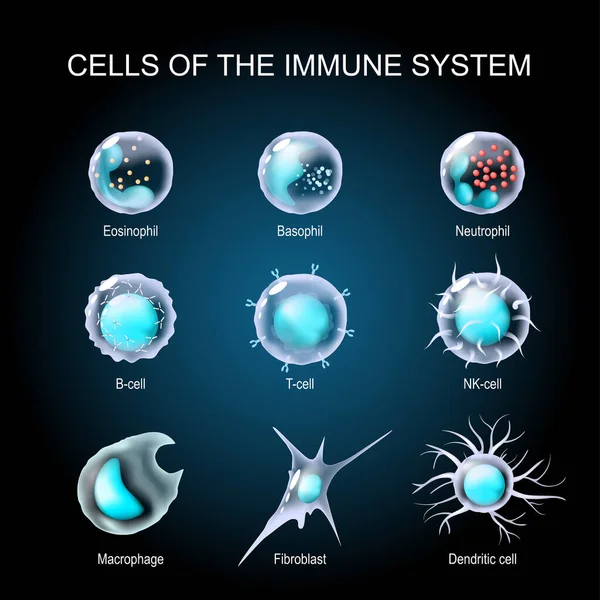
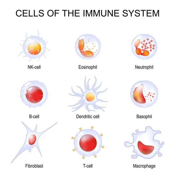
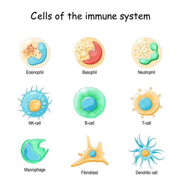
Cells of the immune system. White blood cells or leukocytes Eosinophil, Neutrophil, Basophil, Macrophage, Fibroblast, and Dendritic cell. Vector illustration
Cells Immune System White Blood Cells Leukocytes Eosinophil Neutrophil Basophil Macrophage Fibroblast Dendritic Cell Vector Illustration
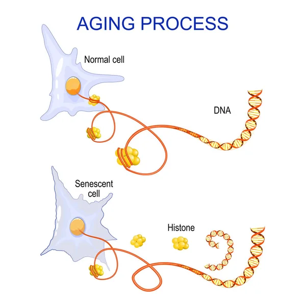
Aging process into cells. chromatin, DNA and histones change in ageing and senescent cells. Vector
Aging Process Cells Chromatin Dna Histones Change Ageing Senescent Cells Vector
Vector diagram with schemes of two types of skin, for cosmetological illustrations
Vector diagram with schemes of two types of skin, for cosmetological and healthcare illustrations
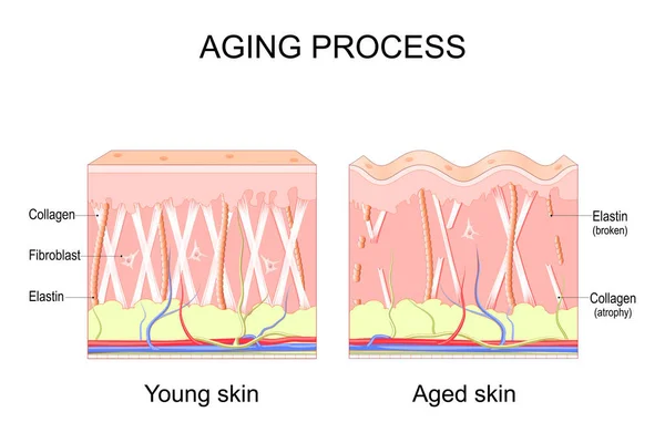
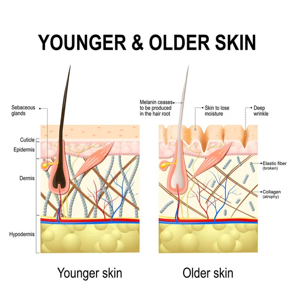
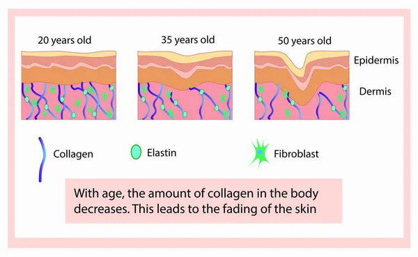
Aging process. comparison of Young and aged skin. Collagen, Elastin and fibroblasts in younger and older skin. age-related changes in the skin when Collagen fibers atrophy, and Elastin broken. Vector illustration
Aging Process Comparison Young Aged Skin Collagen Elastin Fibroblasts Younger Older Skin Age Related Changes Skin Collagen Fibers Atrophy Elastin Broken Vector Illustration
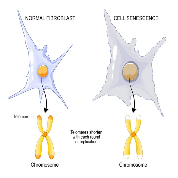
Chromosome of a Normal fibroblast and Cell senescence. Telomeres shorten with each round of replication. aging process. Vector poster
Chromosome Normal Fibroblast Cell Senescence Telomeres Shorten Each Replication Aging Process Vector Poster
Cells of the immune system. White blood cells or leukocytes Plasma cell, Eosinophil, Neutrophil, Basophil, Macrophage, T-cell, NK-cell, B-cell. Isometric Flat vector illustration
Cells Immune System White Blood Cells Leukocytes Plasma Cell Eosinophil Neutrophil Basophil Macrophage Cell Cell Cell Isometric Flat Vector Illustration
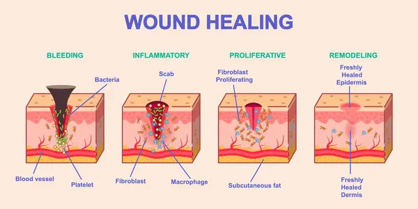
Wound healing process. Medical infographics or diagram with stages or phases of skin regeneration after injury. Bleeding, Inflammation, Proliferation and Remodeling. Cartoon flat vector illustration
Wound Healing Process Medical Infographics Diagram Stages Phases Skin Regeneration Injury Bleeding Inflammation Proliferation Remodeling Cartoon Flat Vector Illustration
Human skin structure with collagen and elastane fibers, hyaluronic acids, fibroblasts. Schematic illustration. Layers of the human skin. Vector diagram
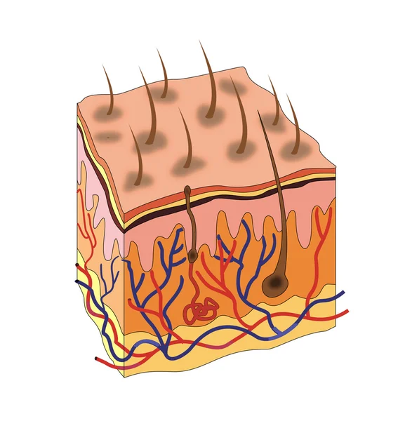
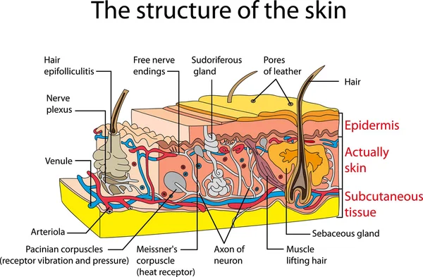
Skin anatomy
Skin anatomy
Human skin changes or ageing skin.
Vector Fibroblast illustration with description. Structure of dermis cell. Connective tissue
Vector Fibroblast Illustration Description Structure Dermis Cell Connective Tissue
Vector scheme of skin, for cosmetological and healthcare illustrations
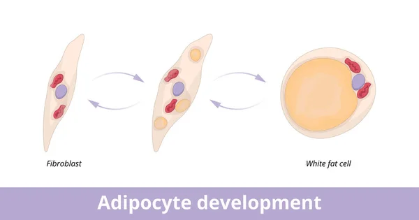
Adipocyte development. Visualization of adipocyte (fat cell) development from fibroblast. Fibroblast as adipocyte progenitor.
Adipocyte Development Visualization Adipocyte Fat Cell Development Fibroblast Fibroblast Adipocyte Progenitor
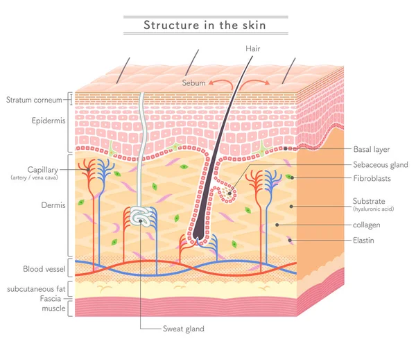
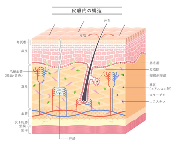
Structure in the skin_no notation
Structure Skin_No Notation
Plasma Lifting or Fibroblast treatments. Blood Plasma Injection Illustration for Rejuvenation. Medical Diagram of Plasma Lifting Procedure PRP therapy
Plasma Lifting Fibroblast Treatments Blood Plasma Injection Illustration Rejuvenation Medical Diagram Plasma Lifting Procedure Prp Therapy
Illustration of young skin layers and components
Illustration Young Skin Layers Components
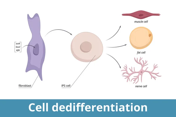
Cell dedifferentiation. Genes encoding three transcription regulators are artificially introduced into fibroblast nucleus, cell divides in culture (iPS cell), induced to de-differentiate.
Cell Dedifferentiation Genes Encoding Three Transcription Regulators Artificially Introduced Fibroblast Nucleus Cell Divides Culture Ips Cell Induced Differentiate
Wound healing process. Tissue repair. Inflammatory, Proliferative, Remodeling phase. Cross section of human skin after trauma. Disruption and damage of tissue and blood vessel. Bleeding. Vector illustration
Wound Healing Process Tissue Repair Inflammatory Proliferative Remodeling Phase Cross Section Human Skin Trauma Disruption Damage Tissue Blood Vessel Bleeding Vector Illustration
Vector Fibroblast illustration with description. Structure of dermis cell. Colorphul scheme of connective tissue on white background
Vector Fibroblast Illustration Description Structure Dermis Cell Colorphul Scheme Connective Tissue White Background
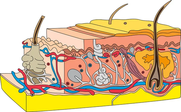
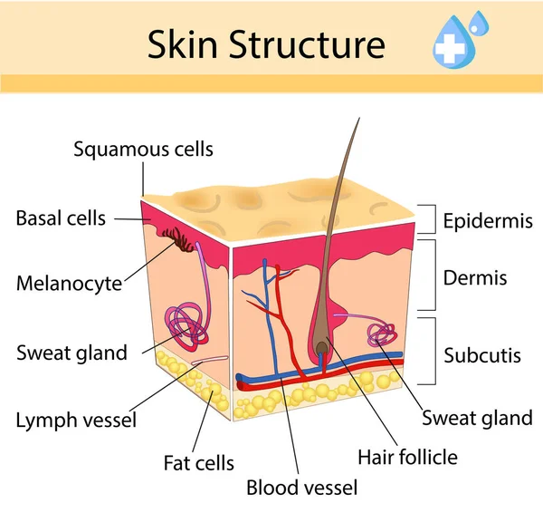
Skin cross section
Skin cross section
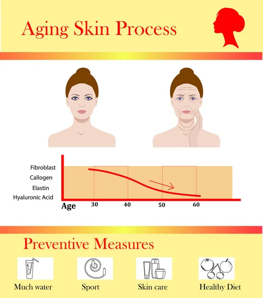
Aging skin process and preventive tipps, vector illustration
Vector illustration with cosmetic filler or Dermal fillers on white background
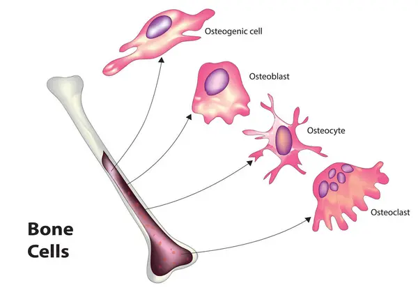
Bone cell classification (Types of bone cell)
Bone Cell Classification Types Bone Cell
Vector scheme of skin, for cosmetological, skin care and healthcare illustrations
Fat grafting structure. Test tube with fat. Close-up of Adipocytes, Fibroblast, Pericyte, Endothelial, and Hematopoietic stem cells. Vector illustration
Fat Grafting Structure Test Tube Fat Close Adipocytes Fibroblast Pericyte Endothelial Hematopoietic Stem Cells Vector Illustration
Cells of the immune system. White blood cells or leukocytes Eosinophil, Neutrophil, Basophil, Macrophage, Fibroblast, and Dendritic cell. Set of transparent realistic cells on a dark background. Vector illustration
Cells Immune System White Blood Cells Leukocytes Eosinophil Neutrophil Basophil Macrophage Fibroblast Dendritic Cell Set Transparent Realistic Cells Dark Background Vector Illustration
3D Isometric Flat Vector Conceptual Illustration of Fibroplast, Connective Tissue Cell
Isometric Flat Vector Conceptual Illustration Fibroplast Connective Tissue Cell
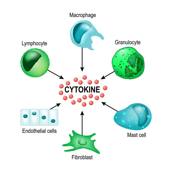
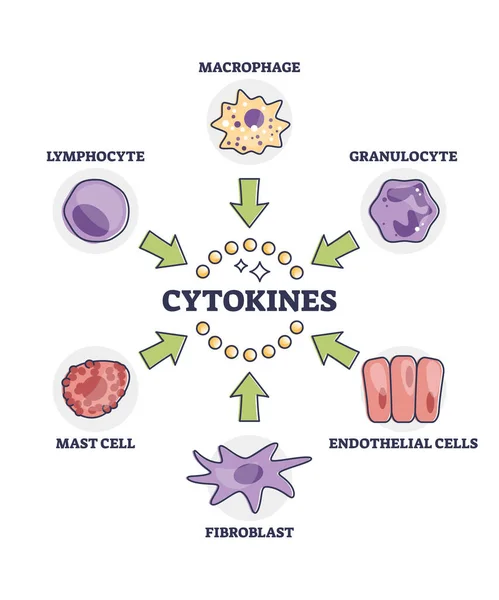
Cytokines releasing cells list for immune system response outline diagram. Labeled educational scheme with macrophage, granulocyte, endothelial, fibroblast, mast and lymphocyte vector illustration.
Cytokines Releasing Cells List Immune System Response Outline Diagram Labeled Educational Scheme Macrophage Granulocyte Endothelial Fibroblast Mast Lymphocyte Vector Illustration
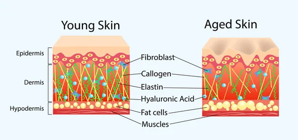
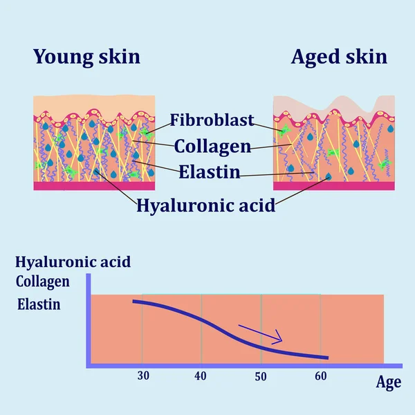
Aging process. Comparison of Young, Adults and aged skin. Collagen, Elastin, Hyaluronic acid and fibroblasts in younger and older skin. age-related changes in the skin when Collagen fibers atrophy, and Elastin broken. Vector illustration
Aging Process Comparison Young Adults Aged Skin Collagen Elastin Hyaluronic Acid Fibroblasts Younger Older Skin Age Related Changes Skin Collagen Fibers Atrophy Elastin Broken Vector Illustration
Aging process. Comparison of Young, Adults and aged skin. Age-related changes in the skin when Collagen fibers atrophy, Elastin broken. Vector illustration
Aging Process Comparison Young Adults Aged Skin Age Related Changes Skin Collagen Fibers Atrophy Elastin Broken Vector Illustration
Skin cross section
Skin cross section
Structure in the skin_English notation
Structure Skin_English Notation
Detailed scheme of human tooth cementum structure, including acellular and cellular cementum, fibers, cementoblasts and fibroblasts
Detailed Scheme Human Tooth Cementum Structure Including Acellular Cellular Cementum Fibers Cementoblasts Fibroblasts
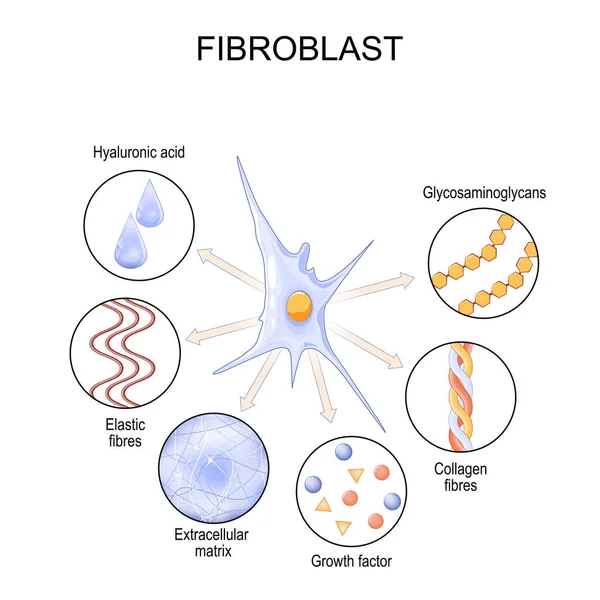
Fibroblast. Cell structure and function. biological cell that synthesizes the extracellular matrix, collagen, and produces the structural framework for tissues. Vector poster
Fibroblast Cell Structure Function Biological Cell Synthesizes Extracellular Matrix Collagen Produces Structural Framework Tissues Vector Poster
Regulatory T cell function. Suppressor T-cells. Prevent autoimmune disease. Immune homeostasis. Adaptive immune response. Vector illustration
Regulatory Cell Function Suppressor Cells Prevent Autoimmune Disease Immune Homeostasis Adaptive Immune Response Vector Illustration
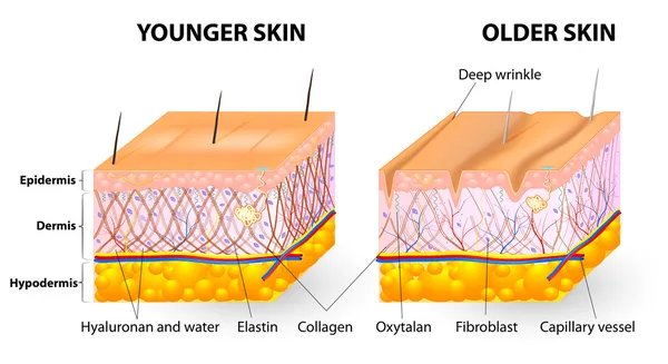
Aging skin chages. Wrinkle formation. Cross section of older and younger skin. Close-up of Elastin and Collagen fibers, Hyaluronic acid, and Fibroblast. Collagen loss. Elastin degradation. Vector illustration. Medical diagram.
Aging Skin Chages Wrinkle Formation Cross Section Older Younger Skin Close Elastin Collagen Fibers Hyaluronic Acid Fibroblast Collagen Loss Elastin Degradation Vector Illustration Medical Diagram
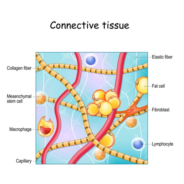
Connective tissue. Structure and anatomy. Extracellular matrix, Elastic and Collagen fibers, blood vessel and cells: lymphocyte, fibroblast, mesenchymal stem cell and Macrophage. Vector illustration

Connective Tissue Structure Anatomy Extracellular Matrix Elastic Collagen Fibers Blood Vessel Cells Lymphocyte Fibroblast Mesenchymal Stem Cell Macrophage Vector Illustration
Cytokines are produced by macrophage, lymphocyte, mast cell, endothelial cells and fibroblast. Cell signaling by Interleukins, Chemokines, Tumor necrosis factor. Cytokine storm. Vector illustration
Cytokines Produced Macrophage Lymphocyte Mast Cell Endothelial Cells Fibroblast Cell Signaling Interleukins Chemokines Tumor Necrosis Factor Cytokine Storm Vector Illustration
Vector illustration of structure cells with collagen, elastin and fibroblast, aging process
Collagen induction therapy. Skin and Collagen fibers before and after Microneedling procedure. Skin rejuvenation. Fibroblast activation. Vector poster. Isometric Flat illustration. Schematic diagram
Collagen Induction Therapy Skin Collagen Fibers Microneedling Procedure Skin Rejuvenation Fibroblast Activation Vector Poster Isometric Flat Illustration Schematic Diagram
Vector scheme of skin, for cosmetological and healthcare illustrations
Blood cells macrophage cell drawing. Anatomy, biology, science concept. Scientific Designing of Macrophage. The White Blood Cell Structure.
Blood Cells Macrophage Cell Drawing Anatomy Biology Science Concept Scientific Designing Macrophage White Blood Cell Structure
Cells of the immune system. White blood cells or leukocytes Eosinophil, Neutrophil, Basophil, Macrophage, Fibroblast, and Dendritic cell. Set of transparent realistic cells on a white background. Vector illustration
Cells Immune System White Blood Cells Leukocytes Eosinophil Neutrophil Basophil Macrophage Fibroblast Dendritic Cell Set Transparent Realistic Cells White Background Vector Illustration
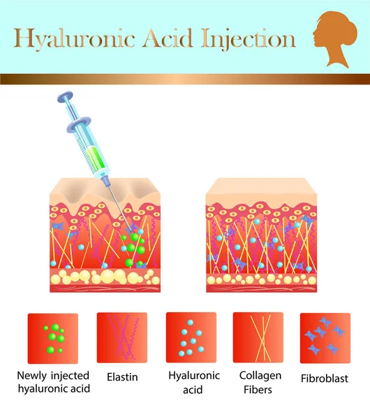
Hyaluronic acid injection, before and affect , vector illustration
Human anatomy disease. medulloblastoma vector illustration on white background. eps 10
Human Anatomy Disease Medulloblastoma Vector Illustration White Background Eps
Fibroblast functions. Extracellular matrix production. Collagen synthesis. Wound Healing and Repair. Tissue Homeostasis. Fibroblast communication with immune cells during inflammation. Fibroblast cell and aging processes. Detailed vector illustration
Fibroblast Functions Extracellular Matrix Production Collagen Synthesis Wound Healing Repair Tissue Homeostasis Fibroblast Communication Immune Cells Inflammation Fibroblast Cell Aging Processes Detailed Vector Illustration
Structure in the skin_Japanese notation
Structure Skin_Japanese Notation
Cells of the immune system. White blood cells or leukocytes: Eosinophil, Neutrophil, Basophil, Macrophage, Fibroblast, and Dendritic cell. Vector diagram
Cells Immune System White Blood Cells Leukocytes Eosinophil Neutrophil Basophil Macrophage Fibroblast Dendritic Cell Vector Diagram
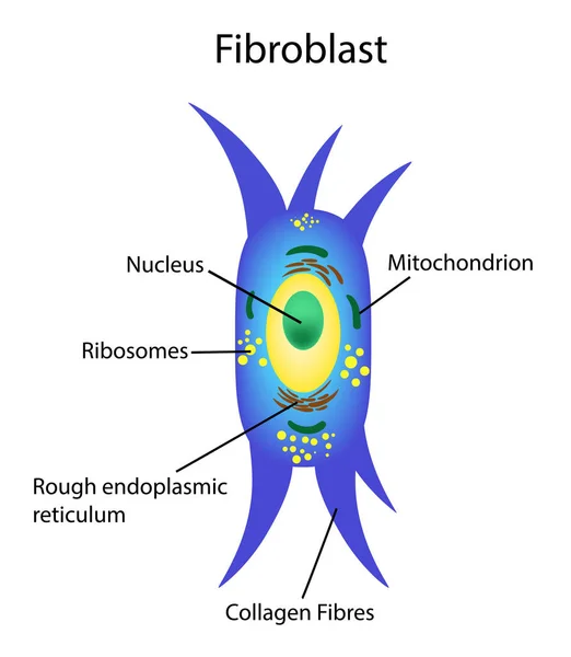
Fibroblast. Cell structure and anatomy. Collagen fibers and skin cell. Vector illustration
Fibroblast Cell Structure Anatomy Collagen Fibers Skin Cell Vector Illustration
Histology of the pulp. Tissue structure includes fibroblasts, nerves, capillary, and reticular fiber. Pulp cell organization. Cell free and cell rich zone formations and pariental layer.
Histology Pulp Tissue Structure Includes Fibroblasts Nerves Capillary Reticular Fiber Pulp Cell Organization Cell Free Cell Rich Zone Formations Pariental Layer
Fibroblast cell structure. Connective tissue cell. Vector illustration
Cytokine receptor. signal transduction between cells.
Illustration showing layers and components of aged skin
Illustration Showing Layers Components Aged Skin
Depiction of aged skin layers and components
Depiction Aged Skin Layers Components
Aged skin layer. Human skin structure with collagen and elastane fibers, hyaluronic acids, fibroblasts. Schematic illustration. Vector diagram
Aged skin layer. Structure human aged skin with collagen and elastin fibers, fibroblasts. Vector diagram
Exosome Serum Skin Care Cosmetic, Skin Rejuvenation
Exosome Serum Skin Care Cosmetic Skin Rejuvenation
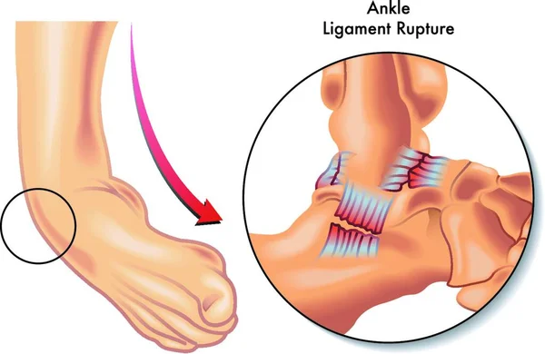
Medical Illustration of ankle ligament rupture
Medical Illustration Ankle Ligament Rupture
Exosome Serum Skin Care Cosmetic, Skin Rejuvenation
Exosome Serum Skin Care Cosmetic Skin Rejuvenation
Both of these illustrations show the difference between a liver with fat buildup or fatty liver and a healthy liver.
Both Illustrations Show Difference Liver Fat Buildup Fatty Liver Healthy Liver
Biological Brochure vector
Medical illustration comparing young and aged human skin structure. Cross section shows collagen and elastin fibers, fibroblasts, dermis, epidermis, wrinkles and age-related changes in the extracellular matrix.
Medical Illustration Comparing Young Aged Human Skin Structure Cross Section Shows Collagen Elastin Fibers Fibroblasts Dermis Epidermis Wrinkles Age Related Changes Extracellular Matrix
Different types of cardiac fibrosis labeled diagram, educational medical poster design healthcare scheme, heart failure, normal myocardium, reactive and replacement fibrosis, interstitium.
Different Types Cardiac Fibrosis Labeled Diagram Educational Medical Poster Design Healthcare Scheme Heart Failure Normal Myocardium Reactive Replacement Fibrosis Interstitium
Fibroblast is a dermis cell. Structure of Fibroblast cell.
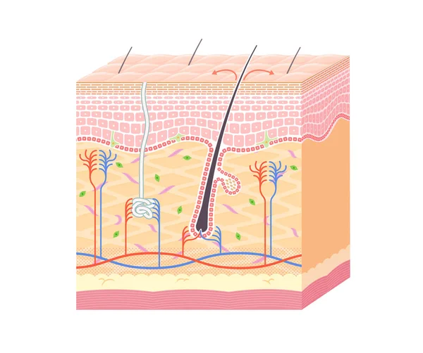
Human skin and hair structure. Anatomical sign. Beauty care isolated illustration
Platelet-Rich Plasma PRP with Microneedling for Skin Rejuvenation and Scar Reduction Medical Illustration. PRP Therapy Before and After Skin Regeneration Cross Section
Platelet Rich Plasma Prp Microneedling Skin Rejuvenation Scar Reduction Medical Illustration Prp Therapy Skin Regeneration Cross Section
Medical illustration of fibroblast cell anatomy. Educational diagram of a connective tissue cell involved in extracellular matrix and collagen production.
Medical Illustration Fibroblast Cell Anatomy Educational Diagram Connective Tissue Cell Involved Extracellular Matrix Collagen Production
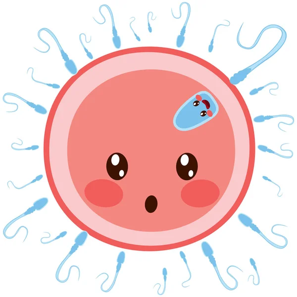
Sperm On Egg Cell Fertilizin
Seamless pattern hyaluronic acid injection medical syringe and containers skin care and medical intervention flat vector illustration on white background
Collagen flask icon isometric vector. Skin drop. Cell age
Collagen Flask Icon Isometric Vector Skin Drop Cell Age
Fibroblast structure. Fibroblast cell. Vector illustration isolated
Vector illustration with cosmetic filler or Dermal fillers on green background
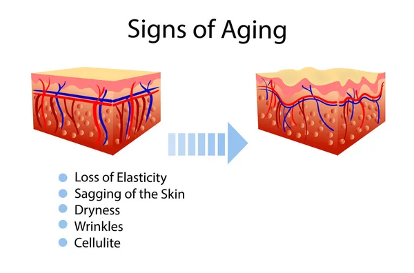
Vector diagram with signs of aging, two types of skin, for cosmetological and healthcare illustrations
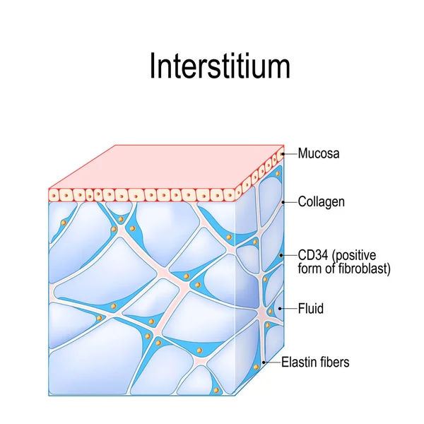
Structure of Interstitium. new organ. interstitial is a reservoir and transportation system for nutrients and solutes distributing among organs, and cells. immune regulation. Human Tissues
Structure Interstitium New Organ Interstitial Reservoir Transportation System Nutrients Solutes Distributing Organs Cells Immune Regulation Human Tissues
Gray-orange Immunoglobulin Round Icon
Synthesizer mixer icon outline vector. Dj music. Audio instrument color flat
Synthesizer Mixer Icon Outline Vector Music Audio Instrument Color Flat
Page