Mitocondrio Stock Vector Images

294 Mitocondrio vector art & graphics are available under a royalty-free license
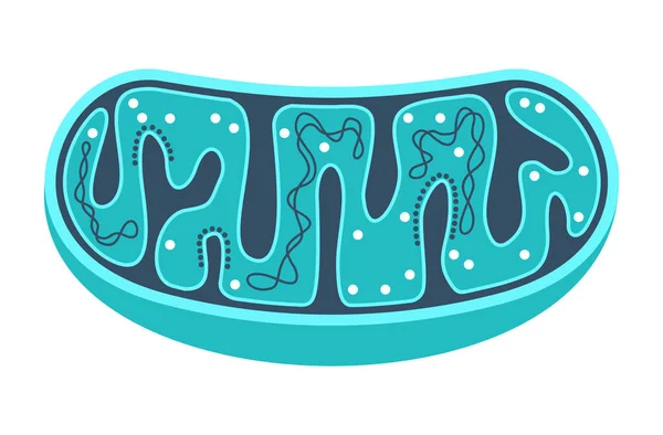
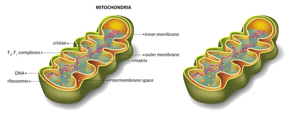
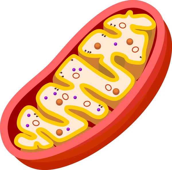
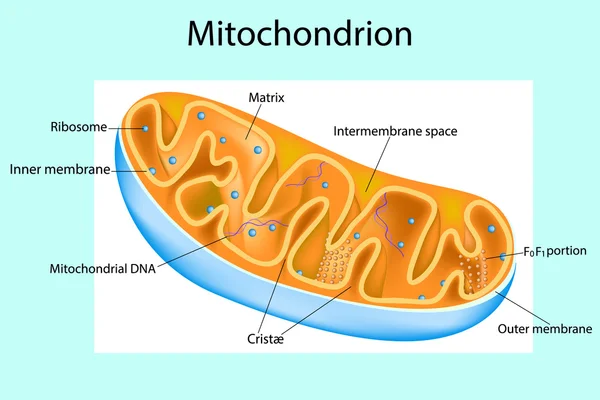
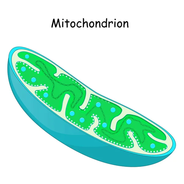
Mitochondria slice with ctructure and components
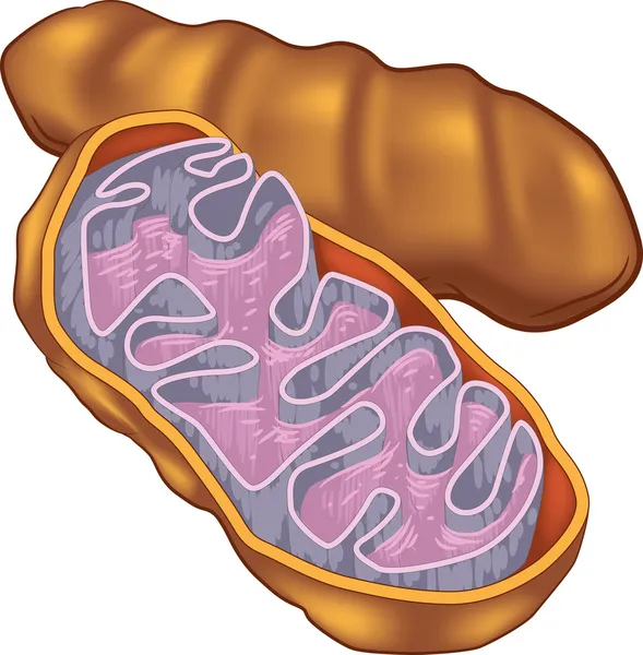
Mitochondria organelle section
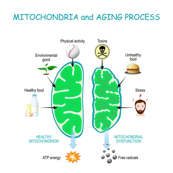
Mitochondria and aging process. Healthy Mitochondrion are produce of Atp energy, cell organelles with Dysfunction produce of Free radicals.
Mitochondria Aging Process Healthy Mitochondrion Produce Atp Energy Cell Organelles Dysfunction Produce Free Radicals
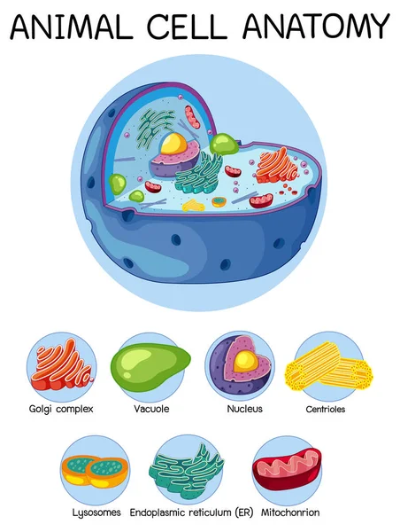
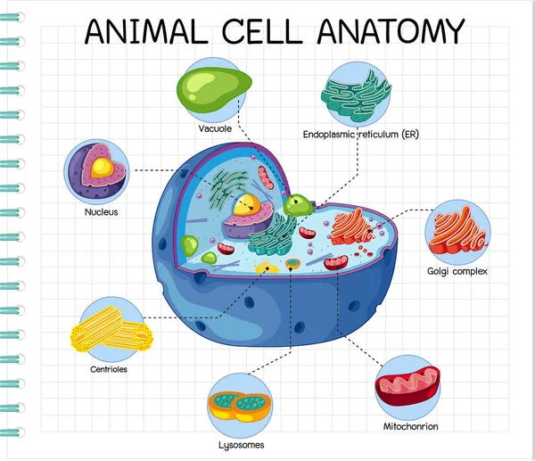
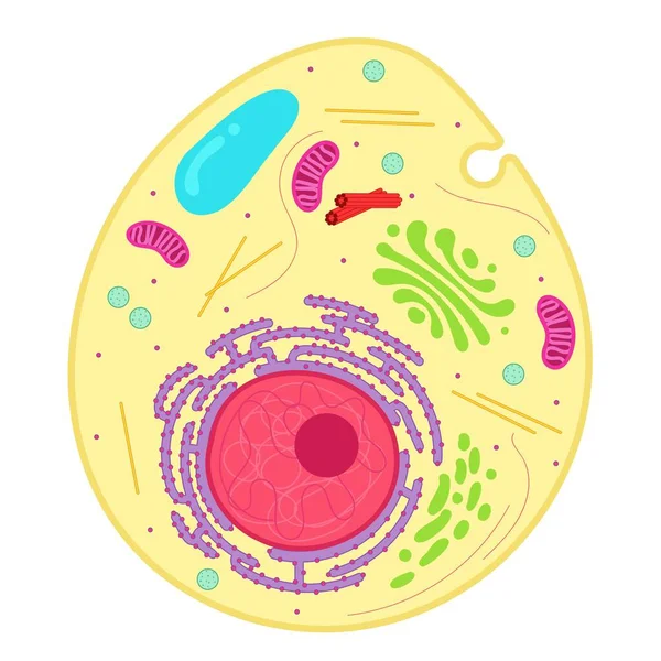
Anatomy of animal cell (Biology Diagram) illustration
Anatomy Animal Cell Biology Diagram Illustration
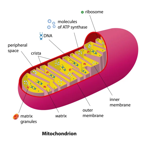
Mitochondrion scheme
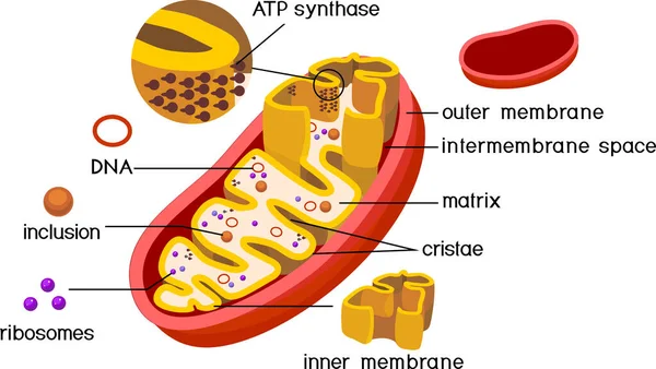
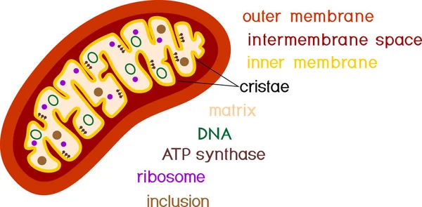
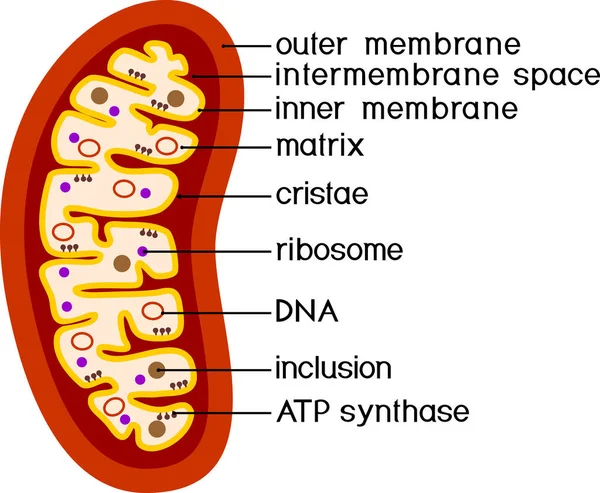
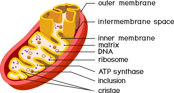
Structure of mitochondrion with ATP synthase on inner membrane and components isolated on white background
Structure Mitochondrion Atp Synthase Inner Membrane Components Isolated White Background
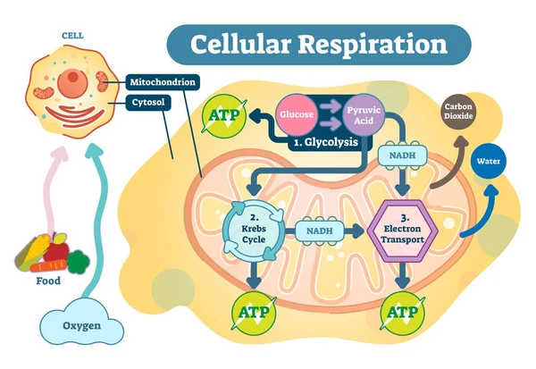
Cellular respiration medical vector illustration diagram, respiration process scheme.
Structure of mitochondrion isolated on white background
Structure Mitochondrion Isolated White Background
Diagram showing cellular respiration
Mitochondrion
Mitochondrion
Mitochondrion. Components of a typical mitochondrion. Structure. Interactive diagram
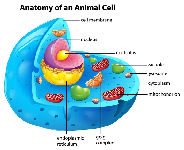
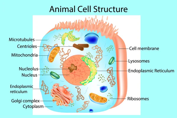
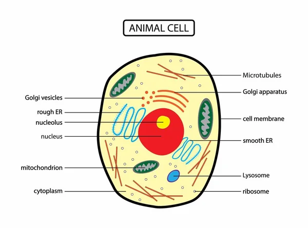
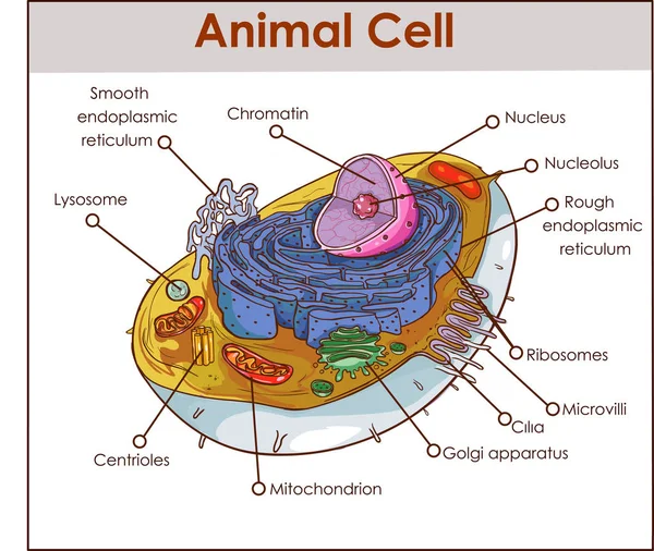
Anatomy of an animal cell
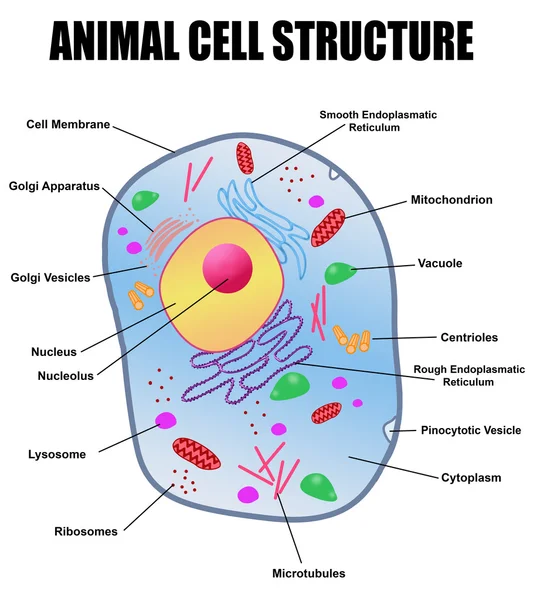
Animal cell structure
Cell icon
Cell icon
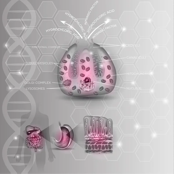
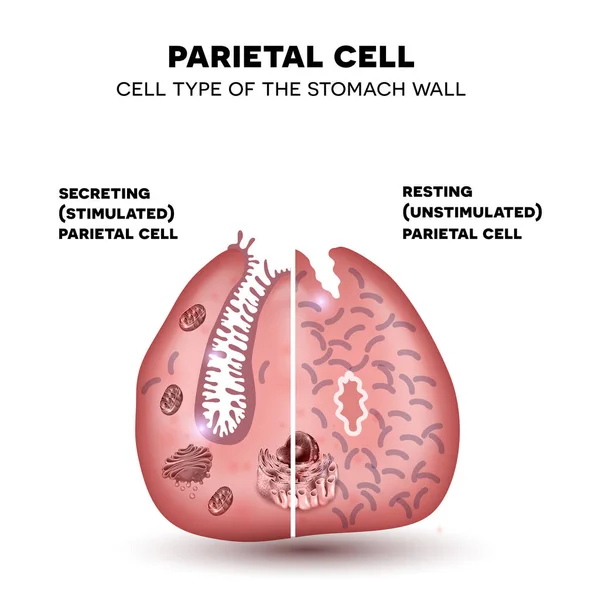
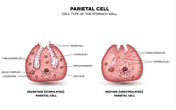
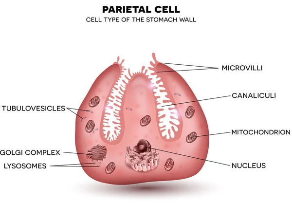
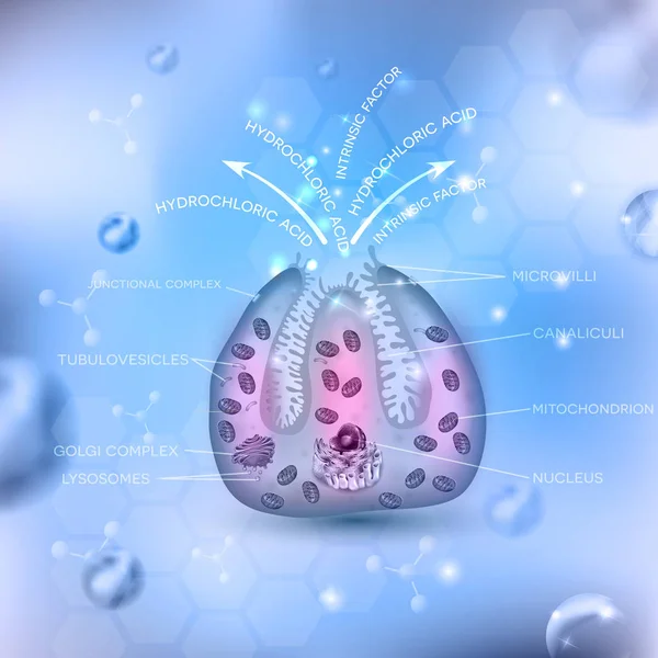
Parietal cell of stomach wall, located in the gastric glands
Parietal cell of stomach wall,  located in the gastric glands
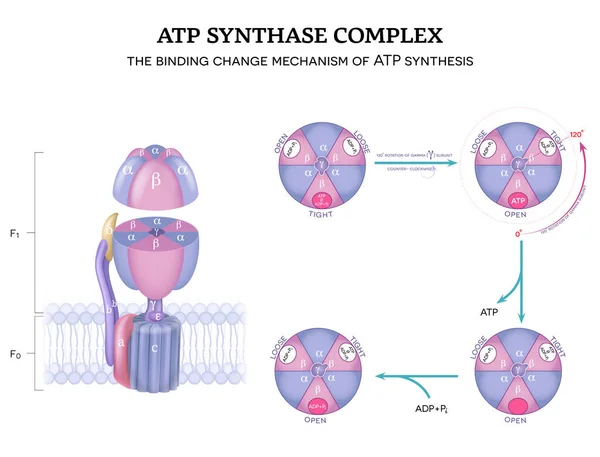
ATP synthase complex structure and mechanism of ATP synthase. The binding change mechanism. 120-degree rotation of gamma subunit counter-clockwise.
Atp Synthase Complex Structure Mechanism Atp Synthase Binding Change Mechanism 120 Degree Rotation Gamma Subunit Counter Clockwise
Fibroblast anatomy. structure of cell. Diagram with golgi apparatus, nucleus, mitochondrion and ribosomes. vector illustration. Poster
Fibroblast Anatomy Structure Cell Diagram Golgi Apparatus Nucleus Mitochondrion Ribosomes Vector Illustration Poster
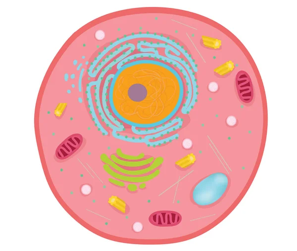
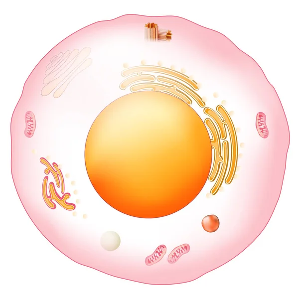
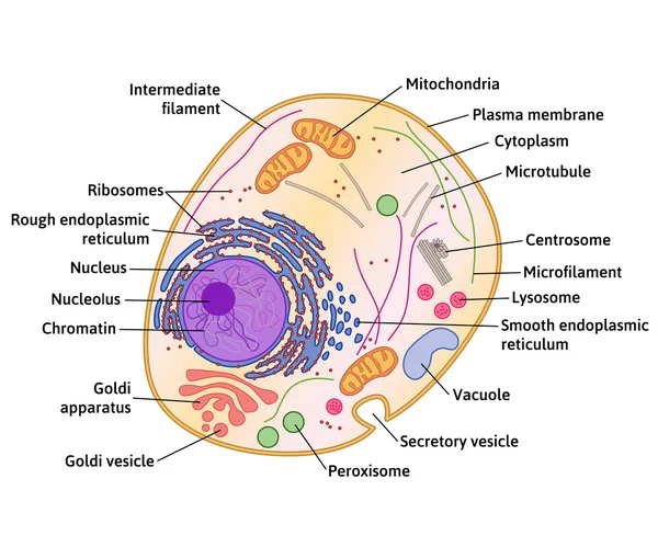
Illustration of the anatomy of an animal cell. Structure of a Eukaryotic cell.
Animal cells are the basic unit of life.
Animal Cells Basic Unit Life
Structure of mitochondrion isolated on white background
Structure Mitochondrion Isolated White Background
Mitochondria minimal vector icon drawn with brush line. Blue linear medical pictogram isolated on transparent background
Mitochondria Minimal Vector Icon Drawn Brush Line Blue Linear Medical Pictogram Isolated Transparent Background
Structure of mitochondrion isolated on white background
Structure Mitochondrion Isolated White Background
Detailed vector illustration of a mitochondrion with vibrant colors and clear structure, highlighting its internal components
Detailed Vector Illustration Mitochondrion Vibrant Colors Clear Structure Highlighting Its Internal Components
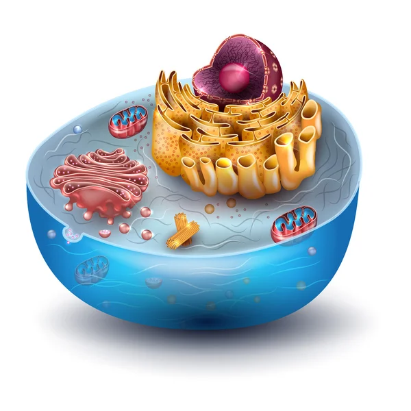
Cell structure cross section
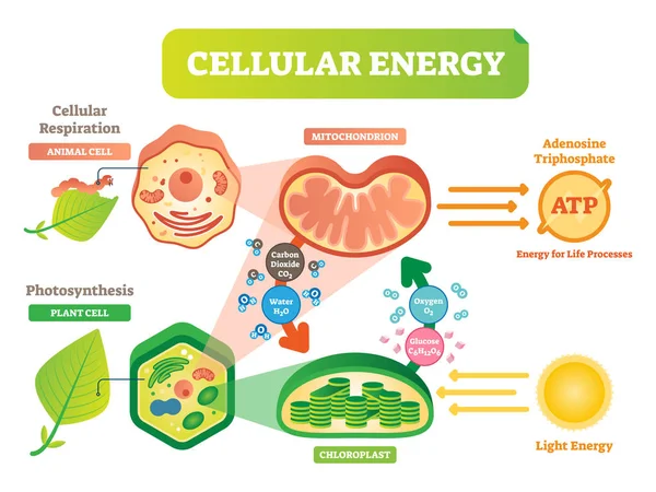
Animal and plant cell energy cycle vector illustration diagram with mitochondrion and chloroplast.
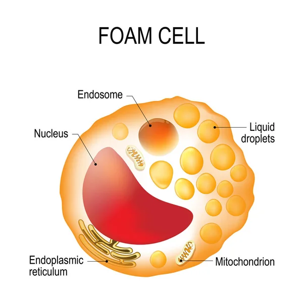
Foam cell. Cell structure
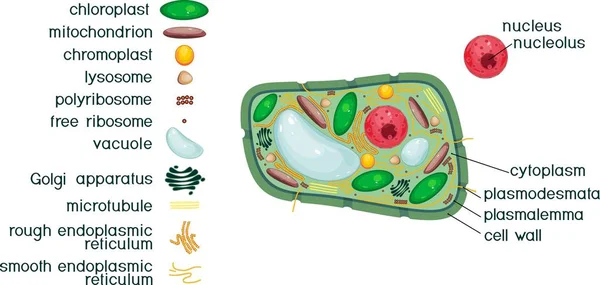
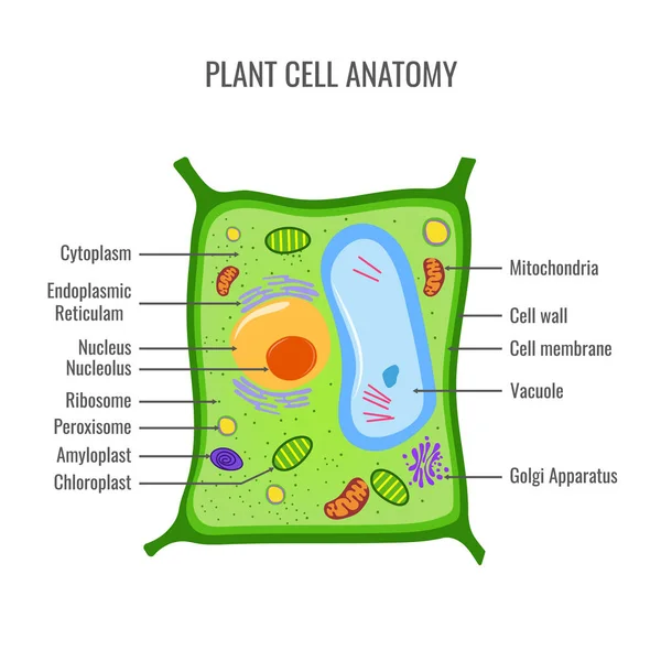
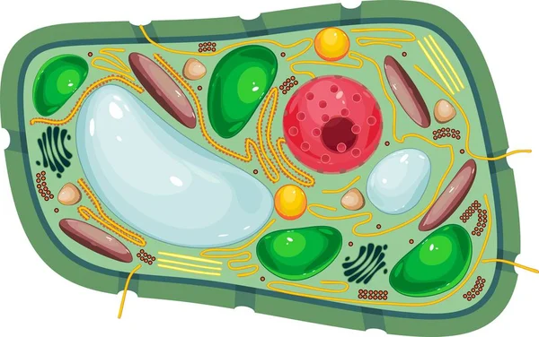
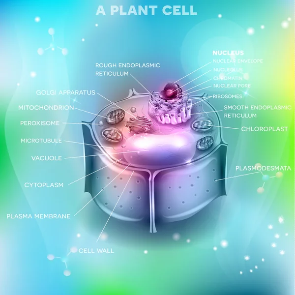
Plant cell structure with titles and different organelles
Plant Cell Structure Titles Different Organelles
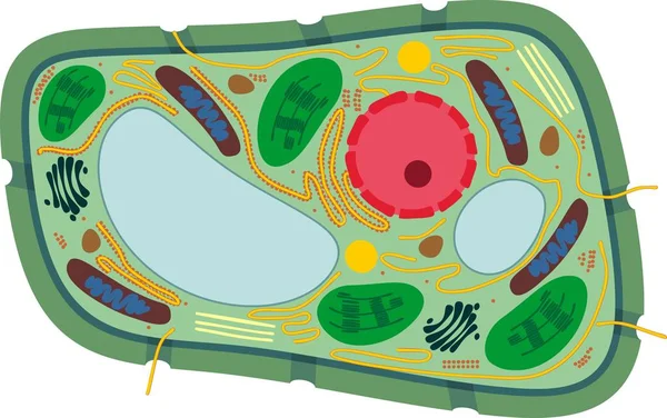
Structure of plant cell with different organelles
Structure Plant Cell Different Organelles
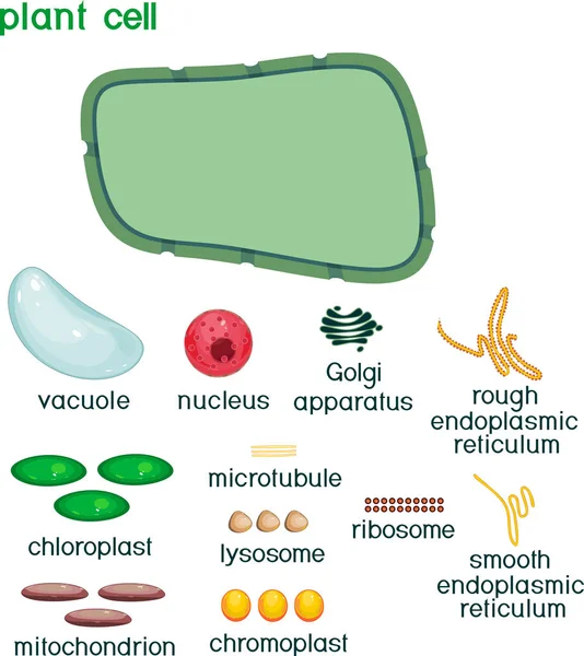
Educational game: assembling cells from ready-made components in form of stickers. Plant cell structure with titles
Educational Game Assembling Cells Ready Made Components Form Stickers Plant Cell Structure Titles
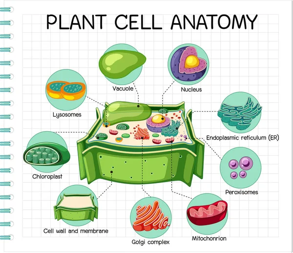
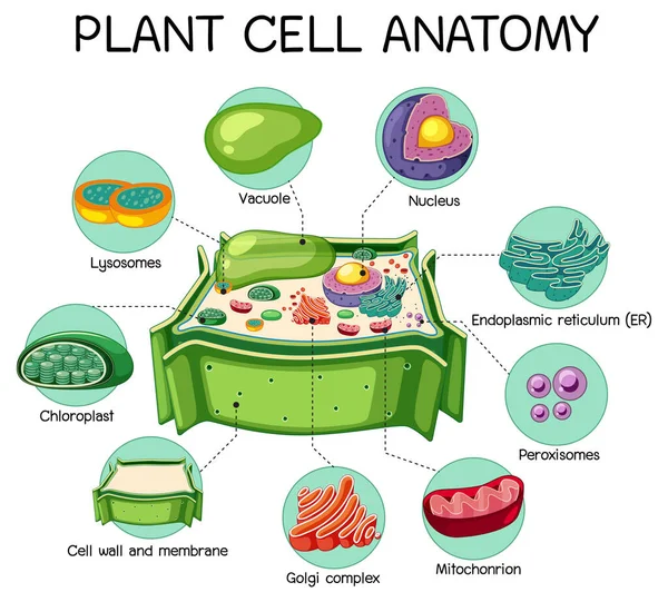
Anatomy of plant cell (Biology Diagram) illustration
Anatomy Plant Cell Biology Diagram Illustration
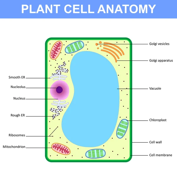
Plant cell anatomy vector illustration
Plant Cell Anatomy Vector Illustration
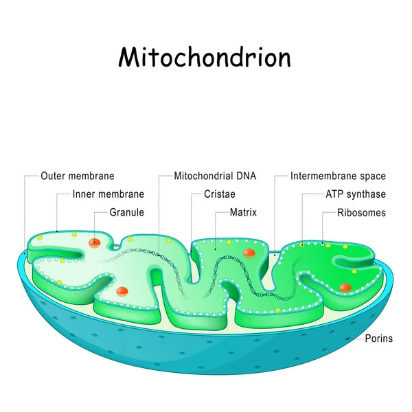
Mitochondrion anatomy. Structure, components and organelles. cross-section of mitochondria. Vector illustration
Mitochondrion Anatomy Structure Components Organelles Cross Section Mitochondria Vector Illustration
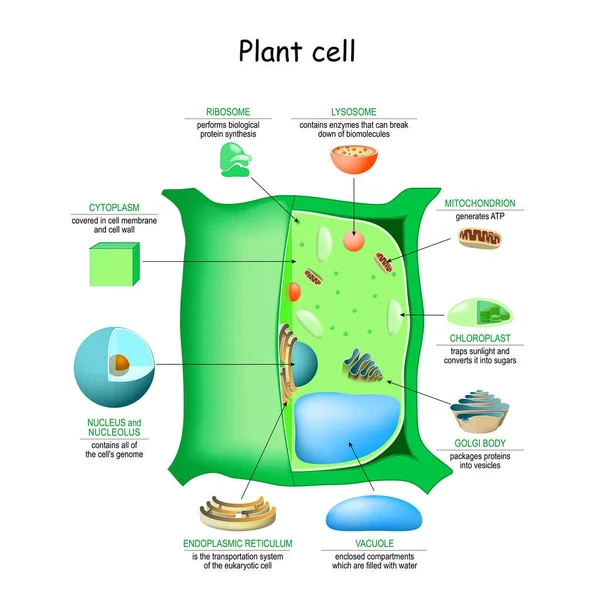
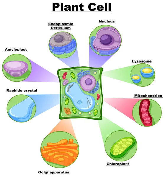
Plant cell anatomy structure. Close-up of Organelles of a plant cell. Educational infographic. Vector illustration
Plant Cell Anatomy Structure Close Organelles Plant Cell Educational Infographic Vector Illustration
Cell anatomy. Structure and organelles of human's cell. Cross sections of animal cell: nucleus, nucleolus, mitochondria, centrosome, golgi apparatus, endoplasmic reticulum, ribosome and membrane. poster biological diagram. vector illustration
Cell Anatomy Structure Organelles Human Cell Cross Sections Animal Cell Nucleus Nucleolus Mitochondria Centrosome Golgi Apparatus Endoplasmic Reticulum Ribosome Membrane Poster Biological Diagram Vector Illustration
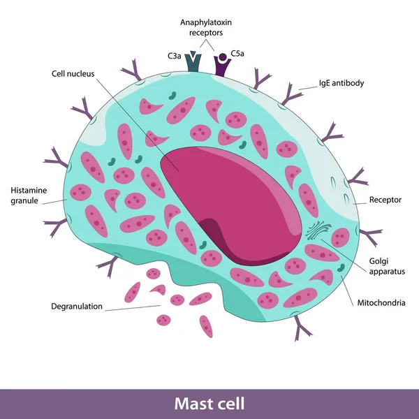
Mast cell structure. Vector image of cell elements, including Golgi apparatus, mitochondria, degranulation process, cell nucleus, C3a, C5a anaphylatoxin receptors, lgE antibody
Mast Cell Structure Vector Image Cell Elements Including Golgi Apparatus Mitochondria Degranulation Process Cell Nucleus C3A C5A Anaphylatoxin Receptors Lge Antibody
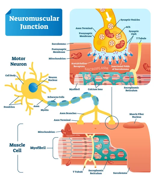
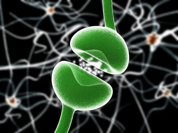
Neuromuscular junction vector illustration scheme. Labeled cell infographic
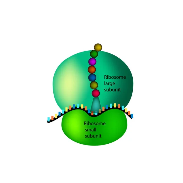
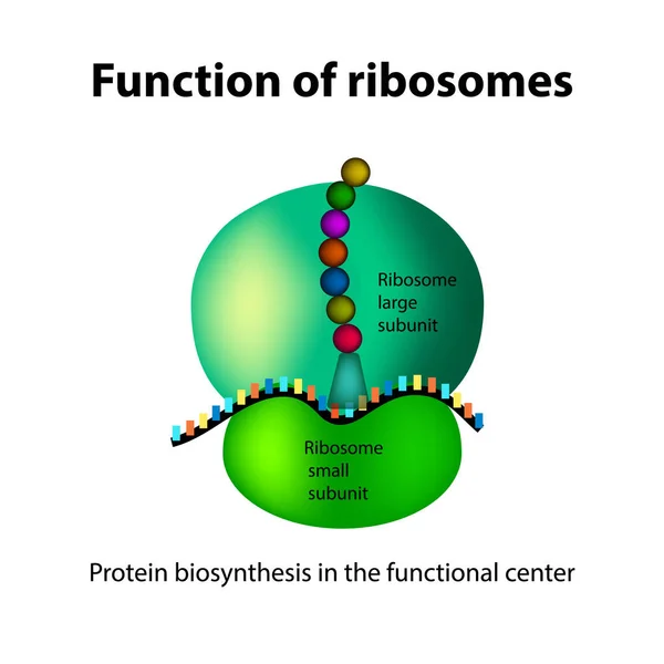
The structure of the ribosome. Infographics. Vector illustration on isolated background
Animal human cell structure educational science. Microscope 3d eukaryotic nucleus organelle medicine technology analysis. Glowing colored biology poster template isolated line vector illustration
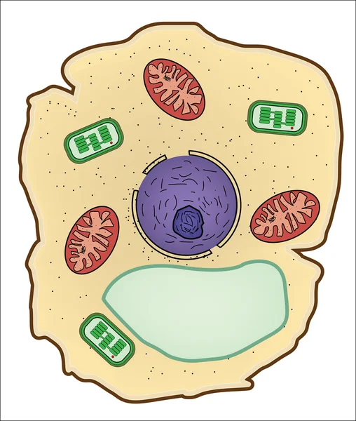
Plant cell isolated on white photo-realistic vector illustration
Plant Cell Isolated White Photo Realistic Vector Illustration
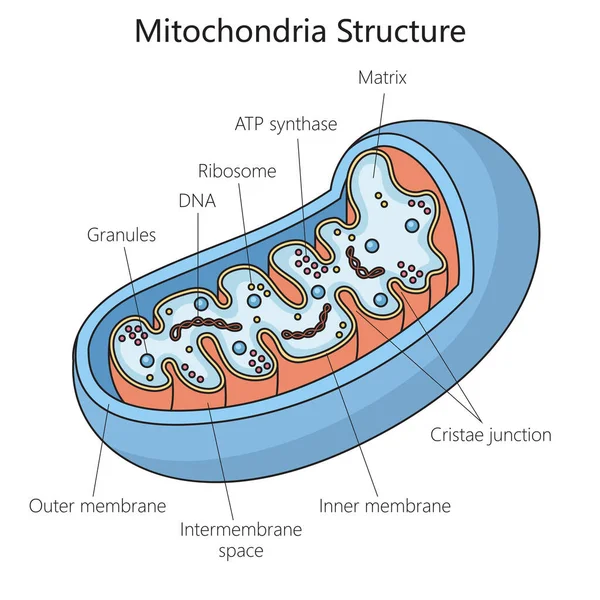
Human mitochondria structure diagram schematic vector illustration. Medical science educational illustration
Human Mitochondria Structure Diagram Schematic Vector Illustration Medical Science Educational Illustration
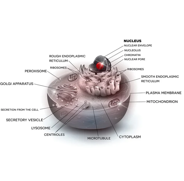
Cell structure beautiful color illustration
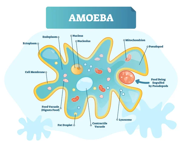
Amoeba labeled vector illustration. Single cell animal structure scheme.
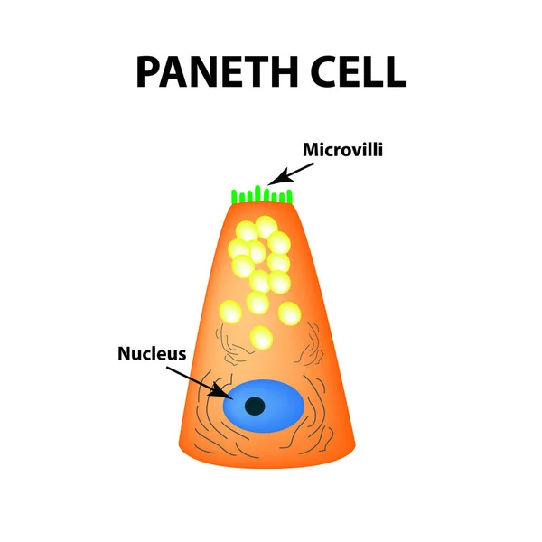
The structure of Paneth cells. Davidoff's cell. fographics. Vector illustration on isolated background.
Vector illustration of cellular respiration showing glycolysis, Krebs cycle, and electron transport chain. Glucose breakdown, ATP production, oxygen intake, and water and carbon dioxide release
Vector Illustration Cellular Respiration Showing Glycolysis Krebs Cycle Electron Transport Chain Glucose Breakdown Atp Production Oxygen Intake Water Carbon Dioxide Release
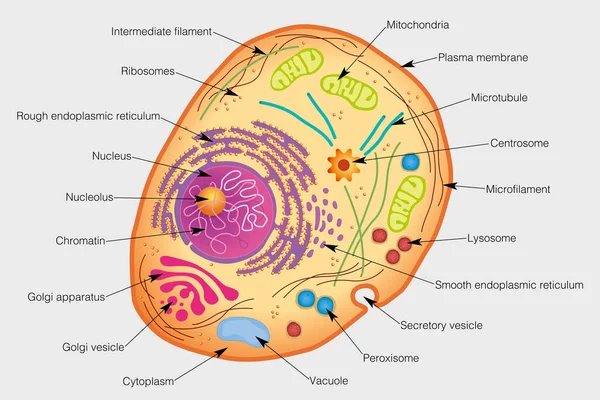
The graphic shows the elements of a human cell. Vector image
Graphic Shows Elements Human Cell Vector Image
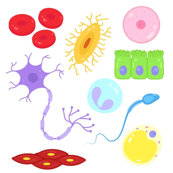
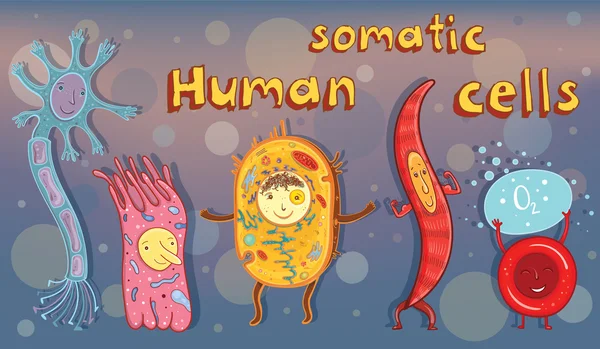
Types of cells in the human body.
Types Cells Human Body
Illustration of biology, Anatomy of animal cell, Animal cell anatomical structure with all parts including cell membrane nucleus nucleolus vacuole lysosome ribosome golgi body cytoplasm
Illustration Biology Anatomy Animal Cell Animal Cell Anatomical Structure All Parts Including Cell Membrane Nucleus Nucleolus Vacuole Lysosome Ribosome Golgi Body Cytoplasm
Animal Cell Anatomy Diagram Structure with all parts nucleus smo
Anatomy of animal cell (Biology Diagram) illustration
Anatomy Animal Cell Biology Diagram Illustration
Plant cell structure with titles and different organelles
Plant Cell Structure Titles Different Organelles
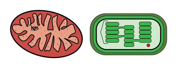
Vector illustration of mitochondria and chloroplast
Mitochondria. structure and anatomy of a mitochondrion. vector icon
Mitochondria Structure Anatomy Mitochondrion Vector Icon
Autophagy of mitochondria. Diagram of the process of autophagy from Forming a membrane and autophagosome to fuse phagosome and lysosome when contents of the vesicles are degraded and recycled. Autophagy defects linked to various diseases and cancer d
Autophagy Mitochondria Diagram Process Autophagy Forming Membrane Autophagosome Fuse Phagosome Lysosome Contents Vesicles Degraded Recycled Autophagy Defects Linked Various Diseases Cancer
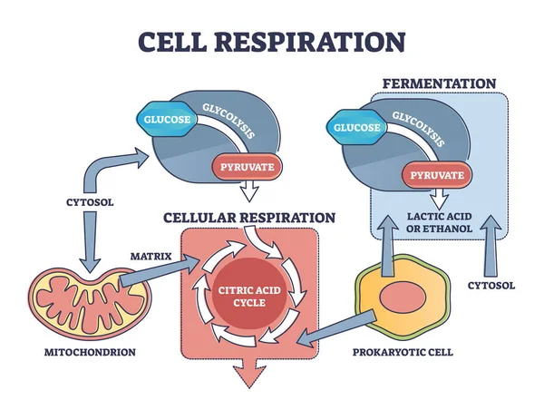
Cell respiration process explanation with biological stages outline diagram
Anatomy of plant cell (Biology Diagram) illustration
Anatomy Plant Cell Biology Diagram Illustration
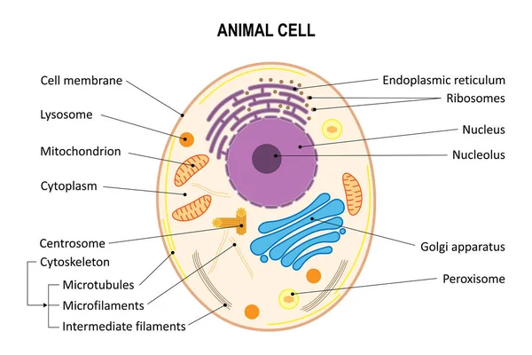
Animal cell anatomy
Animal cell anatomy
Vector illustration of human somatic cells
Plant Cell Anatomy Diagram illustration
Plant Cell Anatomy Diagram Illustration
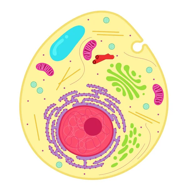
An animal cell is a type of eukaryotic cell.
Animal Cell Type Eukaryotic Cell
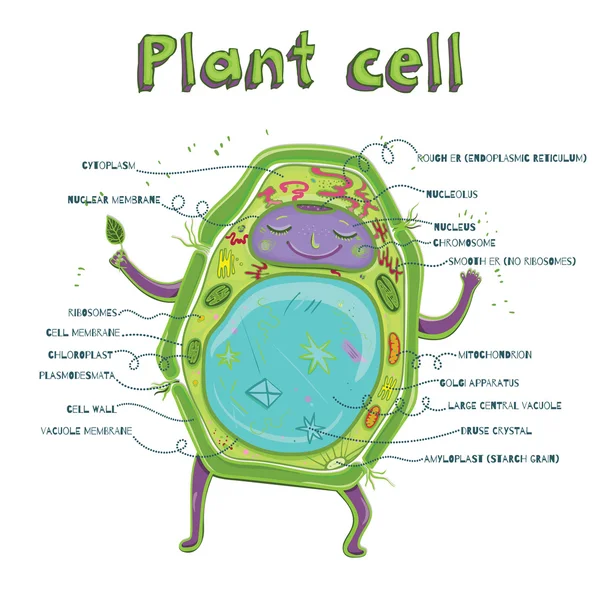
Plant cell anatomy
Plant cell anatomy
Animal cell. Diagram. Animal cell organelles.
Animal Cell Diagram Animal Cell Organelles
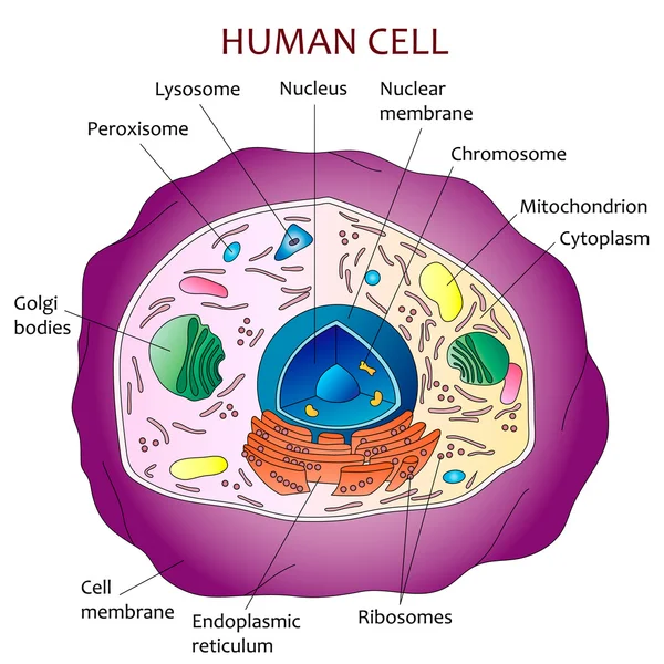
Human cell diagram
Human cell diagram
Structure of mitochondrion isolated on white background
Structure Mitochondrion Isolated White Background
Colorful vector illustration of plant cell structure with labeled organelles, highlighting cellular components and functions in a clear layout
Colorful Vector Illustration Plant Cell Structure Labeled Organelles Highlighting Cellular Components Functions Clear Layout
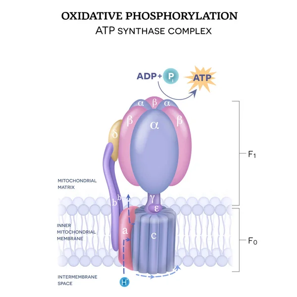
The ATP synthase structure (complex V) consists of two components F0 and F1. The formation of ATP using adenosine diphosphate (ADP) and inorganic phosphate (Pi)
Atp Synthase Structure Complex Consists Two Components Formation Atp Using Adenosine Diphosphate Adp Inorganic Phosphate
Human cell anatomy. Structure of a eukaryotic cell. All organelles: Nucleus, Ribosome, Rough endoplasmic reticulum, Golgi apparatus, mitochondrion, cytoplasm, lysosome, Centrosome. Animal cell on blue background. vector illustration
Human Cell Anatomy Structure Eukaryotic Cell All Organelles Nucleus Ribosome Rough Endoplasmic Reticulum Golgi Apparatus Mitochondrion Cytoplasm Lysosome Centrosome Animal Cell Blue Background Vector Illustration
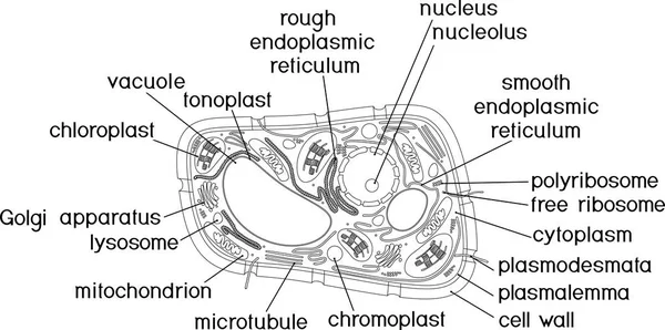
Coloring page. Structure of plant cell with titles
Coloring Page Structure Plant Cell Titles
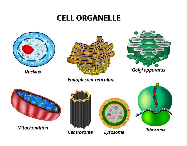
Set the cell organelles. Nucleus, endoplasmic reticulum, Golgi apparatus, mitochondria, centrosome, lysosome, the ribosome. Infographics. Vector illustration on isolated background
Plant cell anatomy. Vacuole structure. Vector illustration. poster. Schematic diagram
Plant Cell Anatomy Vacuole Structure Vector Illustration Poster Schematic Diagram
Parietal cell of stomach wall, located in the gastric glands se
Parietal cell of stomach wall,  located in the gastric glands se
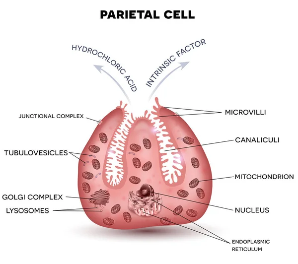
Parietal cell secreting hydrochloric acid and intrinsic factor
Structure of plant cell with different organelles
Structure Plant Cell Different Organelles
Structure of plant cell with different organelles
Structure Plant Cell Different Organelles
Parietal cell of stomach wall, located in the gastric glands sec
Close up diagram of plant cell
Vector illustration of the basic structure of the plant cell.
Vector Illustration Basic Structure Plant Cell
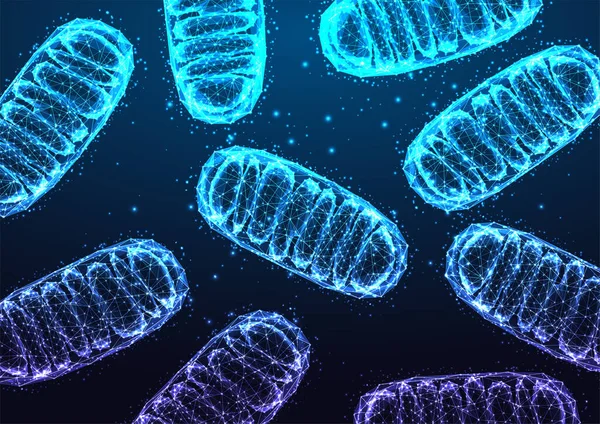
Mitochondria under microscope on dark blue backgound in futuristic glowing low polygonal style. Medical research concept, Science, biology research banner. Modern abstract design vector illustration
Mitochondria Microscope Dark Blue Backgound Futuristic Glowing Low Polygonal Style Medical Research Concept Science Biology Research Banner Modern Abstract Design Vector Illustration
Animal cell anatomy. Structure and organelles of Eukaryotic cell. Vector poster for education. illustration
Animal Cell Anatomy Structure Organelles Eukaryotic Cell Vector Poster Education Illustration
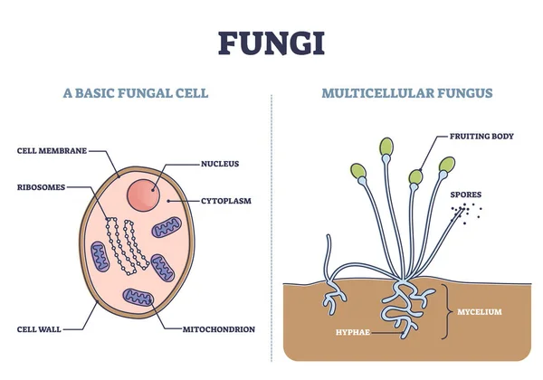
Fungi as basic fungal cell and multicellular fungus structure outline diagram
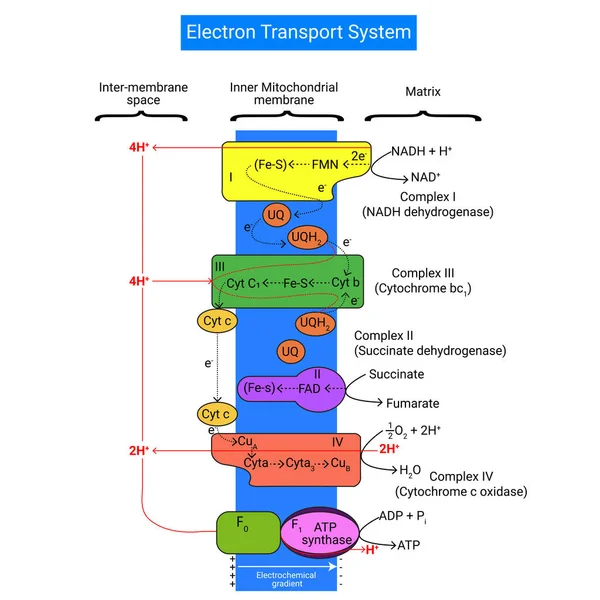
Electron Transport Chain is a series of compounds where it makes use of electrons from electron carrier to develop a chemical gradient
Electron Transport Chain Series Compounds Makes Use Electrons Electron Carrier Develop Chemical Gradient
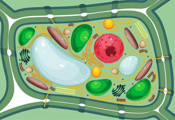
Plant Cell structure, cross section of the cell detailed colorfu
L-carnitine transports fat molecules into the mitochondria
Anatomy of a typical human neuron Vector 4
Plant cell isolated on white photo-realistic vector illustration
Plant Cell Isolated White Photo Realistic Vector Illustration
The structure of the ribosome. Functions. Infographics. Vector illustration on isolated background
Heart valentine light vector background
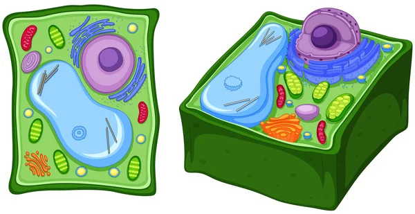
Diagrams of animal and plant cells
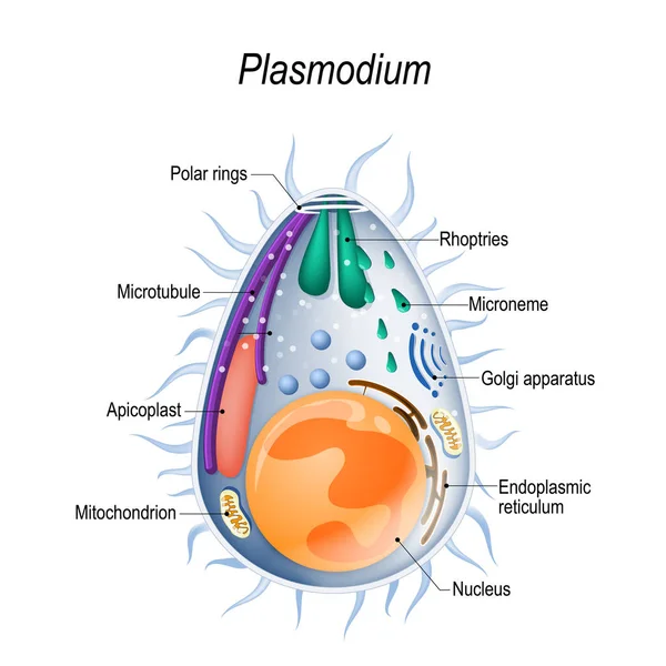
Plasmodium is the malaria parasite, is a large genus of parasitic protozoa. Infection with these protozoans is known as malaria, a deadly disease. Diagram of Plasmodium merozoites structure. vector illustration for medical, educational and science
Plasmodium Malaria Parasite Large Genus Parasitic Protozoa Infection Protozoans Known Malaria Deadly Disease Diagram Plasmodium Merozoites Structure Vector Illustration Medical Educational Science
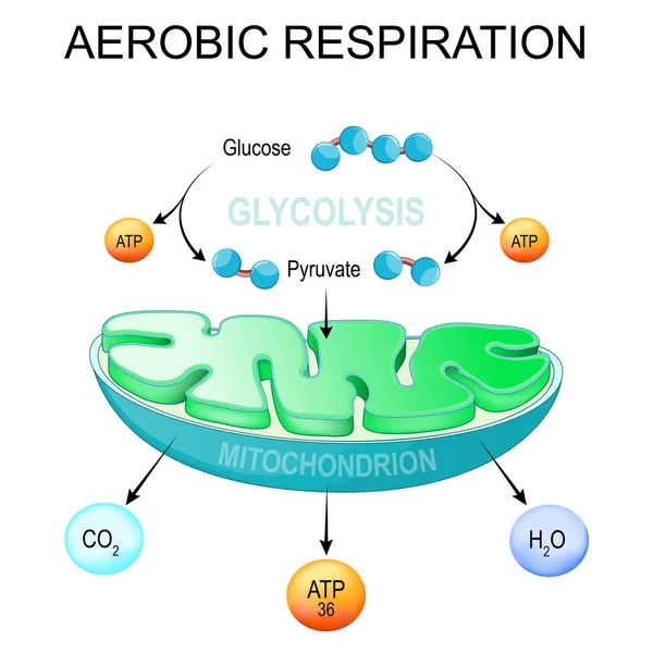
Aerobic respiration. Glycolysis and ATP Synthesis in mitochondria. converting glucose into pyruvate in cells. metabolic pathway. Vector poster
Aerobic Respiration Glycolysis Atp Synthesis Mitochondria Converting Glucose Pyruvate Cells Metabolic Pathway Vector Poster
Diagram showing plant cell
Structure of plant cell with different organelles
Structure Plant Cell Different Organelles
Vector illustration of Plant cell structure, anatomy of cell.
Animal cell anatomy structure illustration
Animal Cell Anatomy Structure Illustration
Animal cell anatomy structure illustration
Animal Cell Anatomy Structure Illustration
Structure of a plant cell
Parietal cell of stomach wall, located in the gastric glands se
Parietal cell of stomach wall,  located in the gastric glands se
Structure of mitochondrion isolated on white background
Structure Mitochondrion Isolated White Background
Parietal cell secreting hydrochloric acid (HCl) and intrinsic f
Parietal cell secreting  hydrochloric acid (HCl) and intrinsic f
Plant Cell anatomy scientific background
An animal cell is a type of eukaryotic cell.
Animal Cell Type Eukaryotic Cell
Page