Current page requires JavaScript, this web browser either does not support JavaScript, or scripts are being blocked.
Please turn on JavaScript or use different browser.
Photos
Vectors
Illustrations
Free Pictures
Videos
Music & SFX
AI Assistant
AI Image Generator
Free Background Remover
Free Image Upscaler
Reverse Image Search
AI training data
Pricing
Vectors
Log In
Sign Up
Optisk nerve Stock Vector Images
33,969,911 Optisk nerve vector art & graphics are available under a royalty-free license
Create with AI
Landscape
Happiness
Nature
Lifestyle
Science
Animals
Business
Food
Architecture
Sport
Best Match
Fresh
Popular
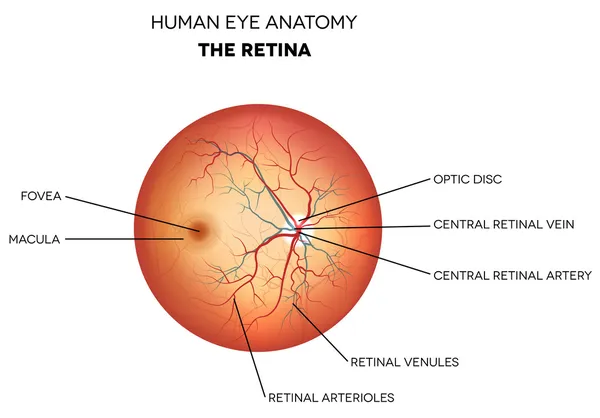
Human eye anatomy, retina
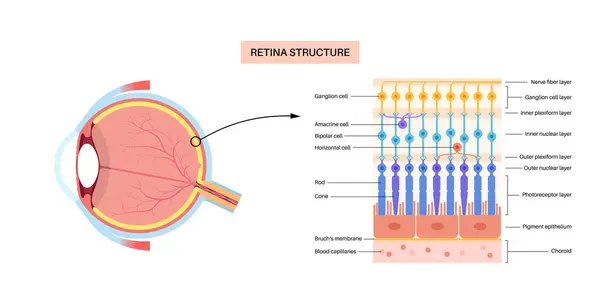
Retina structure anatomical poster. Scheme of the innermost layer in the eye. Posterior portion of the eyeball. Photoreceptor, neuronal and glial cells, cones and rods diagram, vector illustration
Illustration of eye disease, age-related macular degeneration
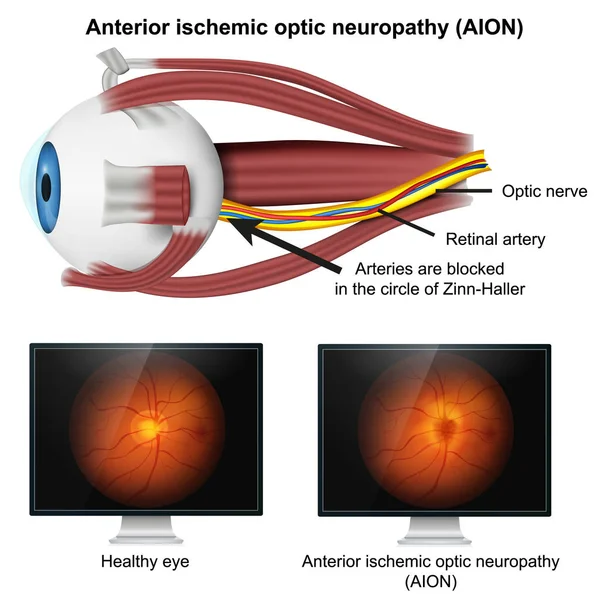
Ishemic optic neuropathy medical 3d vector illustration on white background
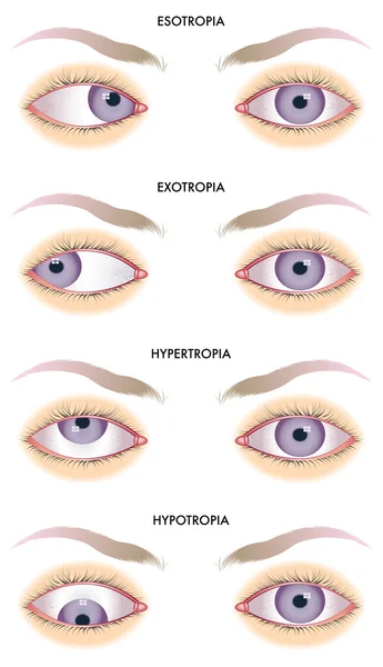
Strabismus
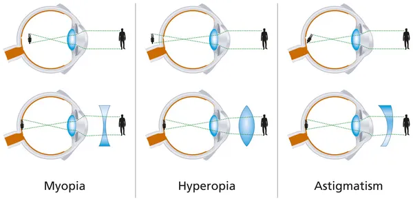
Vision Defects - Myopia, Hyperopia And Astigmatism
How eye work medical illustration, eye - brain diagram
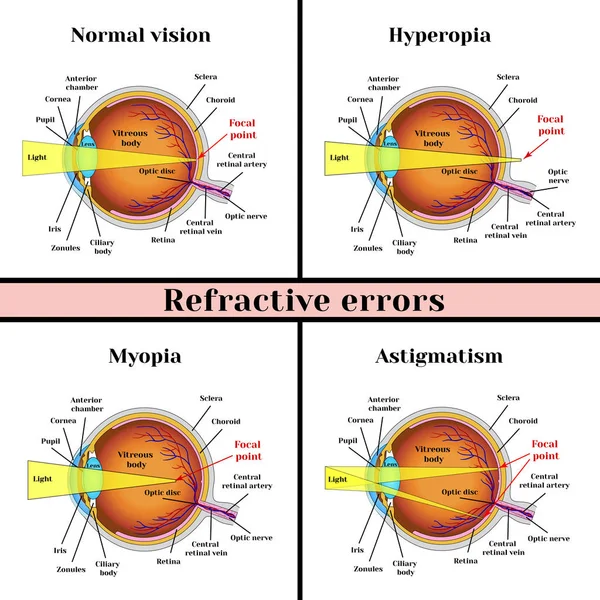
Refractive errors eyeball: hyperopia, myopia, astigmatism.
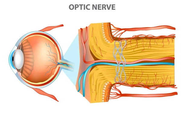
The Optic Nerve. Anatomy of the Eye
Eye system. Vector design.
The human eye. The structure of the eyeball. Medical didactic allowance for study. Vector illustration isolated on a white background.
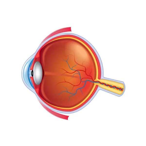
Eye anatomy isolated on white vector
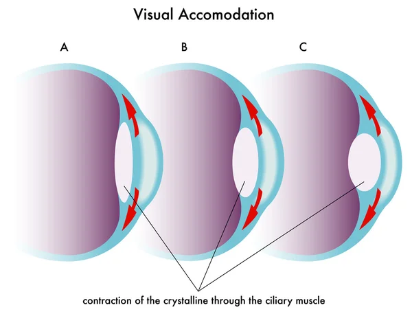
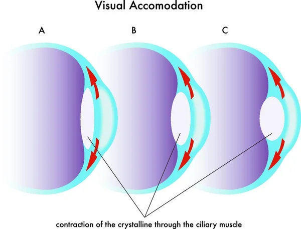
Visual accomodation
Human eye anatomy. Educational material
Anatomy of the eye. the eyeball, iris,pupil
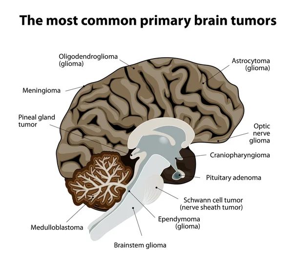
The most common primary brain tumors
Abstract Brain Network Background
Sizeable green eye ball
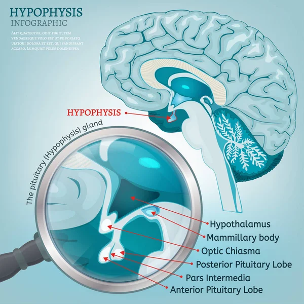
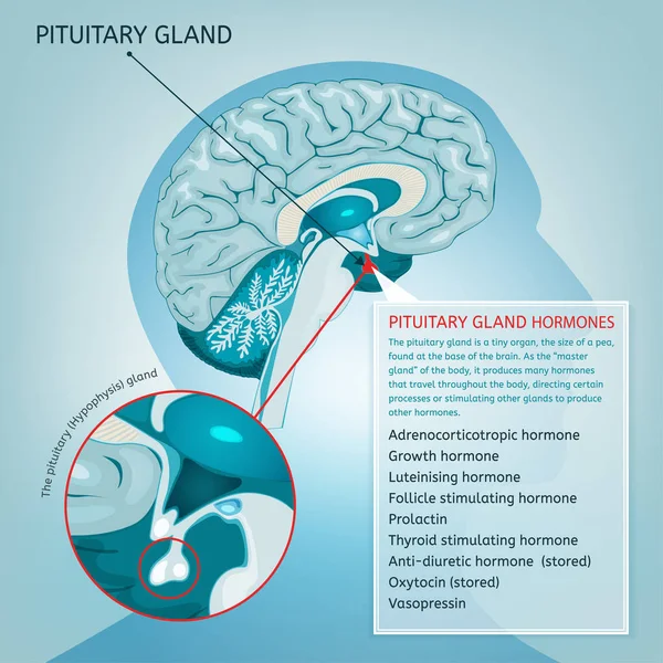
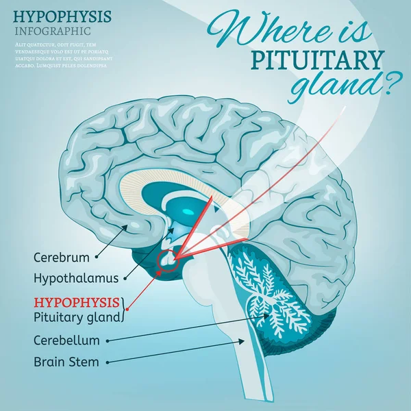
Pituitary gland vector
Human anatomy disease. medulloblastoma vector illustration on white background. eps 10
Illustration of how glaucoma works, glaucoma eye, medical illustration
An eye disease manifested by inflammation of the optic nerve with accompanying pain in the eyeballs. Drawn optic neuritis for use in the medical field. Vector illustration.
Medical illustration of the effects of the strabismus
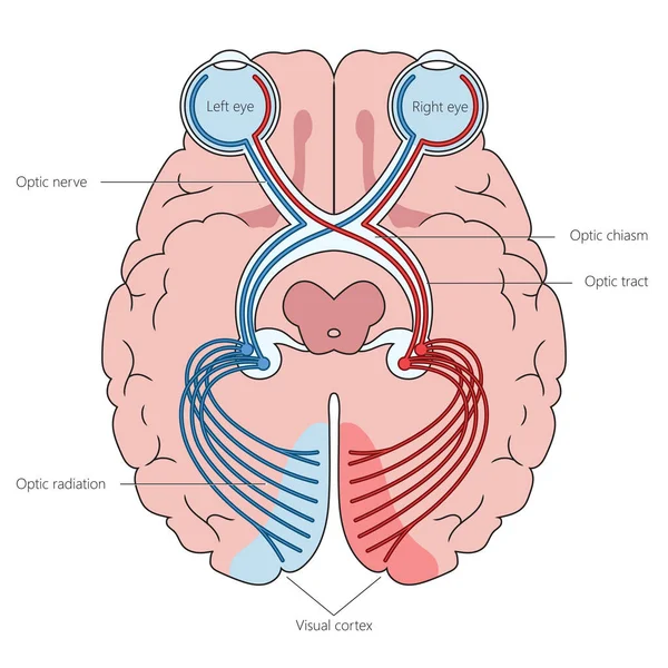
Human optic nerves in brain structure diagram hand drawn schematic vector illustration. Medical science educational illustration
Illustration, Glaucoma of the eye, Medical Illustrations.
Eye Disease, Atrophic Age-Related Macular Degeneration Illustrated
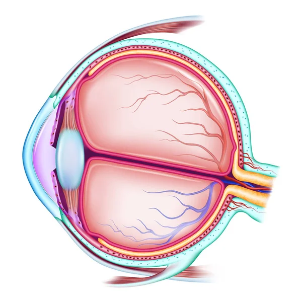
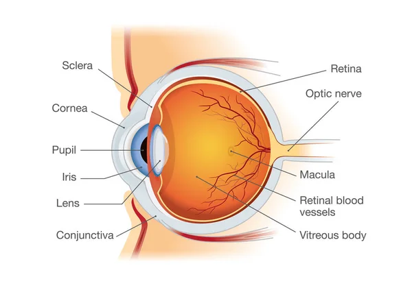
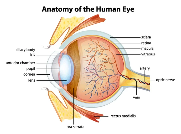
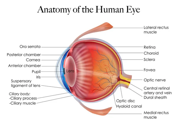
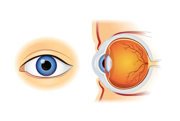
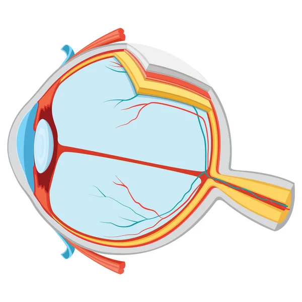
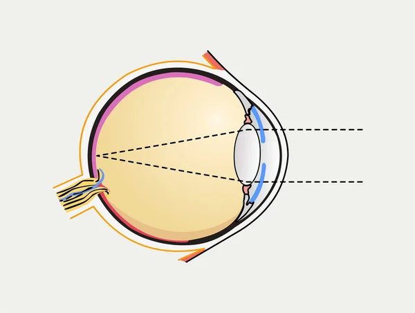
Human eye anatomy in side view.
Illustration of how glaucoma works, normal and glaucomatous eyes, medical illustration
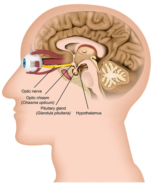
Optic nerve anatomy 3d medical vector illustration on white background
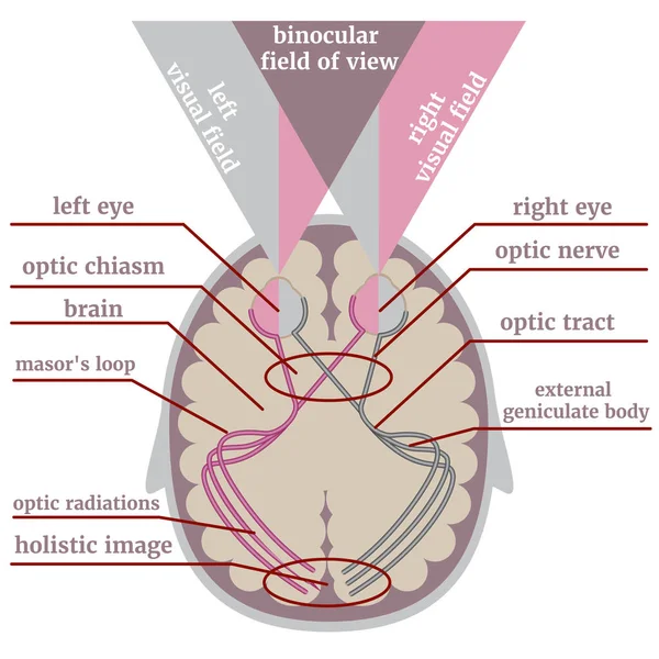
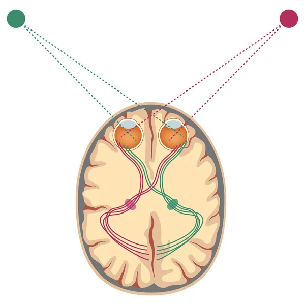
Visual system, path. Infographic with a diagram of how the brain perceives an image. Medical poster. Vector illustration
Pituitary gland vector
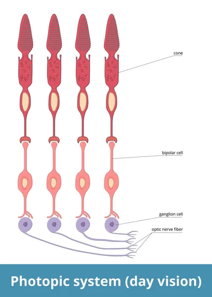
Photopic system (day vision). Each cone is directly connected to the brain through bipolar and ganglion cells, so each optic nerve fiber represents area of retina, and vision in bright light is sharp.
Human anatomy disease. medulloblastoma vector illustration on white background. eps 10
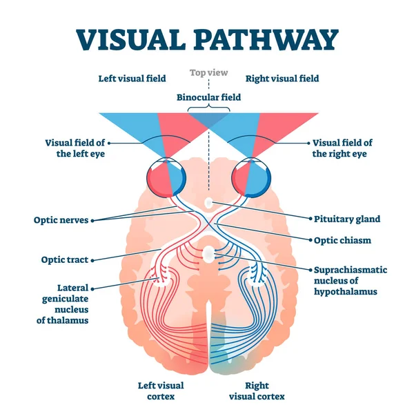
Visual pathway medical vector illustration diagram
Medical diagram of eye work. Anatomical infographics with structure of visual system, connection with brain and function of eyes. Cartoon flat vector illustration isolated on white background
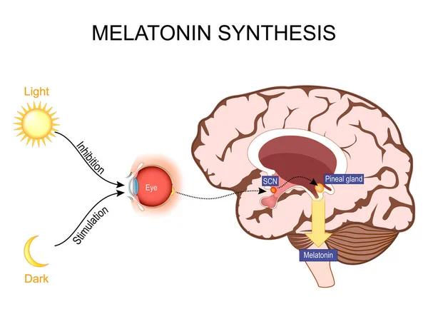
Melatonin and Circadian rhythm regulation. Brain with pineal gland and suprachiasmatic nucleus. sleep-wake cycle. Human anatomy. vector illustration. What does melatonin do?
Glaucoma is a group of eye diseases that lead to damage of the optic nerve. Intraocular pressure in the back of the eye
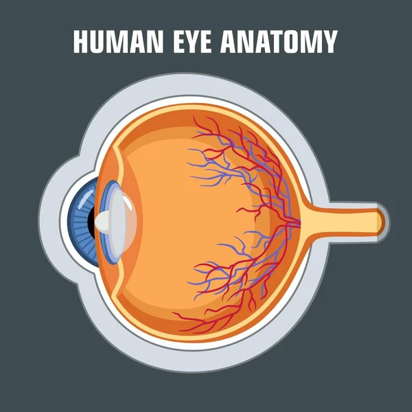
Human eye anatomy
Eyes are the organs of vision. They detect light and convert it into electro-chemical impulses in neurons. Illustration anatomy body.
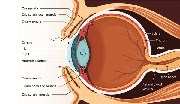
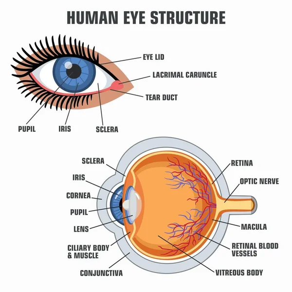
Vector Education poster eye structure. Image human eye anatomy poster
Vector Education poster eye structure. Image human eye anatomy poster
Eye disease, illustration of age-related macular degeneration, normal
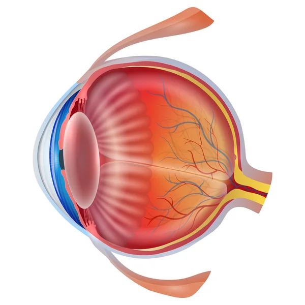
This illustration depicts the detailed structure of the human eye on a white background. It is unlabeled, providing a clear view of the various parts.
Vector cross-section diagram contrasts a healthy human eye with an eye affected by floaters, highlighting vitreous body and visual disturbances in medical style
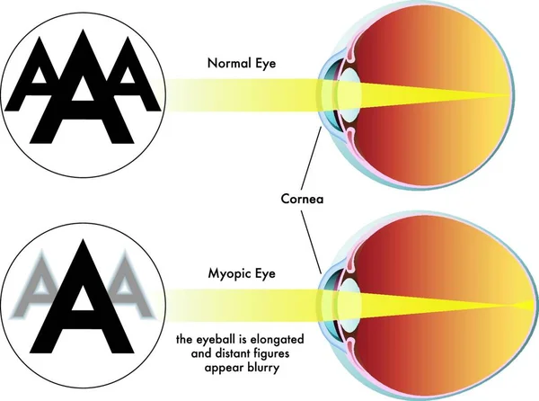
Medical illustration of the symptoms of myopia
Vector Education poster eye structure. Image human eye anatomy poster
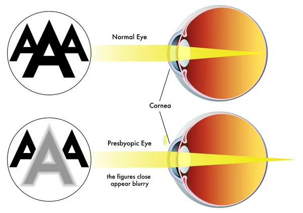
Presbyopia
Detailed illustration of the anatomy and structure of the human eye. The picture shows the iris, pupil, lens, retina, optic nerve, and other significant structures of the eye.
Eye anatomy vector illustration
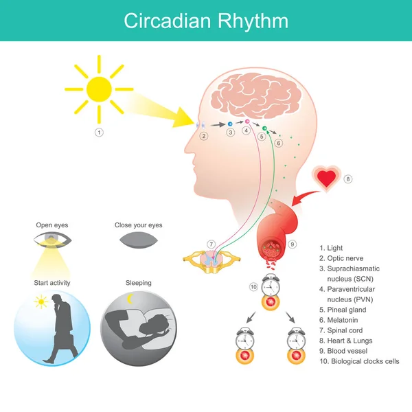
Circadian Rhythm. Diagram human body physical, mental, and behavioural changes that follow a 24-hour cycle
Diagrammatic illustration of the eye (line drawing)
Eye Disease, Exudative Age-Related Macular Degeneration Illustrated
Medical illustration of the cross-sectional structure of the human eye
Human eye anatomy showcasing retinal detachment, including cornea, lens, and optic nerve structure diagram hand drawn schematic vector illustration. Medical science educational illustration
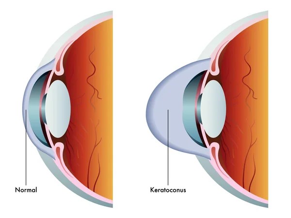
Colorful Keratoconus scheme
Glaucoma. Cross section of a Human eyeball. Intraocular pressure. Vision loss. Eye anatomy. Diagram for education and medical use. Vector illustration
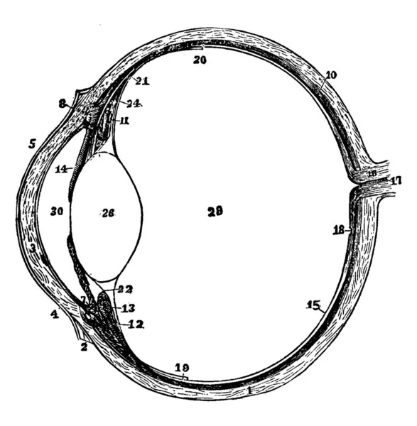
The left eyeball in horizontal section from before back, vintage line drawing or engraving
Illustration of the structure of the eye
Medical illustration of the visual accomodation
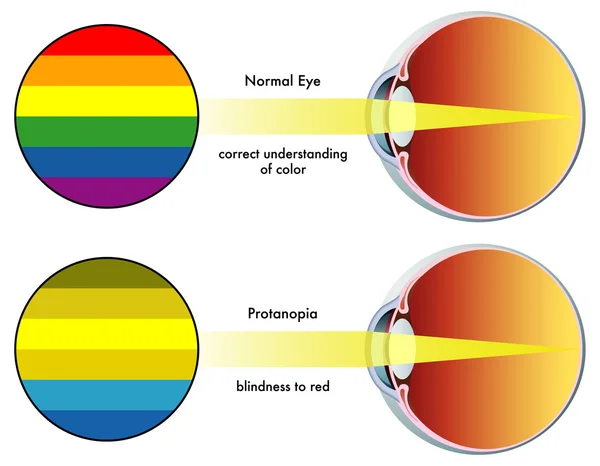
Protanopia
Human eye anatomy in inside and out side view.
Structures Of The Human Eye
Structures Of The Human Eye Labeled
Glaucoma Awareness Month: Vector Design Template for Vision Health. Shed light on eye health with this informative and impactful illustration.
Vector medical illustration of the symptoms of tritanopia
Vector Illustration Of Eye Anatomy
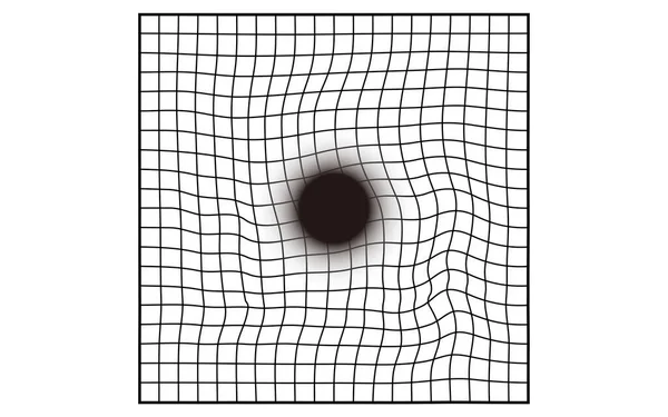
Amsler Chart, Age-related macular degeneration visibility (visual field defects, central darkening)
Glaucoma awareness month is observed every year in January, is a group of eye conditions that damage the optic nerve, the health of which is vital for good vision. Vector illustration
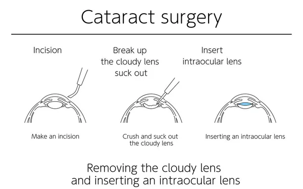
Illustrations, Cataract surgery, Medical Illustrations.
World Glaucoma day is observed every year on March 12, it is a group of eye conditions that damage the optic nerve, the health of which is vital for good vision. Vector illustration
Flow of cataract surgery, illustrated illustration (line drawing)
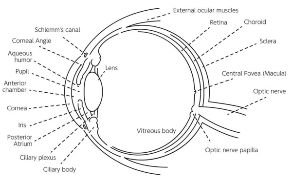
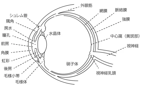
Illustration of the eye - Translation: Schlemm's canal, corner angle, aqueous humor, pupil, anterior chamber, cornea, iris, posterior chamber, ciliary body, ciliary body, lens, vitreous body, external eye muscle, retina, choroid, sclera, central foss
Human Eyeball Anatomy Set
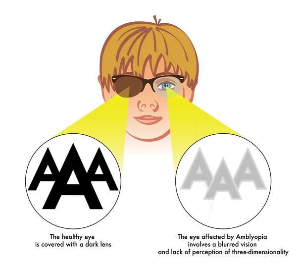
Medical illustration of the symptoms and consequences of amblyopia
Amblyopia (lazy eye)
Human eyeballs Blue eyes Illustration Vector
Diagrammatic illustration of the eye (line drawing)
Body human sight, eye. Biology, anatomy man and human organs.
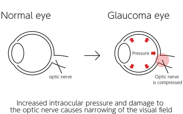
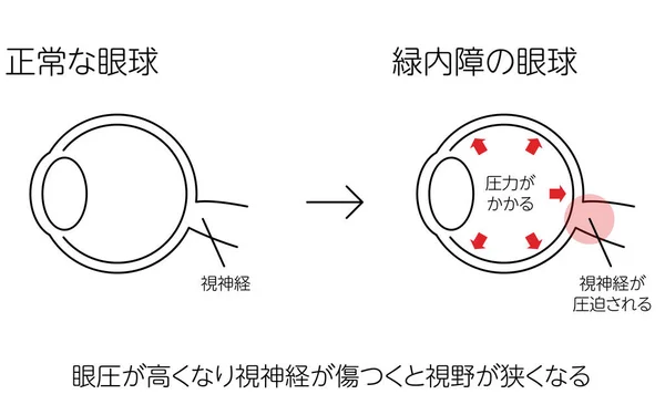
Illustration, Glaucoma of the eye, Medical Illustrations. - Translation: glaucoma in the eye, increased intraocular pressure and damage to the optic nerve, narrowing of the visual field, optic nerve Optic nerve is compressed.
Illustration of how glaucoma works, normal and glaucomatous eyes, medical illustration - Translation: normal eye, glaucoma eye, pressure, optic nerve, optic nerve is compressed, high intraocular pressure and damage to optic nerve causes visual field
Amsler Chart, Age-related macular degeneration visibility (heterophoria, distortion)
Amsler chart, Age-related macular degeneration visibility (visual field defects)
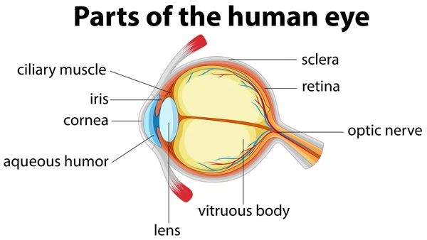
Parts of human eye with name
Pituitary gland vector
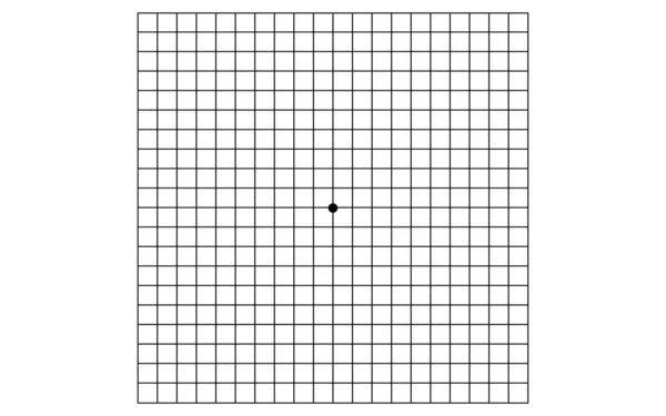
Amsler chart, self-check sheet for visual field abnormalities
Eyeball
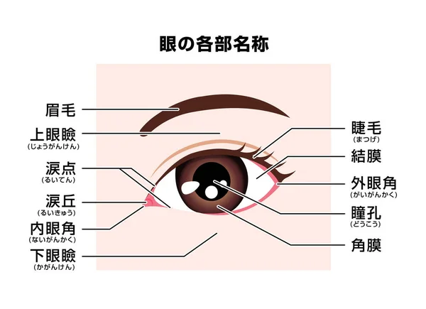
Structure of human eye ( names of parts ) vector illustration / Japanese
World Glaucoma day is observed every year on March 12, it is a group of eye conditions that damage the optic nerve, the health of which is vital for good vision. Vector illustration
World Glaucoma Week is observed every year in March, it is a group of eye conditions that damage the optic nerve, the health of which is vital for good vision. Vector illustration
World Glaucoma Week is observed every year in March, it is a group of eye conditions that damage the optic nerve, the health of which is vital for good vision. Vector illustration
Human Eye Diagram
Illustration of glaucoma, changes in visual field as glaucoma progresses, medical illustrations - Translation: changes in glaucoma vision, normal, early, middle, late
Glaucoma awareness month is observed every year in January, is a group of eye conditions that damage the optic nerve, the health of which is vital for good vision. Vector illustration
World Glaucoma day is observed every year on March 12, it is a group of eye conditions that damage the optic nerve, the health of which is vital for good vision. Vector illustration
Human Eye Anatomy Diagram With Detailed Cross Section Vector Colorful Eye Organ Infographic For Biology And Science Study
Visual system, path. Infographic with a diagram of how the brain perceives an image. Medical poster. Vector illustration
Healthy Eye Versus Cataract Cross Section Medical Vector Illustration
Minimalist Outline Eyeball Cross Section Anatomy Medical Healthcare Line Icon
Happy Cataract Awareness Month Celebration Vector Design Illustration for Background, Poster, Banner, Advertising, Greeting Card
Minimalist Eyeball Anatomy Outline Icon Illustration for Medical Science Healthcare
Next Page
1
2
3
4
5
...
100
Page
Go
Home
Vectors
optisk nerve vector images
Related vector searches
cornea
,
eye anatomy
,
eye
,
contact lens
,
vagus nerve
,
treatment of varicose veins
,
lung abscess
,
glaucoma
,
astigmatism
,
benign kidney cyst
,
neck protector
,
parts of brain
,
rib fracture
,
manual lymphatic drainage
,
painful neck
,
neck braces
,
muscular tissue
,
neck guard
,
round neck