Organell Stock Vector Images

343 Organell vector art & graphics are available under a royalty-free license
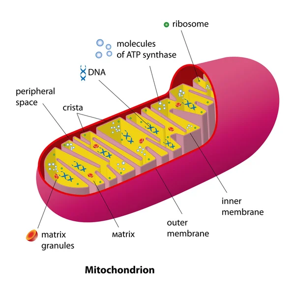
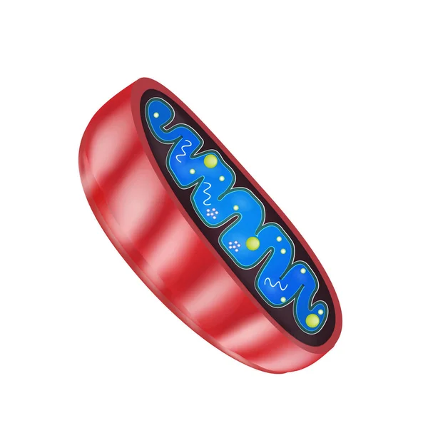
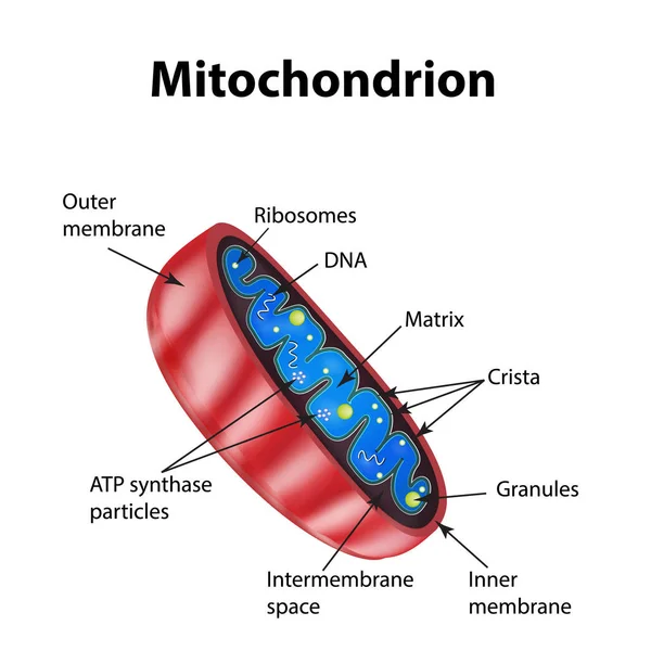
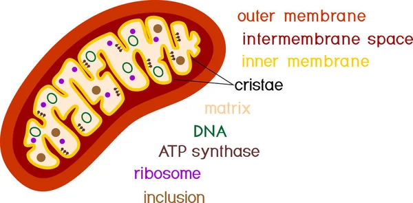
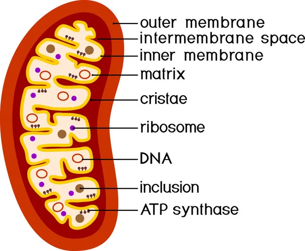
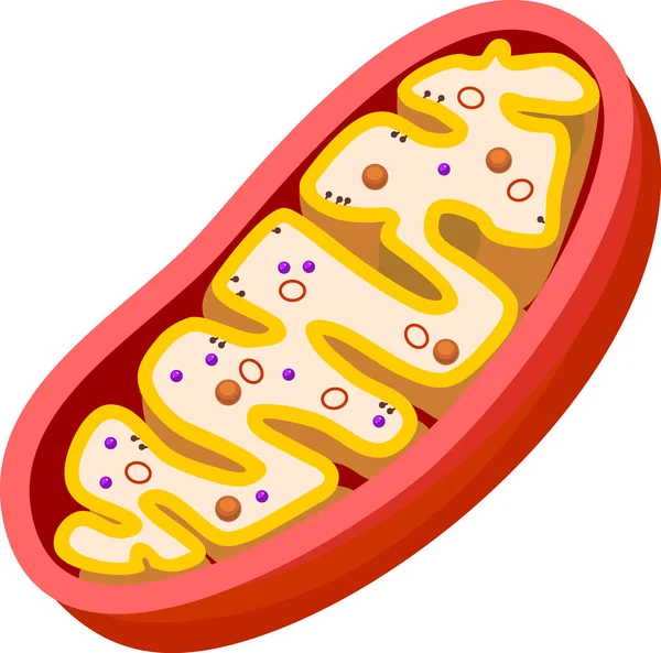
Mitochondrion scheme
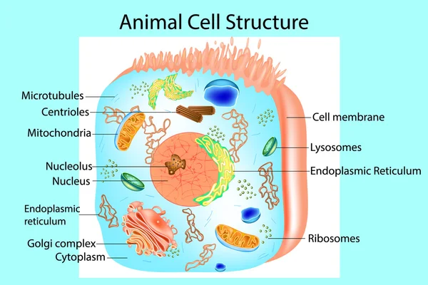
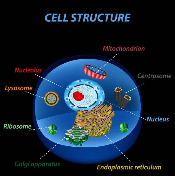
Structure of human cells. Organelles. The core nucleus, endoplasmic reticulum, Golgi apparatus, lysosomes, ribosomes, mitochondria, centriole. Vector illustration
Cell organelles biological vector illustration diagram
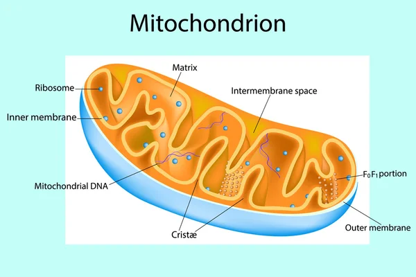
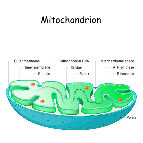
Mitochondrion. Components of a typical mitochondrion. Structure. Interactive diagram
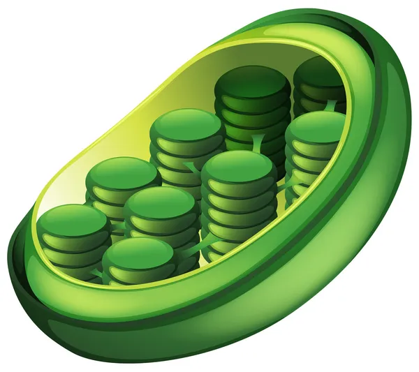
Chloroplast
Chloroplast
Cell structure isolated icon
Mitochondria structure. Vector illustration on isolated background
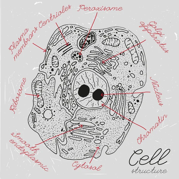
Handdrawn Cell Pattern
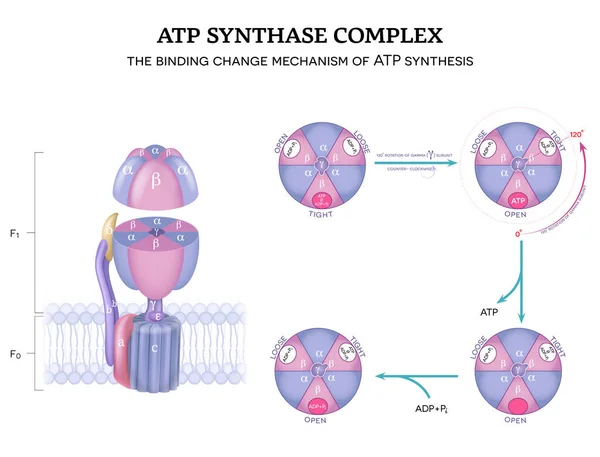
ATP synthase complex structure and mechanism of ATP synthase. The binding change mechanism. 120-degree rotation of gamma subunit counter-clockwise.
Atp Synthase Complex Structure Mechanism Atp Synthase Binding Change Mechanism 120 Degree Rotation Gamma Subunit Counter Clockwise
Mitochondria structure. Vector illustration on isolated background
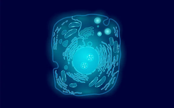
Animal human cell structure educational science. Microscope 3d eukaryotic nucleus organelle medicine technology analysis. Glowing blue biology poster template isolated line art vector illustration
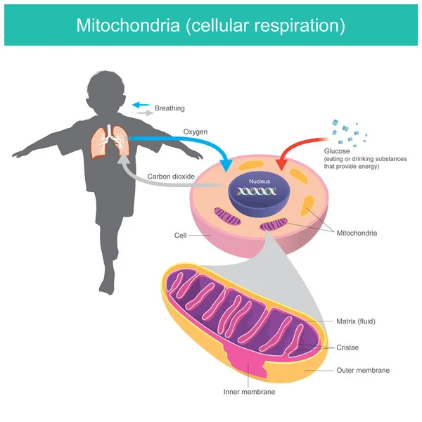
Mitochondria. Illustration explain human body received glucose and oxygen such as eating or drinking after that the cell system changes glucose in a fluid matrix from mitochondria to energy stored and release carbon dioxide gas out
Mitochondria Illustration Explain Human Body Received Glucose Oxygen Eating Drinking Cell System Changes Glucose Fluid Matrix Mitochondria Energy Stored Release Carbon Dioxide Gas Out
Illustration of the anatomy of an animal cell. Structure of a Eukaryotic cell.
Structure of mitochondrion isolated on white background
Structure Mitochondrion Isolated White Background
Mitochondria minimal vector icon drawn with brush line. Blue linear medical pictogram isolated on transparent background
Mitochondria Minimal Vector Icon Drawn Brush Line Blue Linear Medical Pictogram Isolated Transparent Background
Structure of mitochondrion isolated on white background
Structure Mitochondrion Isolated White Background
Detailed vector illustration of a mitochondrion with vibrant colors and clear structure, highlighting its internal components
Detailed Vector Illustration Mitochondrion Vibrant Colors Clear Structure Highlighting Its Internal Components
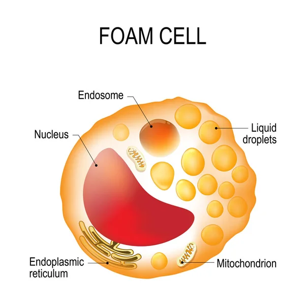
Foam cell. Cell structure
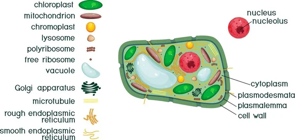
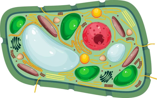
Plant cell structure with titles and different organelles
Plant Cell Structure Titles Different Organelles
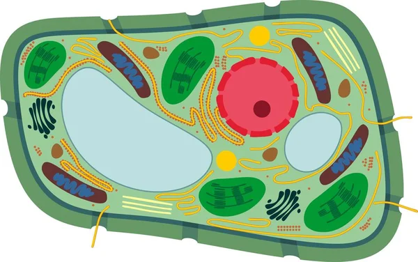
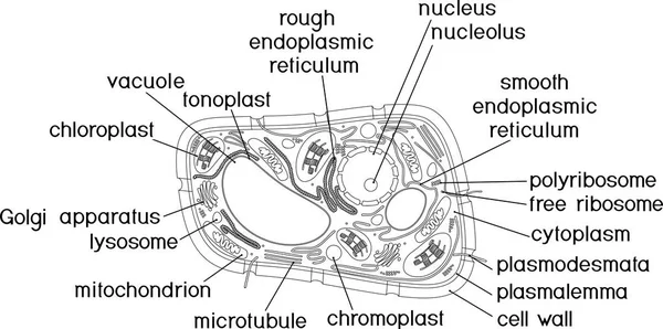
Structure of plant cell with different organelles
Structure Plant Cell Different Organelles
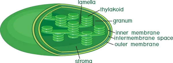
Structure of chloroplast with english titles
Structure Chloroplast English Titles
Coloring page with structure of Chlamydomonas cell
Coloring Page Structure Chlamydomonas Cell
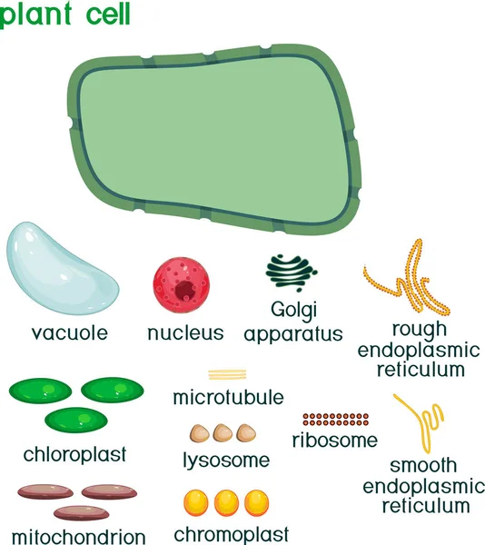
Educational game: assembling cells from ready-made components in form of stickers. Plant cell structure with titles
Educational Game Assembling Cells Ready Made Components Form Stickers Plant Cell Structure Titles
Function of mitochondria. Calcium storage, Lipid metabolism, Regulation of immunity, ATP and Energy production, Steroid synthesis, Regulation of cell apoptosis, Mitochondrial DNA signaling, and Heat production. What mitochondria do. Detailed Vector p
Function Mitochondria Calcium Storage Lipid Metabolism Regulation Immunity Atp Energy Production Steroid Synthesis Regulation Cell Apoptosis Mitochondrial Dna Signaling Heat Production What Mitochondria Detailed Vector
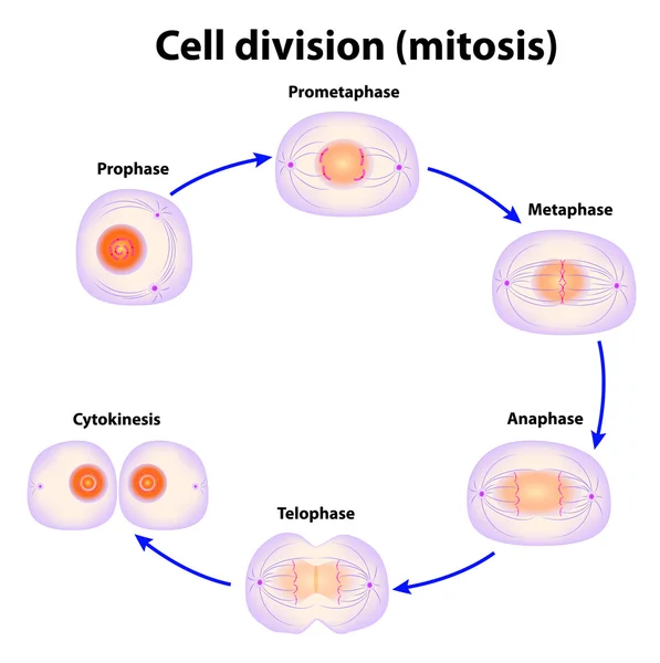
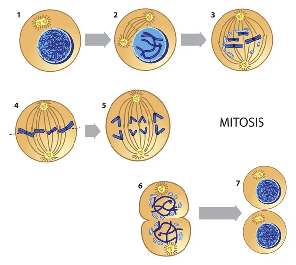
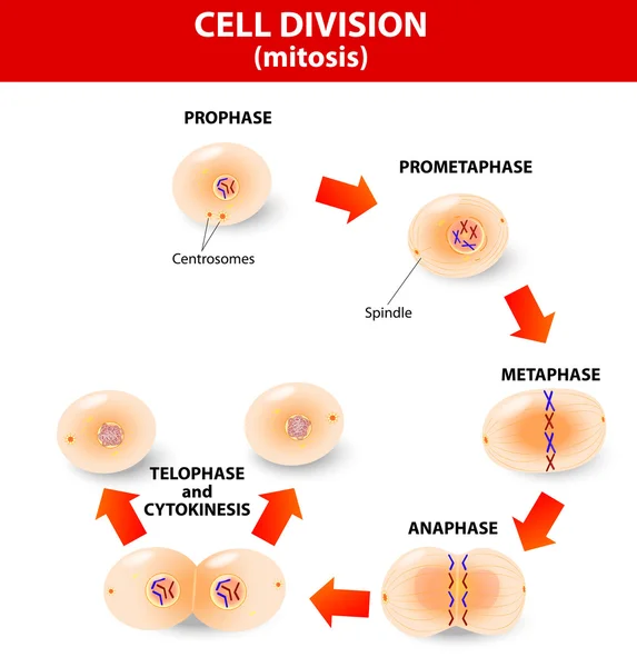
Cell division. Mitosis
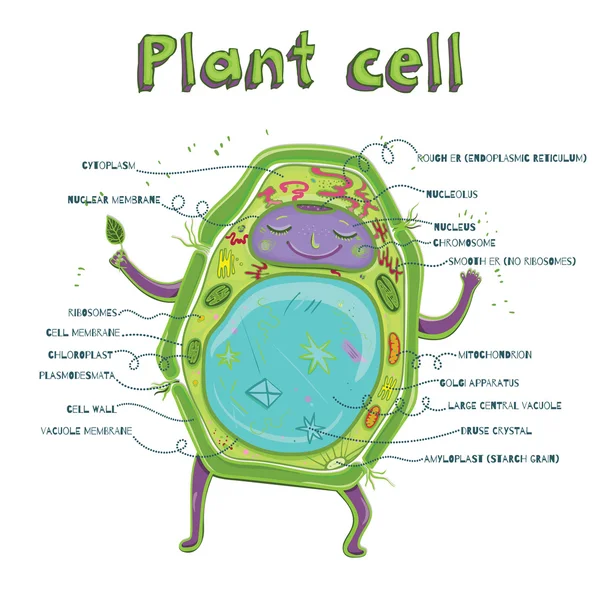
Vector illustration of the Plant cell anatomy structure. Educational infographic.
Vector Illustration Plant Cell Anatomy Structure Educational Infographic
Epithelial Tissue with Goblet Cells. Goblet Cell Structure Illustration. Mucus Secreting. Human Goblet Cell Anatomy Educational Chart
Epithelial Tissue Goblet Cells Goblet Cell Structure Illustration Mucus Secreting Human Goblet Cell Anatomy Educational Chart
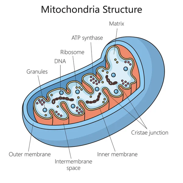
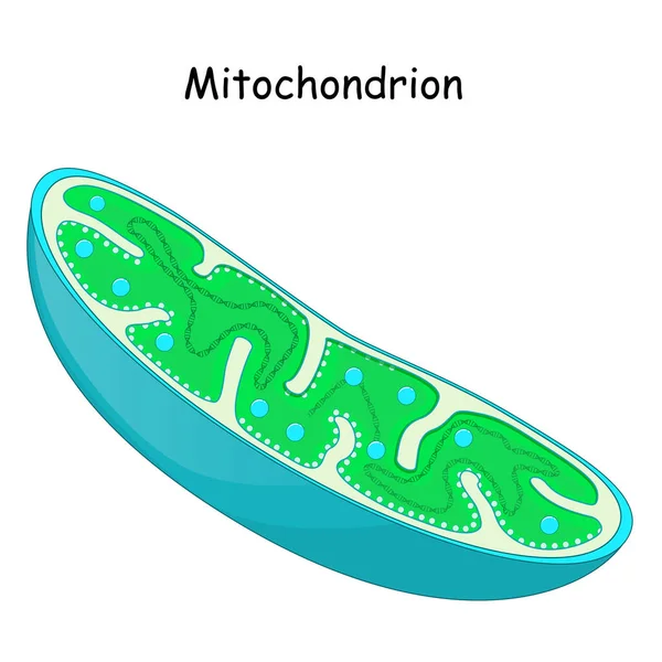
Mitochondrion anatomy. Structure, components and organelles. cross-section of mitochondria. Vector illustration
Mitochondrion Anatomy Structure Components Organelles Cross Section Mitochondria Vector Illustration
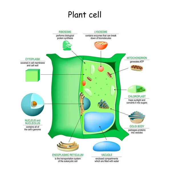
Plant cell anatomy structure. Close-up of Organelles of a plant cell. Educational infographic. Vector illustration
Plant Cell Anatomy Structure Close Organelles Plant Cell Educational Infographic Vector Illustration
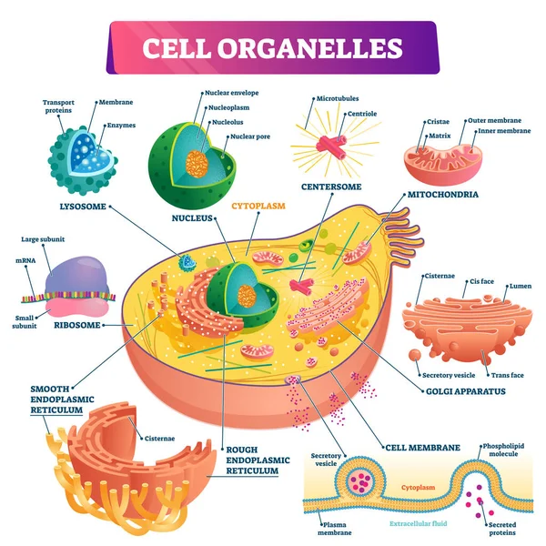
Cell anatomy. Structure and organelles of human's cell. Cross sections of animal cell: nucleus, nucleolus, mitochondria, centrosome, golgi apparatus, endoplasmic reticulum, ribosome and membrane. poster biological diagram. vector illustration
Cell Anatomy Structure Organelles Human Cell Cross Sections Animal Cell Nucleus Nucleolus Mitochondria Centrosome Golgi Apparatus Endoplasmic Reticulum Ribosome Membrane Poster Biological Diagram Vector Illustration
Viral capsids of different shapes: helical, polyhedral, complex shape.
Viral Capsids Different Shapes Helical Polyhedral Complex Shape
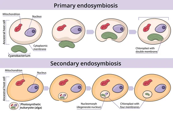
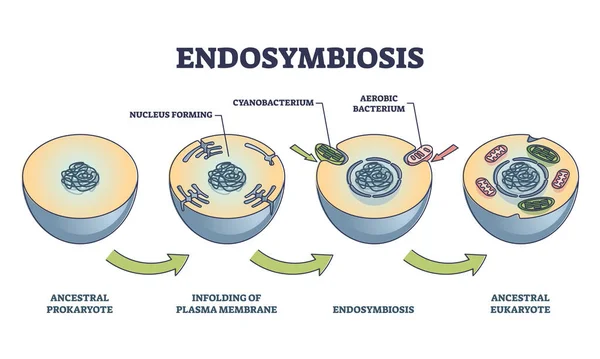
Process of primary and secondary endosymbiosis. Cell engulfs and absorbs a prokaryotic cell. Secondary endosymbiosis occurs when a eukaryotic cell engulfs and absorbs another eukaryotic cell.
Process Primary Secondary Endosymbiosis Cell Engulfs Absorbs Prokaryotic Cell Secondary Endosymbiosis Occurs Eukaryotic Cell Engulfs Absorbs Another Eukaryotic Cell
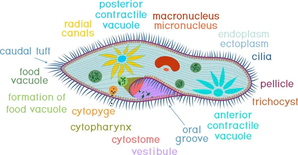
Structure of Paramecium caudatum isolated on white background
Structure Paramecium Caudatum Isolated White Background
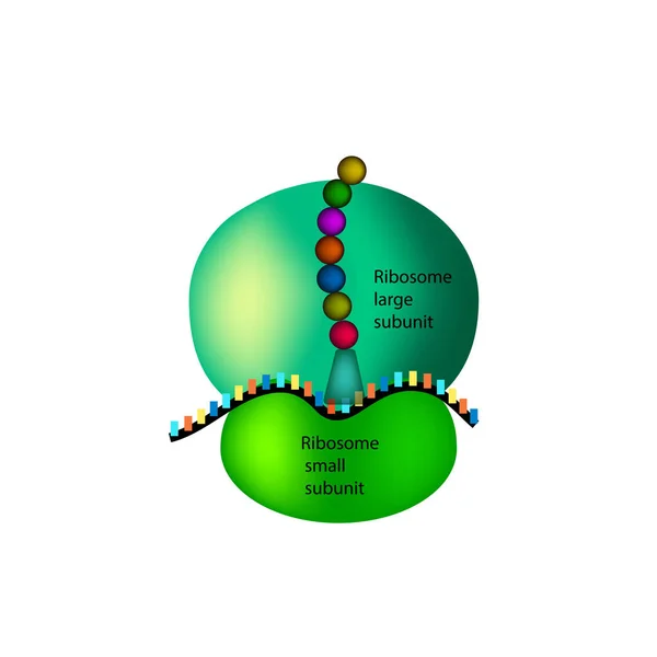
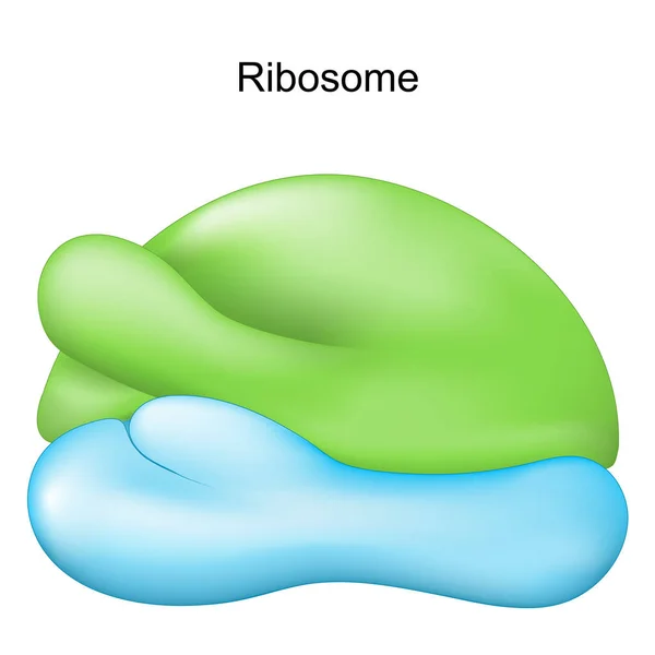
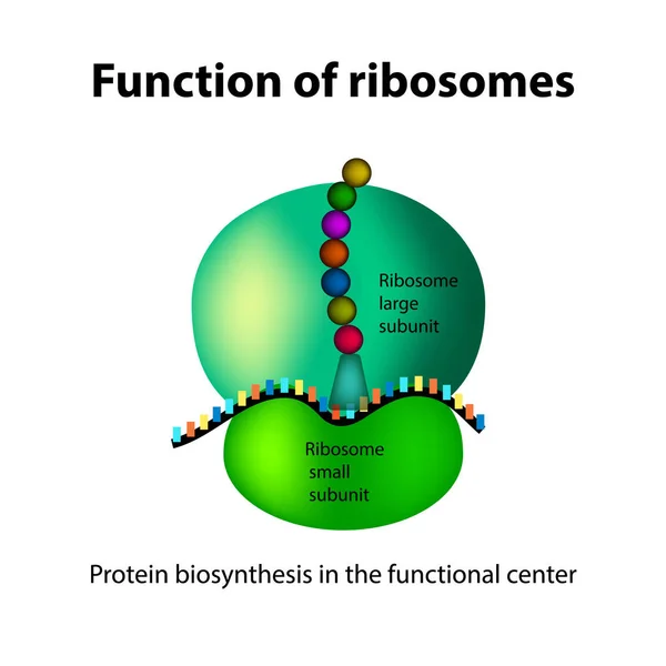
The structure of the ribosome. Infographics. Vector illustration on isolated background
Stylized outline of a mitochondrion, a cellular organelle, with internal circular structures.
Stylized Outline Mitochondrion Cellular Organelle Internal Circular Structures
Animal cell anatomy structure diagram. Educational infographic. Vector illustration
Animal Cell Anatomy Structure Diagram Educational Infographic Vector Illustration
Cell structure isolated icon
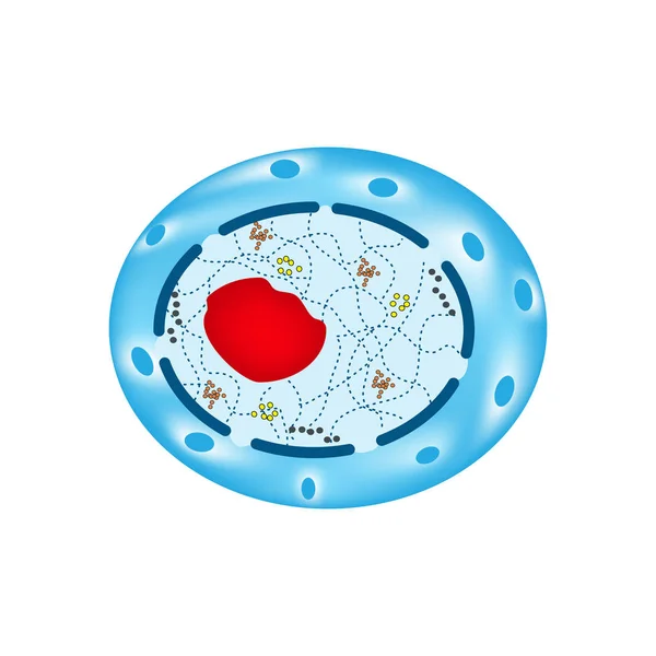
The structure of the human cell nucleus. Infographics. Vector illustration on isolated background
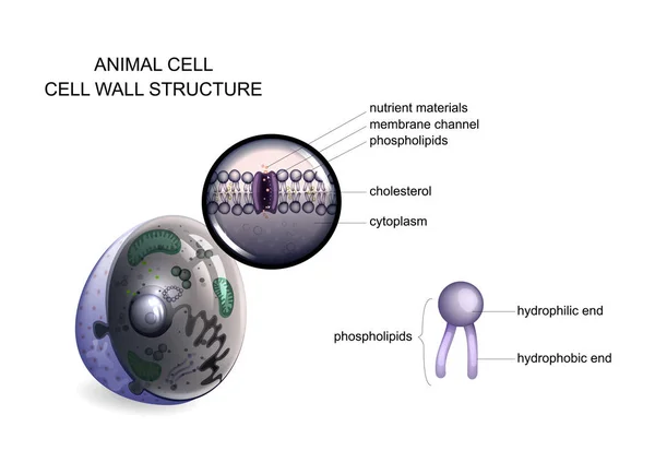
Animal cell. cell wall structure
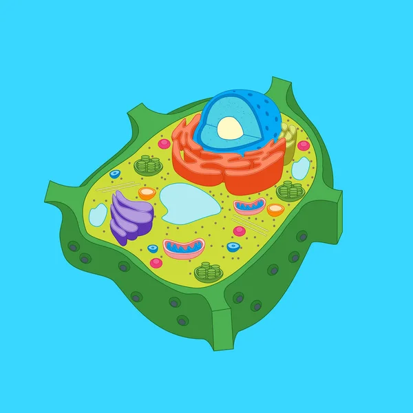
Plant cell anatomy
Plant cell anatomy
Animal human cell structure educational science. Microscope 3d eukaryotic nucleus organelle medicine technology analysis. Glowing colored biology poster template isolated line vector illustration
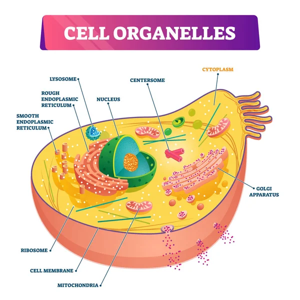
Cell organelles biological anatomy vector illustration diagram
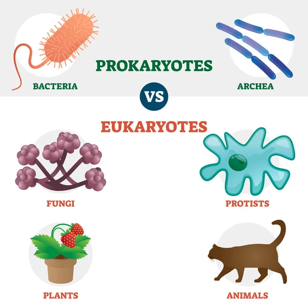
Eukaryote vs Prokaryote cell type organisms educational set
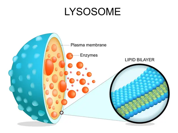
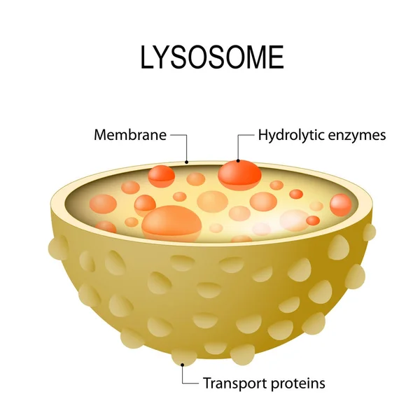
Lysosome anatomy. Cross section of a cell organelle. Close-up of a Lipid bilayer membrane, hydrolytic enzymes, transport proteins. Autophagy. Vector illustration
Lysosome Anatomy Cross Section Cell Organelle Close Lipid Bilayer Membrane Hydrolytic Enzymes Transport Proteins Autophagy Vector Illustration
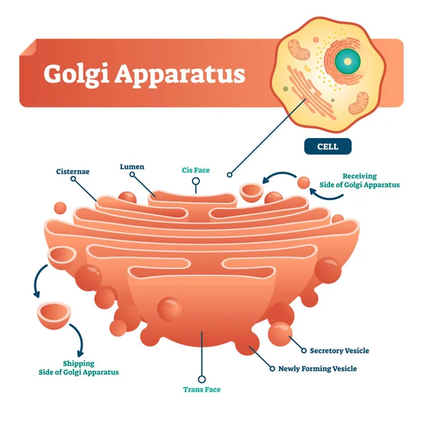
Golgi apparatus vector illustration. Labeled microscopic scheme with cisternae, lumen, secretory and newly forming vesicle. Diagram with receiving and shipping side.
Human mitochondria structure diagram schematic vector illustration. Medical science educational illustration
Human Mitochondria Structure Diagram Schematic Vector Illustration Medical Science Educational Illustration
Structure of Spirogyra (charophyte green algae) with titles isolated on white background
Structure Spirogyra Charophyte Green Algae Titles Isolated White Background
Eukaryotic vs Prokaryotic cells, educational biology vector illustration diagram
Structure of mitochondrion isolated on white background
Structure Mitochondrion Isolated White Background
Chloroplast
Chloroplast
Structure of chloroplast with english titles
Structure Chloroplast English Titles
Plant cell structure with titles and different organelles
Plant Cell Structure Titles Different Organelles
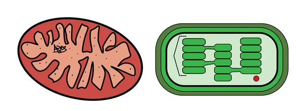
Vector illustration of mitochondria and chloroplast
Mitosis
Mitosis
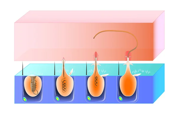
Structure and action cnidocyte
Mitochondria. structure and anatomy of a mitochondrion. vector icon
Mitochondria Structure Anatomy Mitochondrion Vector Icon
Ribosome. Cell organelle for Protein synthesis. Vector illustration
Ribosome Cell Organelle Protein Synthesis Vector Illustration
Autophagy of mitochondria. Diagram of the process of autophagy from Forming a membrane and autophagosome to fuse phagosome and lysosome when contents of the vesicles are degraded and recycled. Autophagy defects linked to various diseases and cancer d
Autophagy Mitochondria Diagram Process Autophagy Forming Membrane Autophagosome Fuse Phagosome Lysosome Contents Vesicles Degraded Recycled Autophagy Defects Linked Various Diseases Cancer
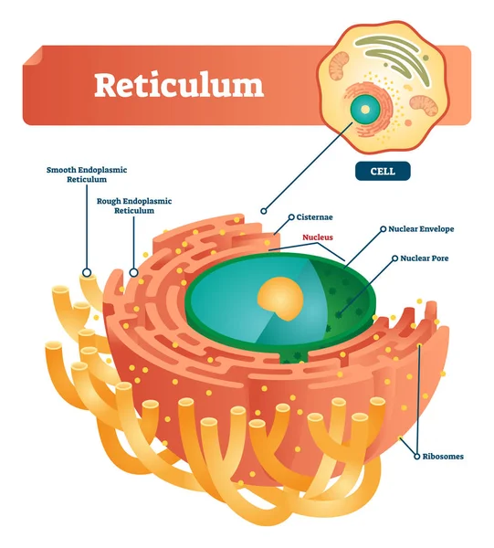
Reticulum labeled vector illustration scheme. Anatomical diagram with endoplasmic reticulum. Closeup with cisternae, nucleus, ribosomes, nuclear envelope and pore.
Simple Cell Biology Icon Outline Circle Diagram
Medical illustration of fibroblast cell anatomy. Educational diagram of a connective tissue cell involved in extracellular matrix and collagen production.
Medical Illustration Fibroblast Cell Anatomy Educational Diagram Connective Tissue Cell Involved Extracellular Matrix Collagen Production
Seamless Vector doodle pattern of Microbes, bacteria and virus
Of the cell
Of the cell
Mitosis. process cell division
Animal cell anatomy
Animal cell anatomy
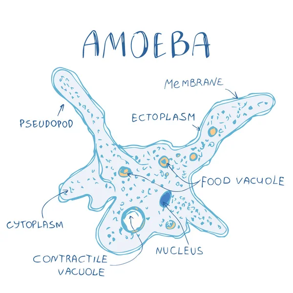
Amoeba structure hand drawn image
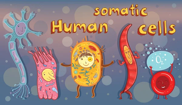
Vector illustration of human somatic cells
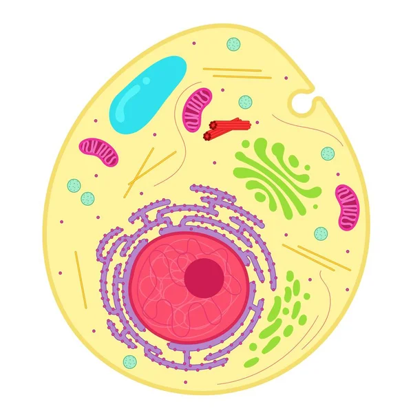
An animal cell is a type of eukaryotic cell.
Animal Cell Type Eukaryotic Cell
Plant cell anatomy
Plant cell anatomy
Structure of mitochondrion isolated on white background
Structure Mitochondrion Isolated White Background
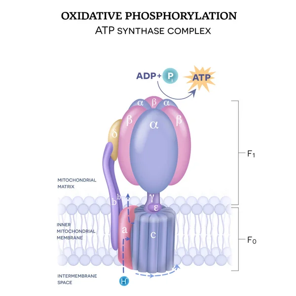
The ATP synthase structure (complex V) consists of two components F0 and F1. The formation of ATP using adenosine diphosphate (ADP) and inorganic phosphate (Pi)
Atp Synthase Structure Complex Consists Two Components Formation Atp Using Adenosine Diphosphate Adp Inorganic Phosphate
Diagram of Euglena. Structure of Euglena viridis with different organelles
Diagram Euglena Structure Euglena Viridis Different Organelles
Coloring page. Structure of plant cell with titles
Coloring Page Structure Plant Cell Titles
Human cell anatomy. Structure of a eukaryotic cell. All organelles: Nucleus, Ribosome, Rough endoplasmic reticulum, Golgi apparatus, mitochondrion, cytoplasm, lysosome, Centrosome. Animal cell on blue background. vector illustration
Human Cell Anatomy Structure Eukaryotic Cell All Organelles Nucleus Ribosome Rough Endoplasmic Reticulum Golgi Apparatus Mitochondrion Cytoplasm Lysosome Centrosome Animal Cell Blue Background Vector Illustration
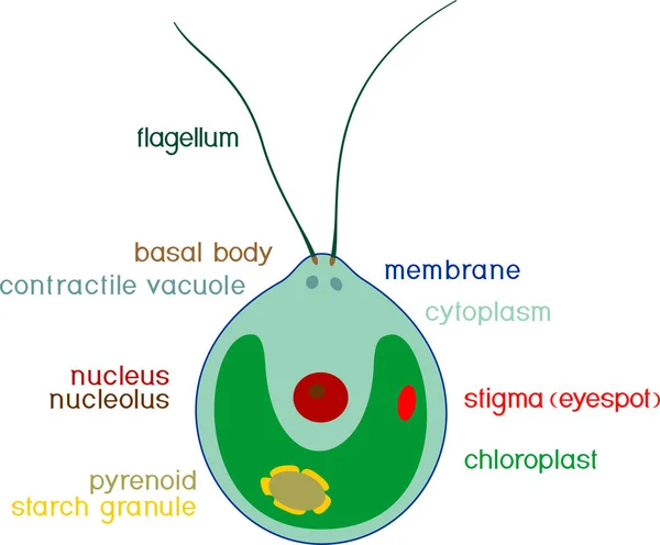
Structure of Chlamydomonas cell with titles isolated on white background
Structure Chlamydomonas Cell Titles Isolated White Background
Structure of plant cell with different organelles
Structure Plant Cell Different Organelles
Structure of plant cell with different organelles
Structure Plant Cell Different Organelles
Endosymbiosis process stages with symbiotic living organisms outline diagram
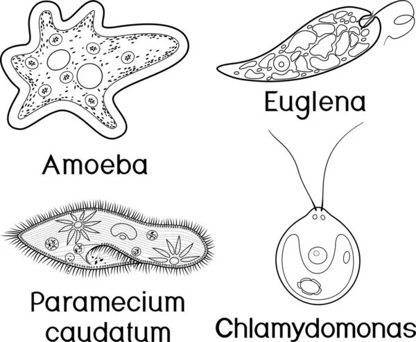
Coloring page. Set of unicellular organisms (protozoa): Paramecium caudatum, Amoeba proteus, Chlamydomonas and Euglena viridis
Coloring Page Set Unicellular Organisms Protozoa Paramecium Caudatum Amoeba Proteus Chlamydomonas Euglena Viridis
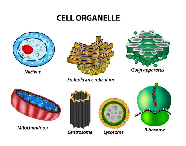
Set the cell organelles. Nucleus, endoplasmic reticulum, Golgi apparatus, mitochondria, centrosome, lysosome, the ribosome. Infographics. Vector illustration on isolated background
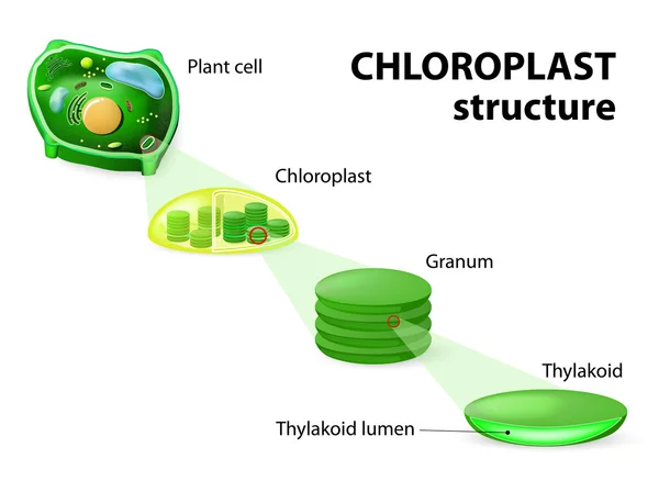
Chloroplast structure
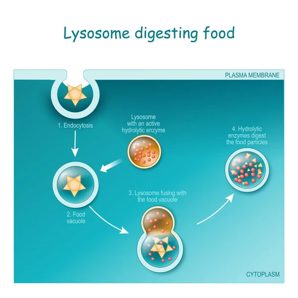
Endocytosis. Lysosome digesting food. Part of cell (plasma membrane, cytoplasm and lysosome), with food vacuole. Lysosome fusing with the food vacuole. Vector illustration
Endocytosis Lysosome Digesting Food Part Cell Plasma Membrane Cytoplasm Lysosome Food Vacuole Lysosome Fusing Food Vacuole Vector Illustration
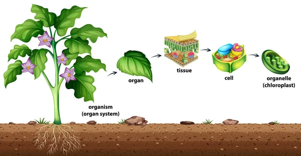
Cell organisation in plants illustration
Cell Organisation Plants Illustration
Animal cell anatomy. Structure and organelles of Eukaryotic cell. Vector poster for education. illustration
Animal Cell Anatomy Structure Organelles Eukaryotic Cell Vector Poster Education Illustration
Stylized outline of a cell with nucleus and organelles, isolated on white.
Stylized Outline Cell Nucleus Organelles Isolated White
Coloring page with structure of Chlorella (single-celled green algae) with titles isolated on white background
Coloring Page Structure Chlorella Single Celled Green Algae Titles Isolated White Background
Structure of human cells. Organelles. The core nucleus, endoplasmic reticulum, Golgi apparatus, lysosomes, ribosomes, mitochondria, centriole. Vector illustration on a black background
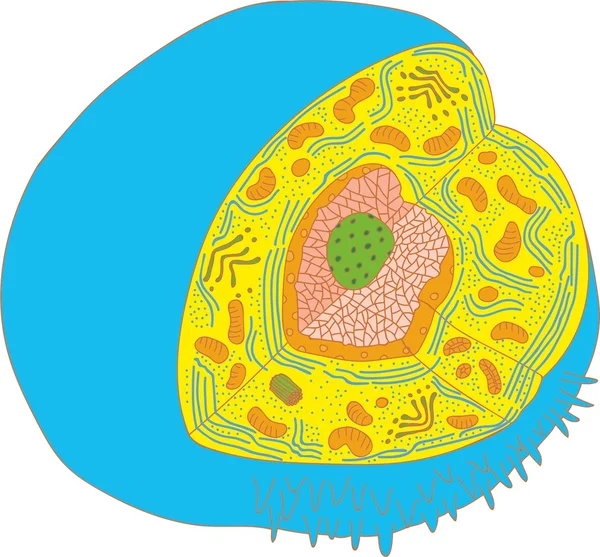
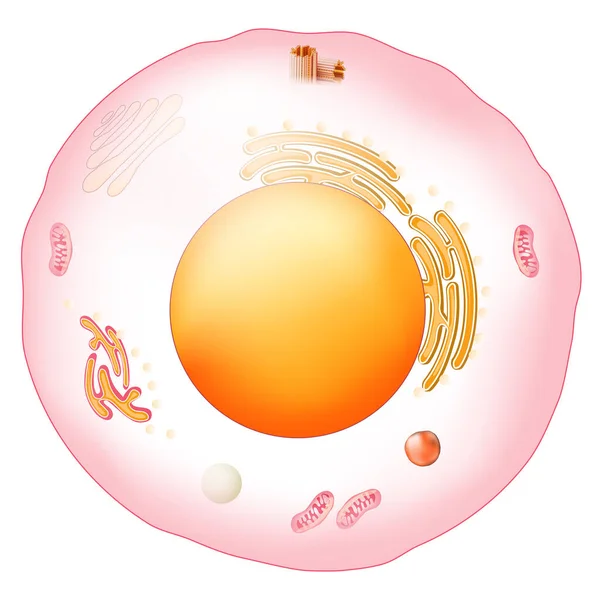
The internal structure of an animal cell
Amoeba under magnifying glass isolated on white background
Amoeba Magnifying Glass Isolated White Background
Hand Drawn Cell
Hand Drawn Cell
An image of a DNA
An image of a DNA
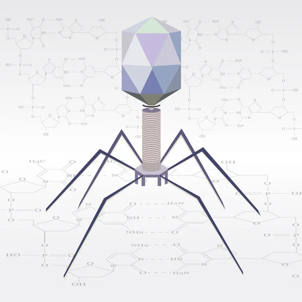
Vector virus bacteriophage model
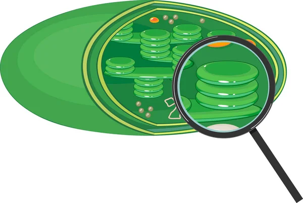
Chloroplast structure under magnifying glass isolated on white background
Chloroplast Structure Magnifying Glass Isolated White Background
Anatomy of the Lysosome.
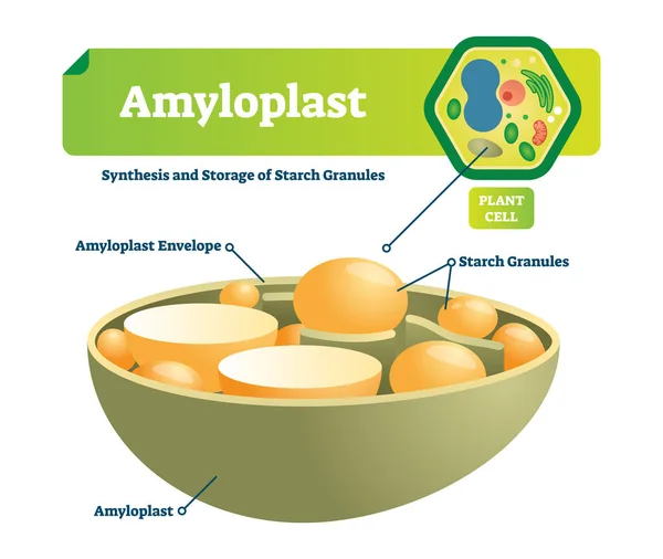
Amyloplast vector illustration. Labeled medical scheme with synhesis and storage of starch granules. Colorful diagram with envelope and plant cell. Microscopic cell structure.
L-carnitine transports fat molecules into the mitochondria
Infographics Of The Biological Hierarchy
Infographics Biological Hierarchy
The structure of the ribosome. Functions. Infographics. Vector illustration on isolated background
Cartoon Plant Cell Anatomy Banner Card Poster. Vector
Structure of Paramecium caudatum with titles
Structure Paramecium Caudatum Titles
Page