Parede celular Stock Vector Images

24,959,285 Parede celular vector art & graphics are available under a royalty-free license
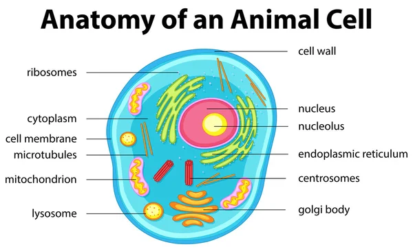
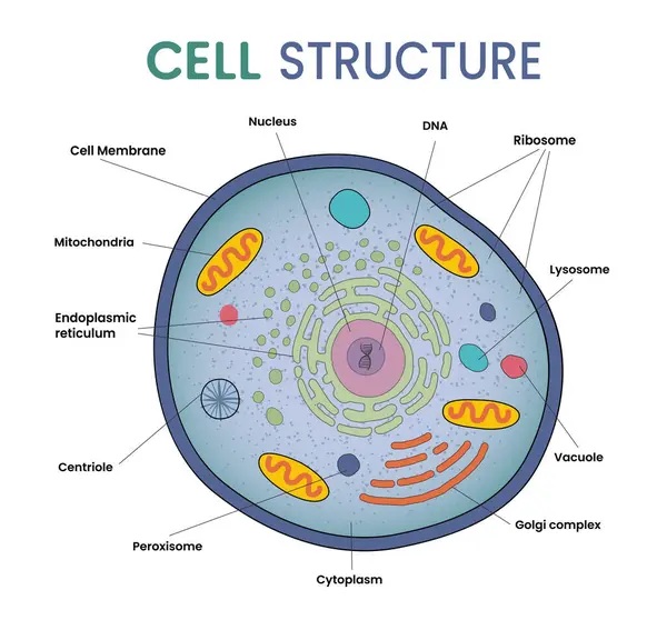
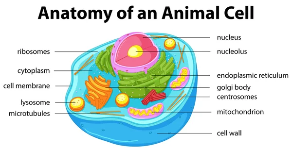
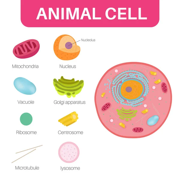
Anatomy of animal cell with words
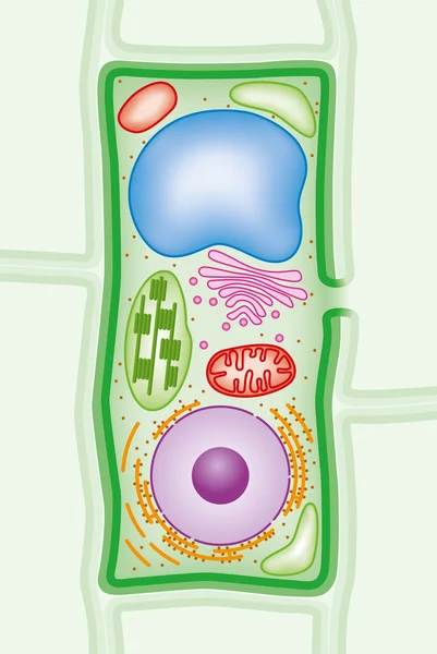
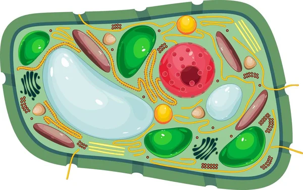
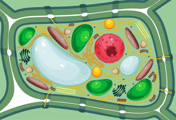
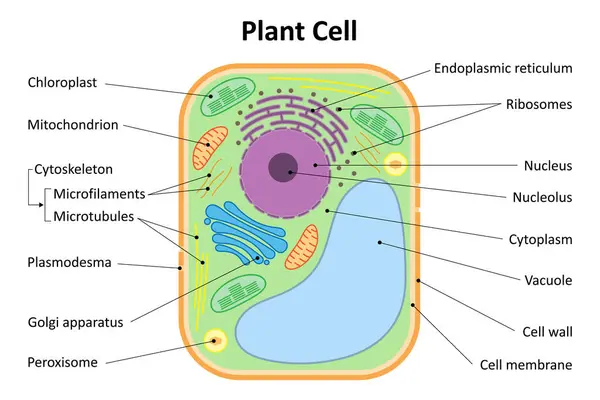
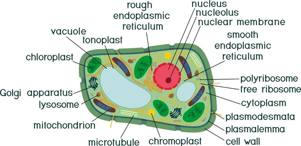
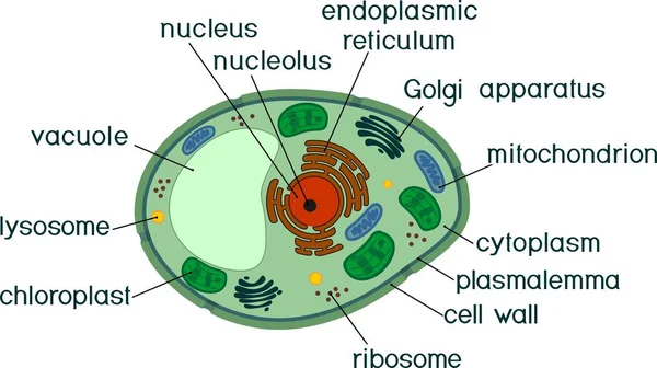
Plant cell
Plant cell
Illustration of cell structure infographic
Illustration Cell Structure Infographic
Plant cell structure cross section illustration
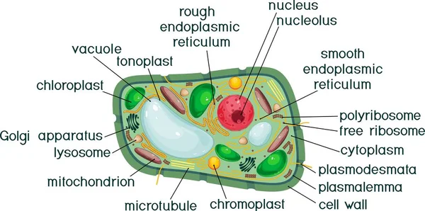
Cell wall structure with plant cellular parts description outline diagram
Structure of plant cell with different organelles
Structure Plant Cell Different Organelles
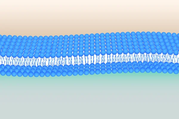
Flexible Phosholipid bilayer
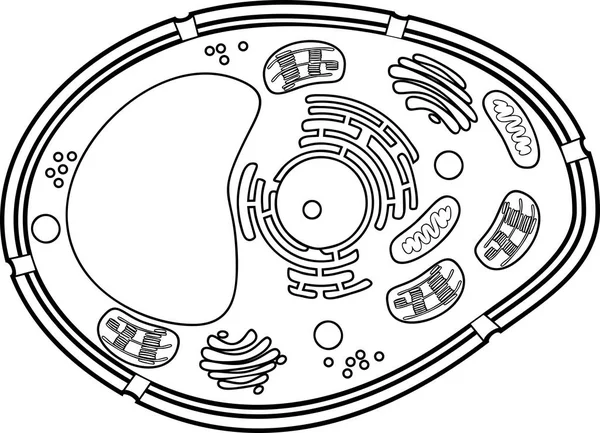
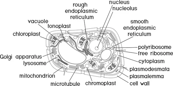
Coloring page. Plant cell structure
Coloring Page Plant Cell Structure
Plant cell isolated on white vector
Green Cells Abstract Vector Background Design
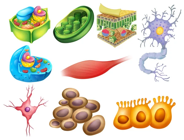
Different biology cells
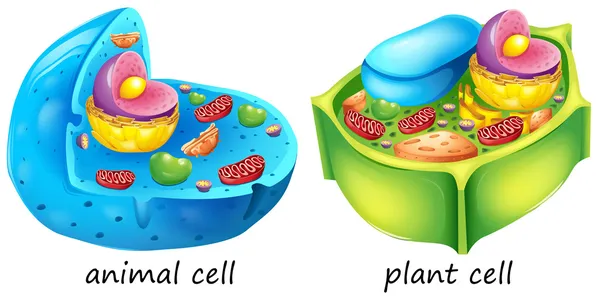
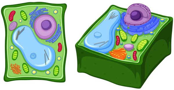
Animal and plant cells
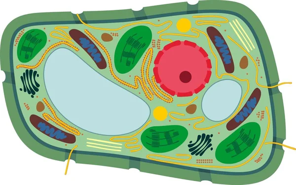
Diagram showing anatomy of plant cell
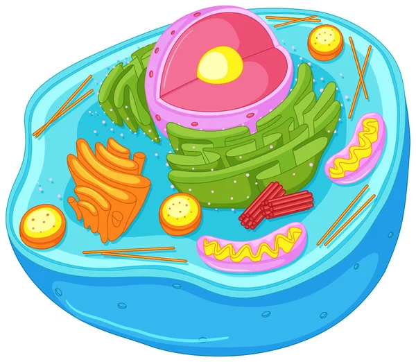
Animal cell in closer look
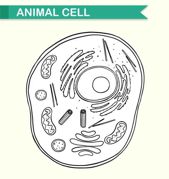
Diagram showing anatomy of animal cell
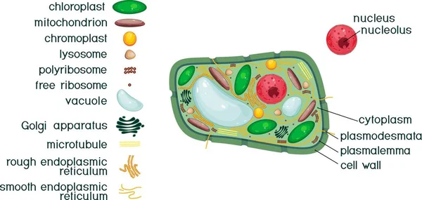
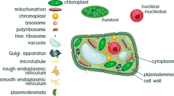
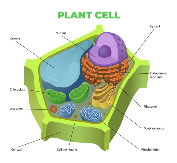
Plant cell structure with titles and different organelles
Plant Cell Structure Titles Different Organelles
Structure of plant cell with different organelles
Structure Plant Cell Different Organelles
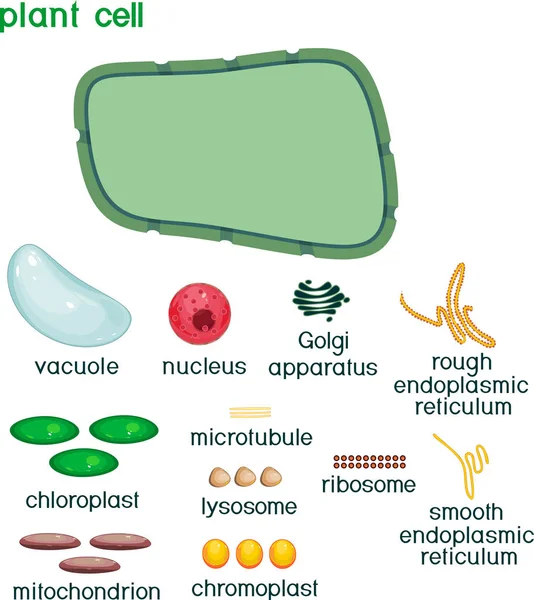
Educational game: assembling cells from ready-made components in form of stickers. Plant cell structure with titles
Educational Game Assembling Cells Ready Made Components Form Stickers Plant Cell Structure Titles
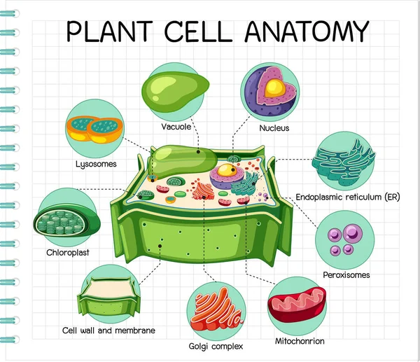
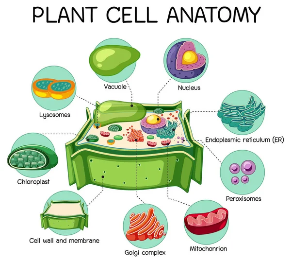
Anatomy of plant cell (Biology Diagram) illustration
Anatomy Plant Cell Biology Diagram Illustration
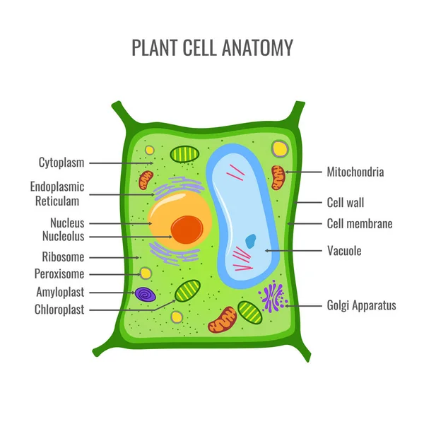
Plant cell anatomy vector illustration
Plant Cell Anatomy Vector Illustration
Colorful vector illustration highlighting plant cell components like nucleus, nucleolus, ribosomes, and endoplasmic reticulum with labeled parts
Colorful Vector Illustration Highlighting Plant Cell Components Nucleus Nucleolus Ribosomes Endoplasmic Reticulum Labeled Parts
Plant cell structure with titles and different organelles
Plant Cell Structure Titles Different Organelles
Anatomy of plant cell (Biology Diagram) illustration
Anatomy Plant Cell Biology Diagram Illustration
Diagram showing animal cell
Colorful vector illustration of plant cell structure with labeled organelles, highlighting cellular components and functions in a clear layout
Colorful Vector Illustration Plant Cell Structure Labeled Organelles Highlighting Cellular Components Functions Clear Layout
Coloring page. Structure of plant cell with titles
Coloring Page Structure Plant Cell Titles
Structure of plant cell with different organelles
Structure Plant Cell Different Organelles
Structure of plant cell with different organelles
Structure Plant Cell Different Organelles
Plant cell anatomy. Vacuole structure. Vector illustration. poster. Schematic diagram
Plant Cell Anatomy Vacuole Structure Vector Illustration Poster Schematic Diagram
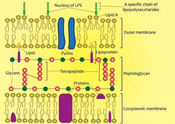
Structure of the cell wall of Gram-negative bacteria (scheme)
Structure Cell Wall Gram Negative Bacteria Scheme
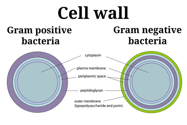
Gram positive versus negative cell wall structure differences outline diagram
Vector illustration of the basic structure of the plant cell.
Vector Illustration Basic Structure Plant Cell
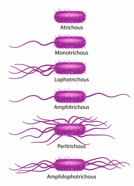
Schematic illustration of bacterial flagella types.
Schematic Illustration Bacterial Flagella Types
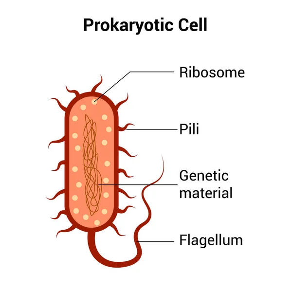
Prokaryotic cell structure diagram vector
Prokaryotic Cell Structure Diagram Vector
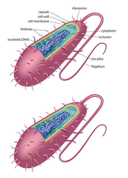
Illustration of bacteria cell diagram
Illustration Bacteria Cell Diagram
Structure of plant cell with different organelles
Structure Plant Cell Different Organelles
Illustration of typical bacterial cell - bacillus type
Illustration Typical Bacterial Cell Bacillus Type
Schematic illustration of difference between bacterial cell wall - gram positive and gram negative.
Schematic Illustration Difference Bacterial Cell Wall Gram Positive Gram Negative
The structure of a plant cell and an animal cell.
Structure Plant Cell Animal Cell
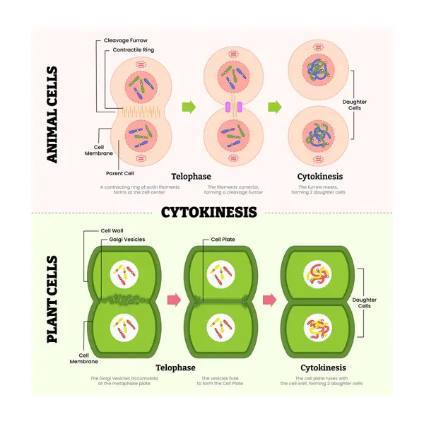
Cytokines in animal cell and plant cell diagram process, produce protein for immune system, labeled telophase, cytokinesis, cleavage furrow, parent and daughter cell division. Vector illustration
Cytokines Animal Cell Plant Cell Diagram Process Produce Protein Immune System Labeled Telophase Cytokinesis Cleavage Furrow Parent Daughter Cell Division Vector Illustration
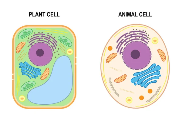
Educational game: assembling cells from ready-made components in form of stickers. Plant cell structure with titles
Educational Game Assembling Cells Ready Made Components Form Stickers Plant Cell Structure Titles
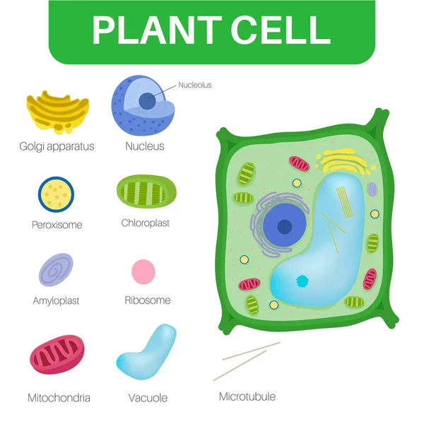
Plant cell structure with titles and different organelles
Plant Cell Structure Titles Different Organelles
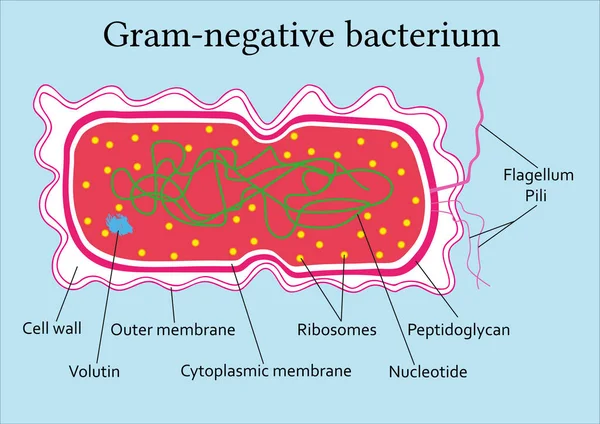
Structure of gram-negative bacterium
Structure Gram Negative Bacterium
Anatomy of plant cell (Biology Diagram) illustration
Anatomy Plant Cell Biology Diagram Illustration
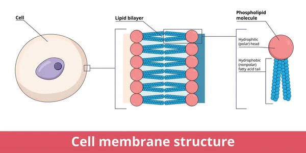
Cell membrane structure, that is represented by lipid bilayer and its phosphatidylcholine (a phospholipid), that is composed of polar hydrophilic head and nonpolar hydrophobic tail.
Cell Membrane Structure Represented Lipid Bilayer Its Phosphatidylcholine Phospholipid Composed Polar Hydrophilic Head Nonpolar Hydrophobic Tail
Diagram showing plant cell
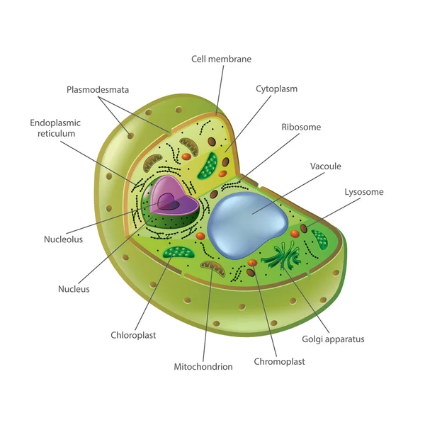
Structure of a plant cell. Plant cell organelles. Diagram.
Structure Plant Cell Plant Cell Organelles Diagram
Plant cell structure with titles
Plant Cell Structure Titles
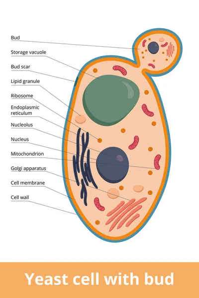
Detailed scheme of yeast cell with bud and scar. Yeast cell as basic representation of fungus cell.
Detailed Scheme Yeast Cell Bud Scar Yeast Cell Basic Representation Fungus Cell
Coloring page. Plant cell structure with titles
Coloring Page Plant Cell Structure Titles
Plant cell structure with titles
Plant Cell Structure Titles
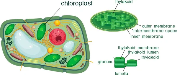
Plant cell and chloroplast structure with titles
Plant Cell Chloroplast Structure Titles
Plant Cell structure, cross section of the cell detailed colorfu
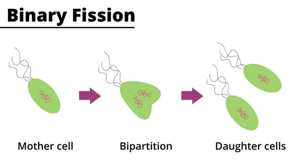
Binary fission. Asexual reproduction of some unicellular organisms. Vector illustration. Didactic illustration.
Binary Fission Asexual Reproduction Some Unicellular Organisms Vector Illustration Didactic Illustration
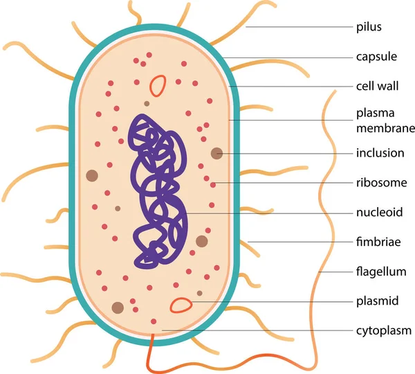
Typical prokaryotic cell. Scheme of typical bacteria
Typical Prokaryotic Cell Scheme Typical Bacteria
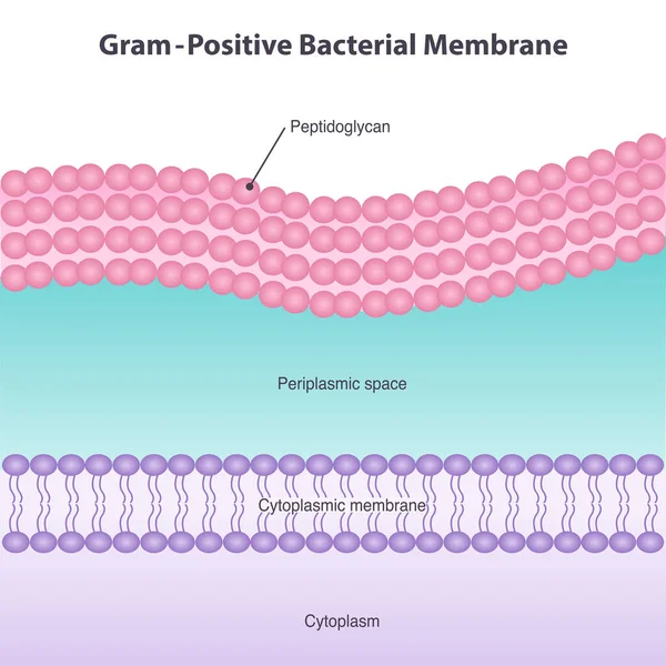
Gram-Positive Bacterial Membrane Diagram Illustration
Gram Positive Bacterial Membrane Diagram Illustration
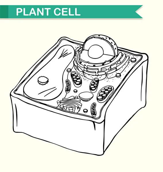
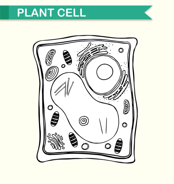
Diagram showing plant cell in black and white
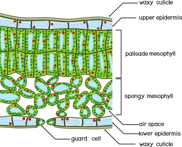
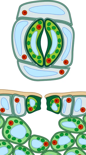
Sectional diagram of plant leaf structure. Cross-section through a leaf
Sectional Diagram Plant Leaf Structure Cross Section Leaf
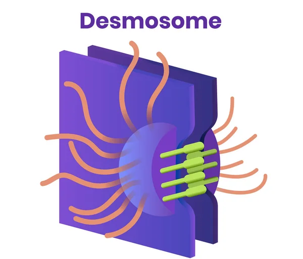
Desmosome vector. Illustration of the tight cell junction
Photoreceptor cell (rod).Anatomy of a rod, including the nucleus, inner and outer segments, and membrane disks.
Photoreceptor Cell Rod Anatomy Rod Including Nucleus Inner Outer Segments Membrane Disks
Structure of gram-positive bacterium
Structure Gram Positive Bacterium
Illustration of plant cell diagram
Illustration Plant Cell Diagram
Plant cells are eukaryotic cells.
Plant Cells Eukaryotic Cells
Difference Bacteria cell or prokaryote structure anatomy versus Archaea or archaebacteria structure anatomy, labeled fimbriae, plasmid, capsule, flagellum, pilus, ribosome, chromosome.
Difference Bacteria Cell Prokaryote Structure Anatomy Archaea Archaebacteria Structure Anatomy Labeled Fimbriae Plasmid Capsule Flagellum Pilus Ribosome Chromosome
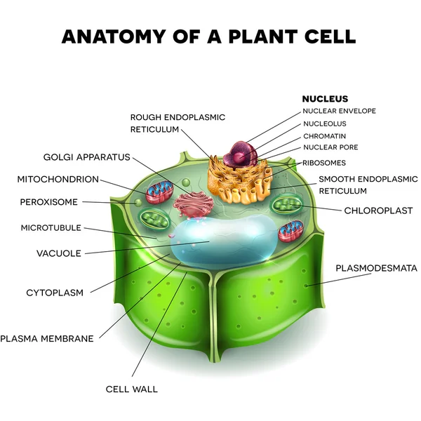
Plant cell anatomy structure with organelles. Anatomy of Plant cell cross section. Cell biology. Nucleus, Smooth and Rough endoplasmic reticulum, Golgi apparatus, Mitochondria and Chloroplast.
Plant Cell Anatomy Structure Organelles Anatomy Plant Cell Cross Section Cell Biology Nucleus Smooth Rough Endoplasmic Reticulum Golgi Apparatus Mitochondria Chloroplast
An animal cell is a type of eukaryotic cell.
Animal Cell Type Eukaryotic Cell
Colorful vector illustration of plant cell organelles with labels, showcasing detailed cellular structures and vibrant colors
Colorful Vector Illustration Plant Cell Organelles Labels Showcasing Detailed Cellular Structures Vibrant Colors
Fungi anatomy. Mushroom, mycelium, hyphae. Chitin and Structure of fungal cell wall. Close-up of Filamentous mycelium. High-quality vector illustration for educational use. Poster. Schematic diagram.
Fungi Anatomy Mushroom Mycelium Hyphae Chitin Structure Fungal Cell Wall Close Filamentous Mycelium High Quality Vector Illustration Educational Use Poster Schematic Diagram
Anatomy of plant cell (Biology Diagram) illustration
Anatomy Plant Cell Biology Diagram Illustration
Anatomy of plant cell (Biology Diagram) illustration
Anatomy Plant Cell Biology Diagram Illustration
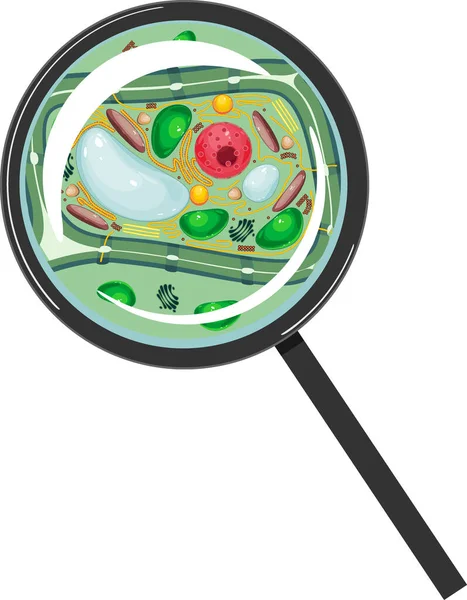
Plant cell under magnifying glass isolated on white background
Plant Cell Magnifying Glass Isolated White Background
Plant Cell Color Diagram of organelles inside the cell wall for science and biology concepts.
Plant Cell Color Diagram Organelles Cell Wall Science Biology Concepts
Colorful vector diagram of plant cell anatomy with labeled organelles for scientific and educational use.
Colorful Vector Diagram Plant Cell Anatomy Labeled Organelles Scientific Educational Use
Bacteria cell wall illustration. Gram positive and gram negative cell wall differents.
Bacteria cell wall  illustration. Gram positive and gram negative cell wall differents.
Section view of stomate and plant leaf structure. Cross-section through a leaf
Section View Stomate Plant Leaf Structure Cross Section Leaf
Plant cell structure with titles
Plant Cell Structure Titles
Plant cell. Structure and Anatomy. Close-up of layers of a cell wall. Vector illustration
Plant Cell Structure Anatomy Close Layers Cell Wall Vector Illustration
Gram negative bacteria do not retain the crystal violet stain. Gram positive bacteria give a positive result in the Gram stain test. comparison and difference type of bacterial cell wall. vector poster
Gram Negative Bacteria Retain Crystal Violet Stain Gram Positive Bacteria Give Positive Result Gram Stain Test Comparison Difference Type Bacterial Cell Wall Vector Poster
Close up diagram of plant cell
Close up diagram of animal cell
Bacteria prokaryote cartoon character speaking on phone. Vector illustration
Bacteria Prokaryote Cartoon Character Speaking Phone Vector Illustration
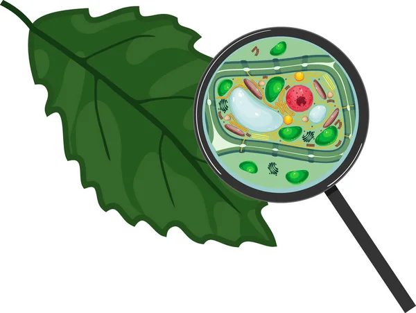
Green leaf and plant cell under magnifying glass isolated on white background
Green Leaf Plant Cell Magnifying Glass Isolated White Background
Plant cell and green leaf
Plant cell in three dimensions
HELICOBACTER BACTERIA Medicine Education Diagram Vector Scheme Human Illness Vector Illustration Set
Helicobacter Bacteria Medicine Education Diagram Vector Scheme Human Illness Vector Illustration Set
Coloring page. Structure of plant cell
Coloring Page Structure Plant Cell
AMOEBA DYSENTERY
AMOEBA DYSENTERY
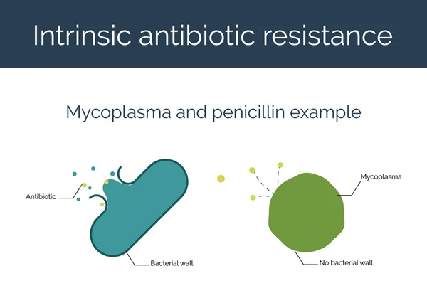
Intrinsic antibiotic resistance vector illustration.
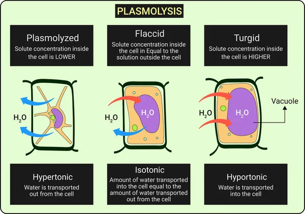
Plasmolysis is the process in which cells lose water in a hypertonic solution. The reverse process, deplasmolysis or cytolysis
Plasmolysis Process Which Cells Lose Water Hypertonic Solution Reverse Process Deplasmolysis Cytolysis
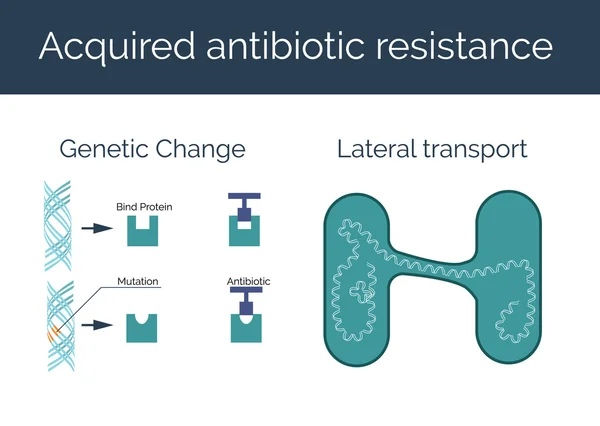
Acquired antibiotic resistance vector illustration.
Stomatal complex and section view of stomate and plant leaf structure
Stomatal Complex Section View Stomate Plant Leaf Structure
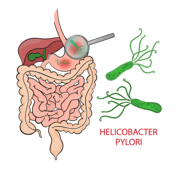
HELICOBACTER PYLORI Medicine Education Diagram Vector Scheme Human Illness Vector Illustration Set
Helicobacter Pylori Medicine Education Diagram Vector Scheme Human Illness Vector Illustration Set
Set of 8 scientific thin outline icons such as atoms, germs, dna, chemistry, cell wall, power, chemistry, cellular
Set Scientific Thin Outline Icons Atoms Germs Dna Chemistry Cell Wall Power Chemistry Cellular
TREE
TREE
Mascot design of smiling Fitness exercise bacteria spirilla lift up barbells. Vector illustration
Mascot Design Smiling Fitness Exercise Bacteria Spirilla Lift Barbells Vector Illustration
Gram Positive and Gram Negative Bacteria Microscopy Comparison Diagram for Microbiology Science Education
Gram Positive Gram Negative Bacteria Microscopy Comparison Diagram Microbiology Science Education
Detailed vector illustration of a prokaryotic bacterium showing internal DNA, ribosomes, and external flagellum.
Detailed Vector Illustration Prokaryotic Bacterium Showing Internal Dna Ribosomes External Flagellum
Educational vector illustration of a fungal cell showing organelles and internal structures on white background
Educational Vector Illustration Fungal Cell Showing Organelles Internal Structures White Background
Step-by-Step Gram Staining Procedure Diagram for Microbiology Laboratory Cell Wall Differentiation
Step Step Gram Staining Procedure Diagram Microbiology Laboratory Cell Wall Differentiation
Minimalist Outline Line Art Plant Cell Structure Diagram With Organelles Biology Education Icon Symbol
Minimalist Outline Line Art Plant Cell Structure Diagram Organelles Biology Education Icon Symbol
Page