Sklera Stock Vector Images

360 Sklera vector art & graphics are available under a royalty-free license
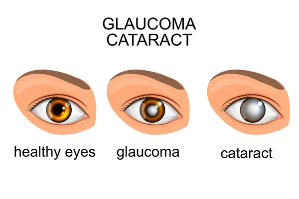
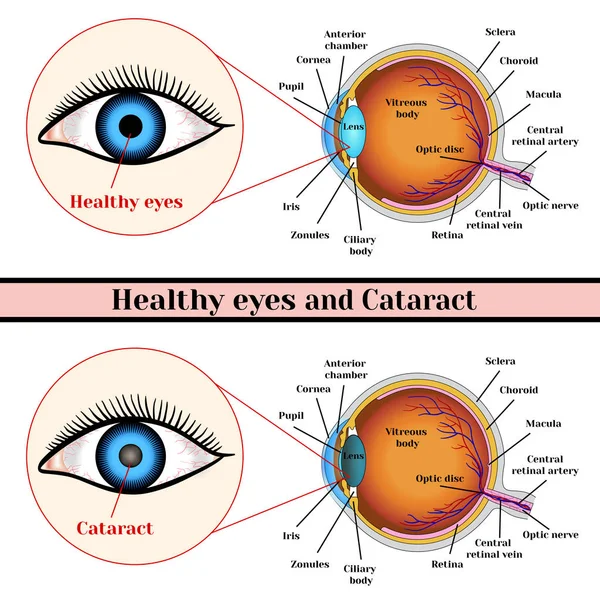
Healthy eyes, glaucoma, cataracts
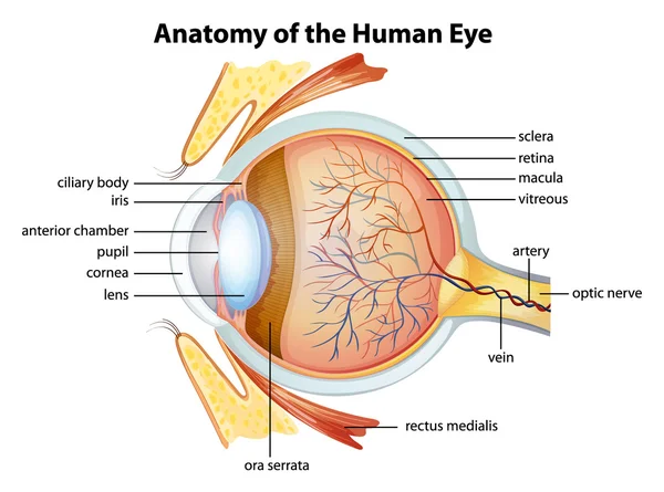
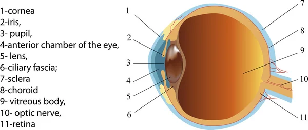
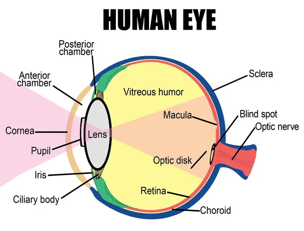
Human eye anatomy
Human eye anatomy
Blepharitis. Purulent, inflammatory eyes disease.
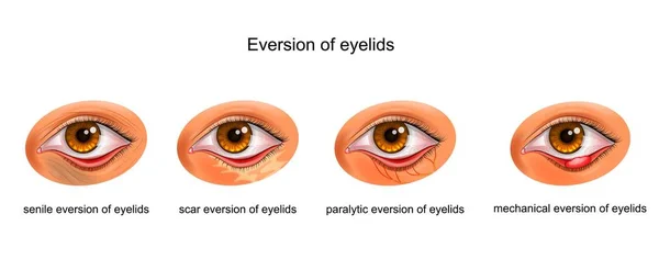
The causes of eversion of eyelids
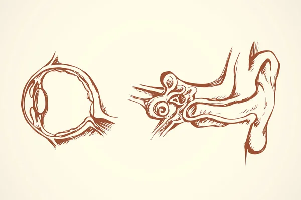
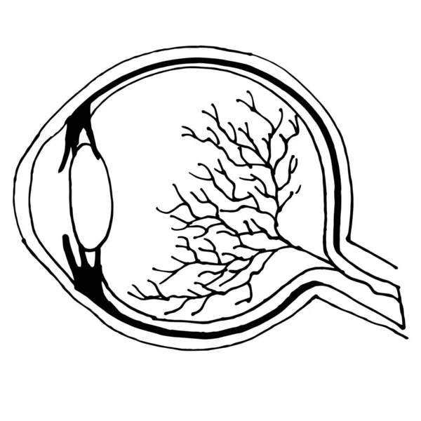
Eye section. Vector drawing
Eyeball x ray vector cartoon illustration
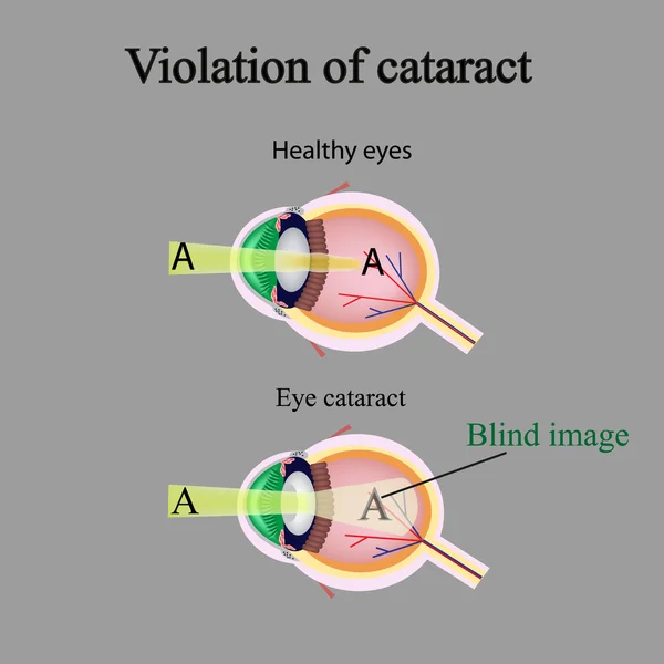
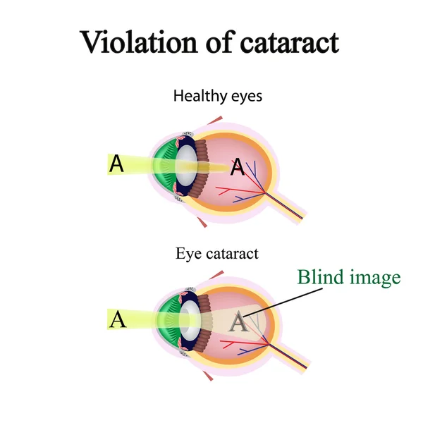
The structure of the eye. Cataracts. As the affected eye sees cataracts. Violation of cataract
Jaundice. The Comparison between Healthy person and person with Icterus. yellowish pigmentation of the skin and sclera due to high bilirubin levels. health problems. vector illustration
Jaundice Comparison Healthy Person Person Icterus Yellowish Pigmentation Skin Sclera Due High Bilirubin Levels Health Problems Vector Illustration
Extraocular muscles anatomy. Structure of the human eye infographic. control the movements of the eyeball and the superior eyelid. Iris, outermost, retina and sclera medical flat vector illustration
Extraocular Muscles Anatomy Structure Human Eye Infographic Control Movements Eyeball Superior Eyelid Iris Outermost Retina Sclera Medical Flat Vector Illustration
Halloween Bloody Eyeball
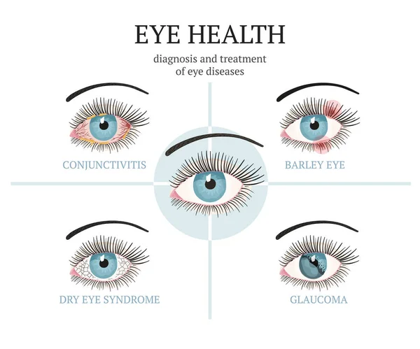
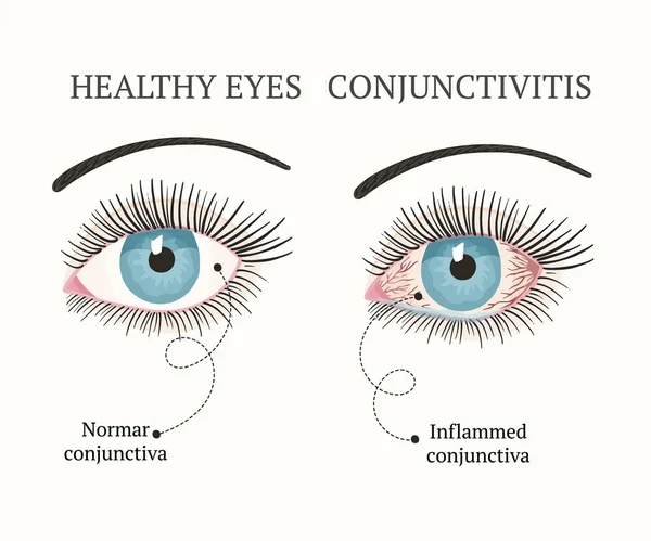
Most common eye problems - conjunctivitis, glaucoma, dry eye syndrome, barley eyes.
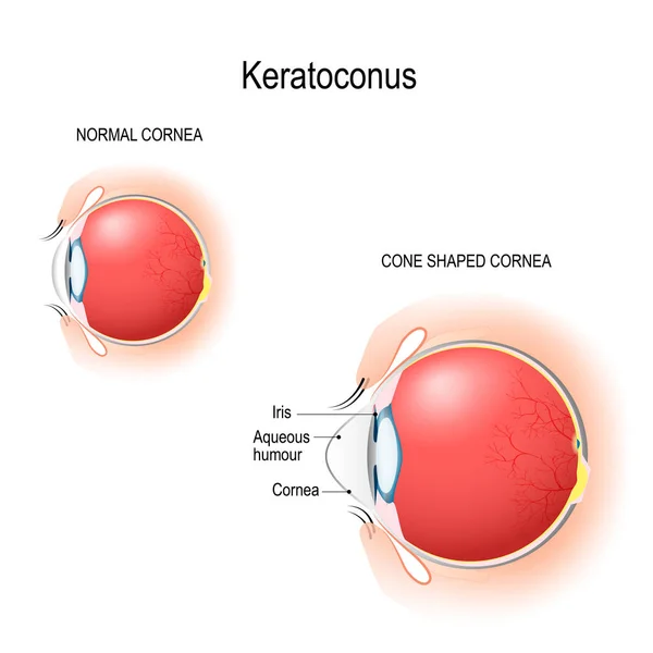
Keratoconus. Normal cornea and cone shaped cornea. Anatomy of the human eye. Vertical section of the eye and eyelids. Schematic diagram. detailed illustration. for biological, science, and medical use.
Keratoconus Normal Cornea Cone Shaped Cornea Anatomy Human Eye Vertical Section Eye Eyelids Schematic Diagram Detailed Illustration Biological Science Medical Use
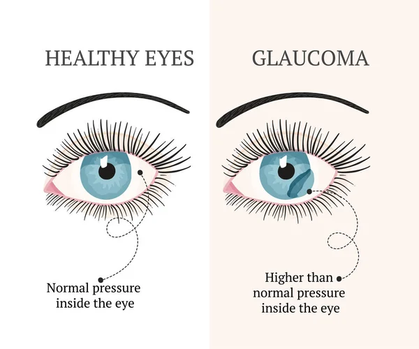
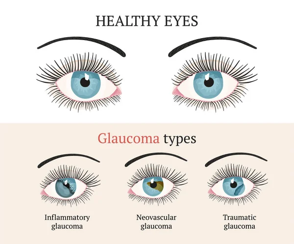
Normal vision. Glaucoma.
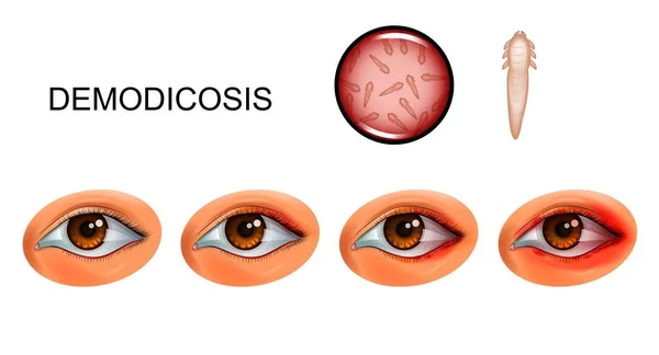
Vector illustration of demodecosis. tick Demodex on eyelashes
Vector Illustration Demodecosis Tick Demodex Eyelashes
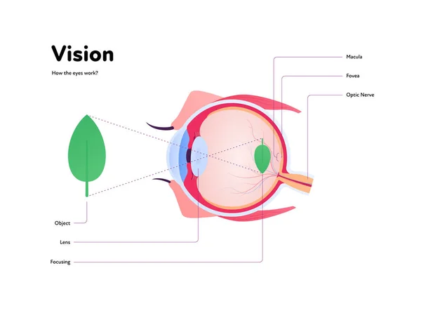
Human eye anatomy and vision medical infographic poster. Vector healthcare illustration. Side view of human eyeball with text isolated on white. Design for ophthalmology
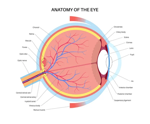
Human Eye Anatomy Vision Medical Infographic Poster Vector Healthcare Illustration Side View Human Eyeball Text Isolated White Design Ophthalmology
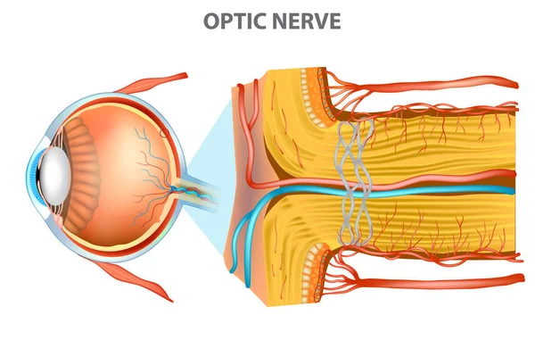
The Optic Nerve. Anatomy of the Eye
Optic Nerve Anatomy Eye
Human eye anatomy illustration. Parts of the eye, labeled vector illustration diagram. Eyelid, eyelashes, pupil, lacrimal gland and other anatomical parts. Realistic 3d vector isolated
Human Eye Anatomy Illustration Parts Eye Labeled Vector Illustration Diagram Eyelid Eyelashes Pupil Lacrimal Gland Other Anatomical Parts Realistic Vector Isolated
Cartoon Bloodshot Eyeball Character with Red Veins and Black Pupil
Cartoon Bloodshot Eyeball Character Red Veins Black Pupil
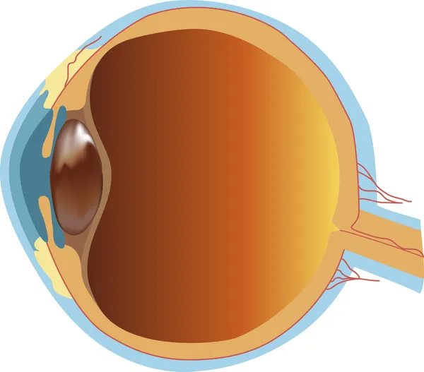
Structure of human eye (section)
Human eye
Human eye
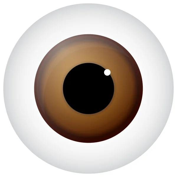
Brown Eye
Brown Eye
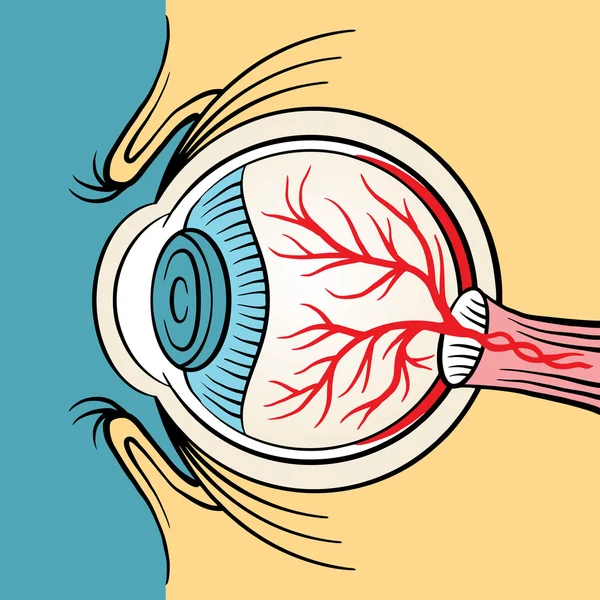
Vector image of eyeball anatomy
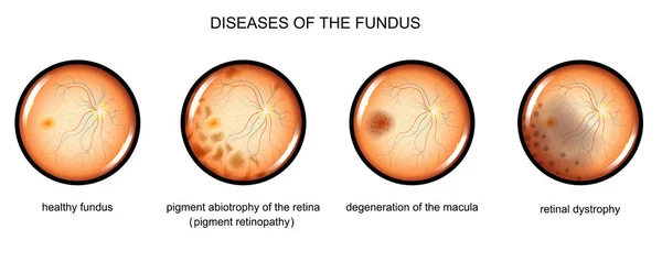
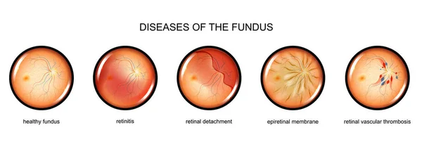
Degenerative diseases of the fundus
Normal eye anatomy background
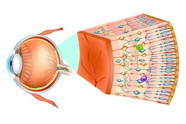
Human eye anatomy. Retina structure. Cross-section of the eye. Cells in section of the retina. Detailed vector poster
Human Eye Anatomy Retina Structure Cross Section Eye Cells Section Retina Detailed Vector Poster
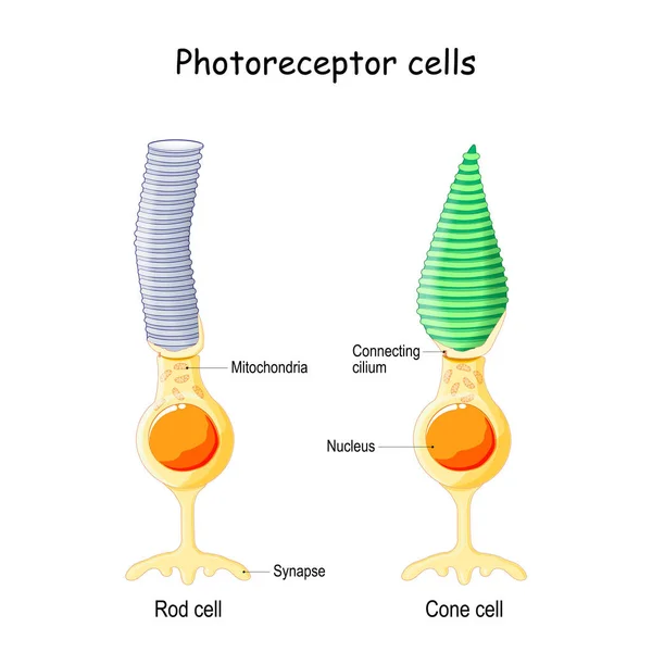
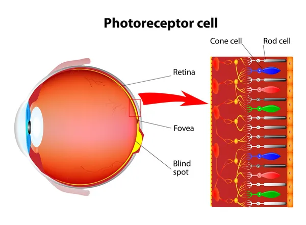
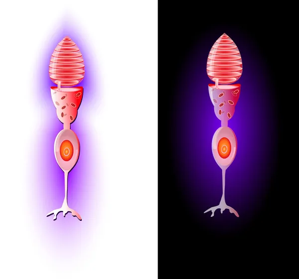
Anatomy of Photoreceptor. cell of a retina in the eye. Cone cells in respond to color vision and send signals to brain. Rod cells are used in peripheral vision
Anatomy Photoreceptor Cell Retina Eye Cone Cells Respond Color Vision Send Signals Brain Rod Cells Used Peripheral Vision
Photoreceptor cells in the retina of the eye
Extraocular muscles anatomy. Structure of the human eye infographic. control the movements of the eyeball and the superior eyelid. Iris, outermost, retina and sclera medical flat vector illustration
Extraocular Muscles Anatomy Structure Human Eye Infographic Control Movements Eyeball Superior Eyelid Iris Outermost Retina Sclera Medical Flat Vector Illustration
Human Eye Anatomy Diagram With Detailed Cross Section Vector Colorful Eye Organ Infographic For Biology And Science Study
Human Eye Anatomy Diagram Detailed Cross Section Vector Colorful Eye Organ Infographic Biology Science Study
Herpes simplex keratitis HSV1 infection detailed infographic poster showing human eyeball cross section anatomy with highlighted focus zone over corneal lesion vector illustration.
Herpes Simplex Keratitis Hsv1 Infection Detailed Infographic Poster Showing Human Eyeball Cross Section Anatomy Highlighted Focus Zone Corneal Lesion Vector Illustration
Human eye anatomy labeled diagram, educational medical poster design healthcare scheme, tarsal plate, ocular structure. Labeled cornea, iris, lens, retina, choroid, optic nerve, sclera, eyelid
Human Eye Anatomy Labeled Diagram Educational Medical Poster Design Healthcare Scheme Tarsal Plate Ocular Structure Labeled Cornea Iris Lens Retina Choroid Optic Nerve Sclera Eyelid
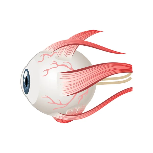
Eyeball muscles symbol. Eye anatomy in side view. Vector illustration in cartoon style isolated on white background
Hyposphagma of eyes
Hyposphagma of eyes
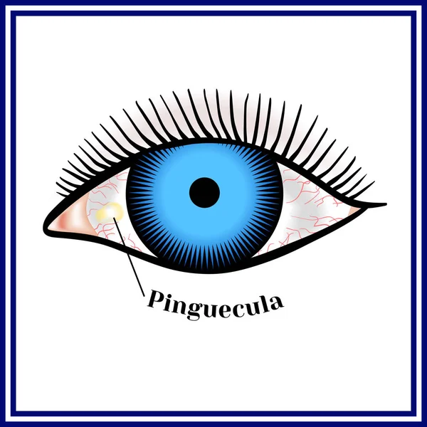
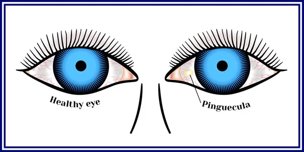
Pinguecula. Benign neoplasm of yellow color.
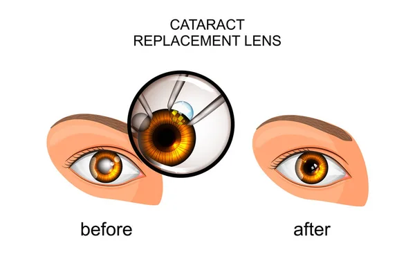
Replacement of the crystalline lens in cataract
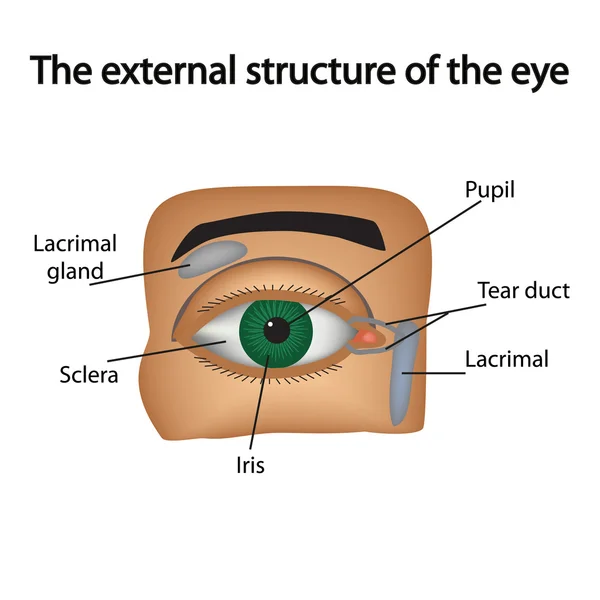
The external structure of the eye. Vector illustration..
Eye anatomy poster. Structure of the human eye infographic. Outermost, retina and sclera. Pigmented choroid, first lens and iris. Extraocular muscles, blood vessels and optic nerve medical flat vector
Eye Anatomy Poster Structure Human Eye Infographic Outermost Retina Sclera Pigmented Choroid First Lens Iris Extraocular Muscles Blood Vessels Optic Nerve Medical Flat Vector
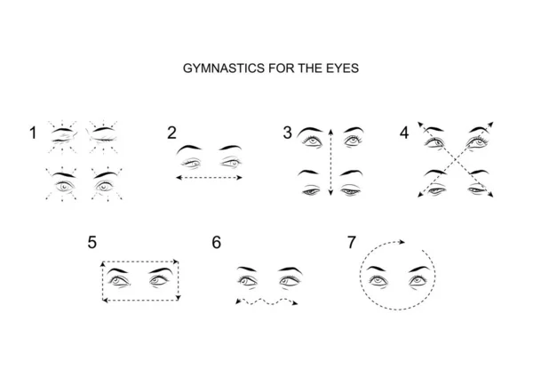
Gymnastics for the eyes
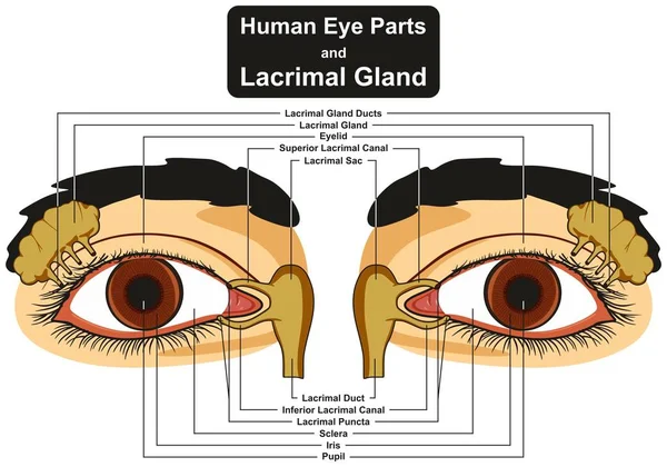
Human Eye Parts and Lacrimal Gland infographic diagram including pupil iris sclera canal duct sac eyelid for medical science education and health care
Human Eye Parts Lacrimal Gland Infographic Diagram Including Pupil Iris Sclera Canal Duct Sac Eyelid Medical Science Education Health Care
Eye. Medicine 3d vector icon
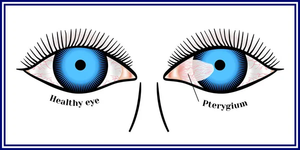
Pterygium. Spreading of the conjunctiva of the eye on the cornea
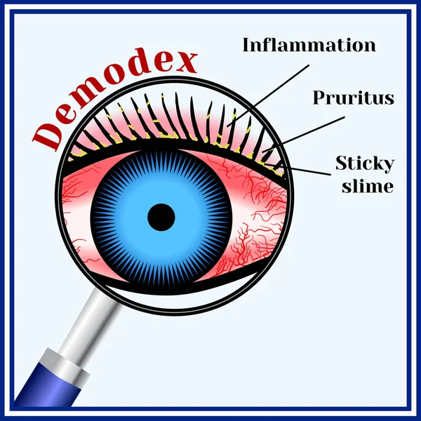
Demodex. Parasitic eye disease.
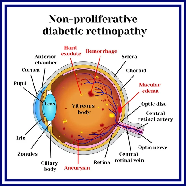
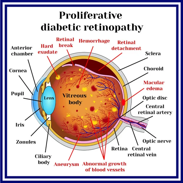
Types of diabetic retinopathy
Extraocular muscles anatomy. Structure of the human eye infographic. control the movements of the eyeball and the superior eyelid. Iris, outermost, retina and sclera medical flat vector illustration
Extraocular Muscles Anatomy Structure Human Eye Infographic Control Movements Eyeball Superior Eyelid Iris Outermost Retina Sclera Medical Flat Vector Illustration
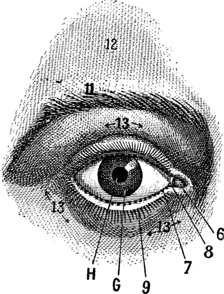
External View of the Human Eye, showing pupil, iris, sclera and eyelid, vintage engraved illustration. Dictionary of Words and Things - Larive and Fleury - 1895
External View Human Eye Showing Pupil Iris Sclera Eyelid Vintage Engraved Illustration Dictionary Words Things Larive Fleury 1895
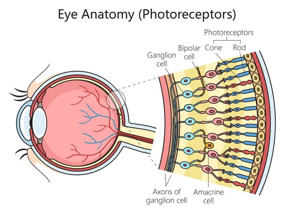
Human eye photoreceptor cell structure scheme diagram schematic vector illustration. Medical science educational illustration
Human Eye Photoreceptor Cell Structure Scheme Diagram Schematic Vector Illustration Medical Science Educational Illustration
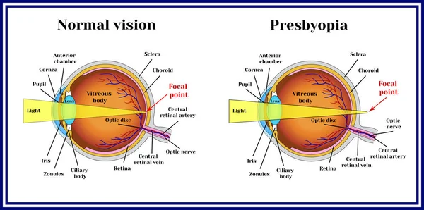
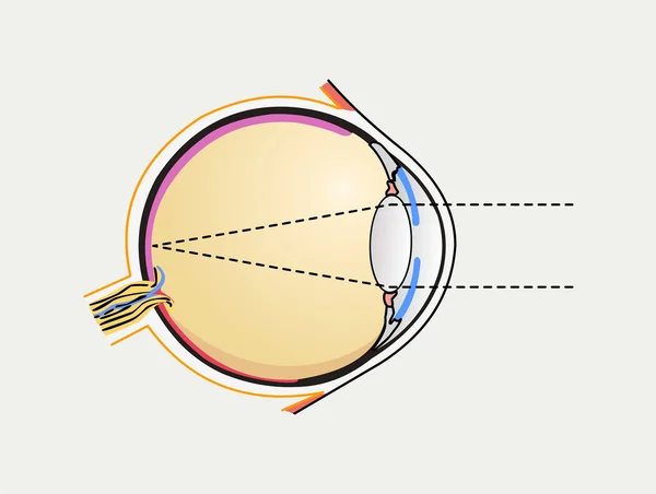
Refractive errors eyeball. Presbyopia.
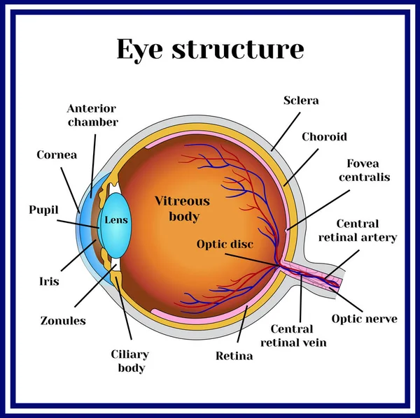
Eyeball structure. Medicine.
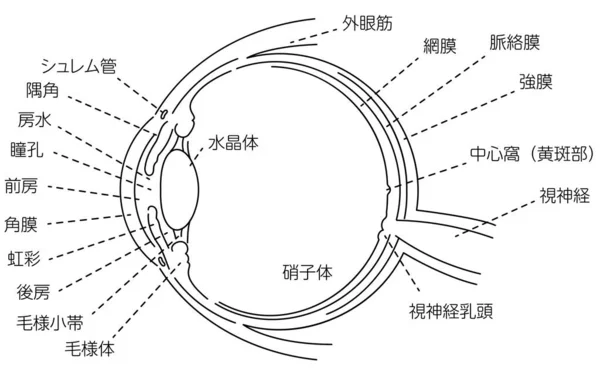
Illustration of the eye - Translation: Schlemm's canal, corner angle, aqueous humor, pupil, anterior chamber, cornea, iris, posterior chamber, ciliary body, ciliary body, lens, vitreous body, external eye muscle, retina, choroid, sclera, central foss
Illustration Eye Translation Schlemm Canal Corner Angle Aqueous Humor Pupil Anterior Chamber Cornea Iris Posterior Chamber Ciliary Body Ciliary Body Lens Vitreous Body External Eye Muscle Retina Choroid Sclera Central Foss
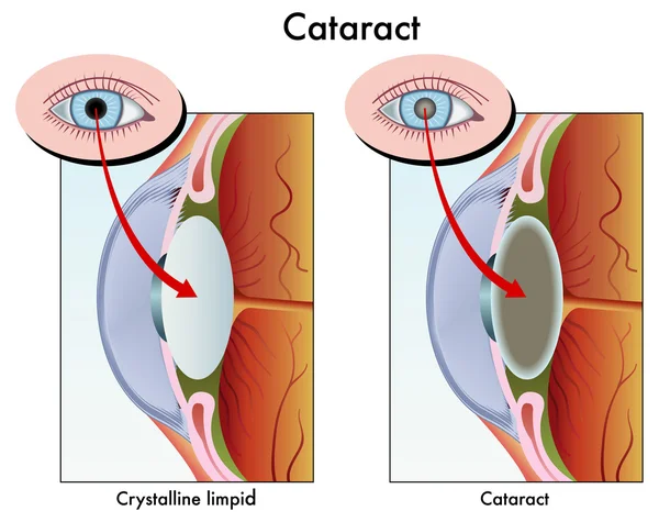
Healthy eyes and cataract (opacity of the eye lens).
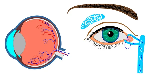
Structure of eyeball and localization lachrymal gland
Structures Of The Human Eye
Human eye anatomy
Human eye anatomy
Jaundice disease vector concept illustration
Jaundice Disease Vector Concept Illustration
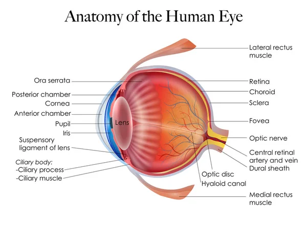
Vector Education poster eye structure. Image human eye anatomy poster
Vector Education Poster Eye Structure Image Human Eye Anatomy Poster
Ophthalmic shingles herpes zoster ophthalmicus infection infographic showing human eye close up view with red blood vessels irritation and eyelid blisters rash vector illustration.
Ophthalmic Shingles Herpes Zoster Ophthalmicus Infection Infographic Showing Human Eye Close View Red Blood Vessels Irritation Eyelid Blisters Rash Vector Illustration
Pinguecula. Benign neoplasm of yellow color.
Inflammatory disease of eyes. itch sensation in the eye
Linear hand drawn illustration of human eye for logotype or banner / design template
Linear Hand Drawn Illustration Human Eye Logotype Banner Design Template
Eye disease. Ophthalmology health illustration.
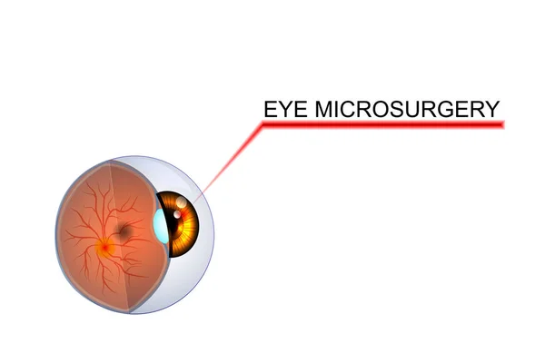
Illustration of the eye, the eyeball. microsurgery in ophthalmol
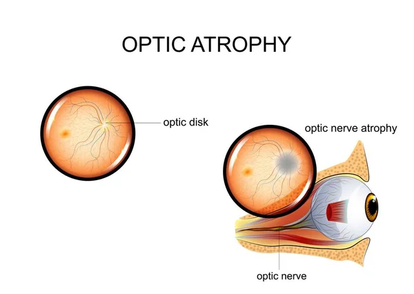
Vector illustration of optic nerve atrophy. eye fundus.
Vector Illustration Optic Nerve Atrophy Eye Fundus
Demodex. Parasitic eye disease.
Eye disease. Ophthalmology health illustration.
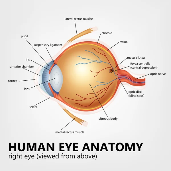
Human eye anatomy, right eye viewed from above
Inflammation of the fundus, retinal detachment
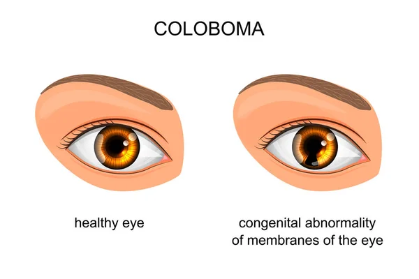
Congenital disorder of membranes of the eye. coloboma
Human Eye Diagram
Human Eye Diagram
Macular degeneration. Age-related dry macular degeneration. AMD. Cross section of human eye. Central vision loss. Close-up of a macula, optic disc, choroid, retina, sclera, and fovea. Vector poster
Macular Degeneration Age Related Dry Macular Degeneration Amd Cross Section Human Eye Central Vision Loss Close Macula Optic Disc Choroid Retina Sclera Fovea Vector Poster
Cataract
Cataract
Colorful Uveitis scheme
Refractive errors eyeball. Astigmatism.
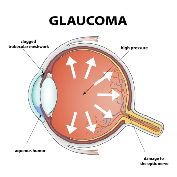
Human eye. Disease glaucoma
Jaundice symptoms infographic in outline style. Yellowing of skin and eyes, caused by bilirubin buildup. Hyperbilirubinemia etiology. Medical concept. Vector illustration isolated on a white
Jaundice Symptoms Infographic Outline Style Yellowing Skin Eyes Caused Bilirubin Buildup Hyperbilirubinemia Etiology Medical Concept Vector Illustration Isolated White
Types of diabetic retinopathy
Human Eyeball Anatomy Set
Diseases of the eye barley. Causes of barley. Demodex mite infestations. Inflammation volosyannoy bulbs
Eye system. Vector design.
Eye Ball Structure. Modern Flat Vector Illustration. Detailed Anatomy of the Healthy Eye and Focal Point. Website Banner. Social Media Template.
Eye Ball Structure Modern Flat Vector Illustration Detailed Anatomy Healthy Eye Focal Point Website Banner Social Media Template
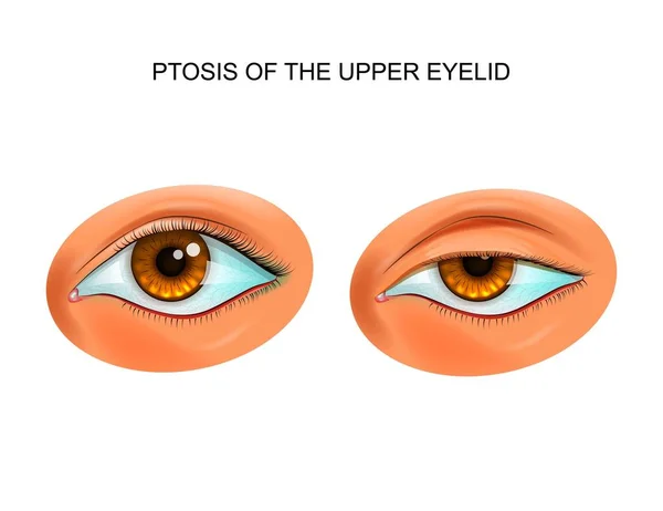
Ptosis of the eyelid
Fundus. retinal tears
Cone cells. Vector
Cone cells. Vector
Funny eye in superhero costume on white background
Funny Eye Superhero Costume White Background
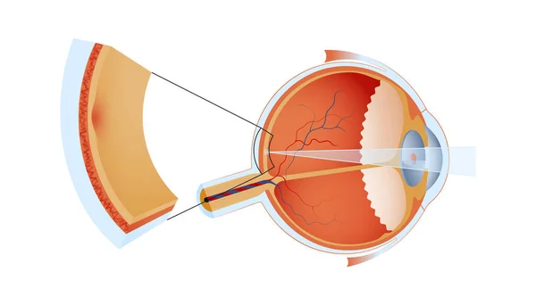
Structure of the human eye and organization of the retina. Optic part of retina.
Structure Human Eye Organization Retina Optic Part Retina
Retinal detachment. Cross section of a normal human eyes and disorder of the eye in which the retina peels. Schematic diagram. Detailed vector illustration
Retinal Detachment Cross Section Normal Human Eyes Disorder Eye Which Retina Peels Schematic Diagram Detailed Vector Illustration
Diagram of human eyeball anatomy illustration
Diagram Human Eyeball Anatomy Illustration
A striking graphic design featuring a stylized, dripping eye with the text ANTI-CONTROL in an arc above it, set against a stark black background. Below the eye, the text CHAOS UNDERGROUND STREETWEAR is visible. The artwork has a grunge, street art ae
Striking Graphic Design Featuring Stylized Dripping Eye Text Control Arc Set Stark Black Background Eye Text Chaos Underground Streetwear Visible Artwork Has Grunge Street Art
Ocular herpes simplex virus infection site infographic layout showing human eyeball cross section anatomy with highlighted red cornea target focus zone vector illustration.
Ocular Herpes Simplex Virus Infection Site Infographic Layout Showing Human Eyeball Cross Section Anatomy Highlighted Red Cornea Target Focus Zone Vector Illustration
Human eye anatomy labeled diagram, educational medical poster design healthcare scheme, cross-sectional ocular structure. Labeled cornea, iris, lens, retina, choroid, optic nerve, sclera, fovea optic
Human Eye Anatomy Labeled Diagram Educational Medical Poster Design Healthcare Scheme Cross Sectional Ocular Structure Labeled Cornea Iris Lens Retina Choroid Optic Nerve Sclera Fovea Optic
Eyeball 3 D icon. Vector sign medical shape.
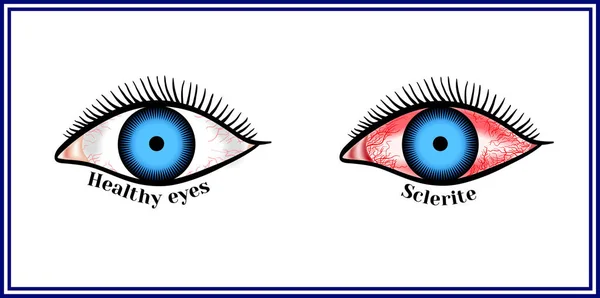
Sclerite. Inflammatory disease of sclera.
Vector eyeballs isolated on white.
Doctor explaining the eyeball
Eye disease. Ophthalmology health illustration.
Eyes scary with veins of halloween
Detailed illustration of the anatomy and structure of the human eye. The picture shows the iris, pupil, lens, retina, optic nerve, and other significant structures of the eye.
Detailed Illustration Anatomy Structure Human Eye Picture Shows Iris Pupil Lens Retina Optic Nerve Other Significant Structures Eye
Sclerite. Inflammatory disease of sclera.
Page