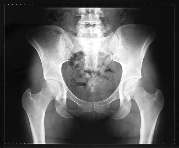
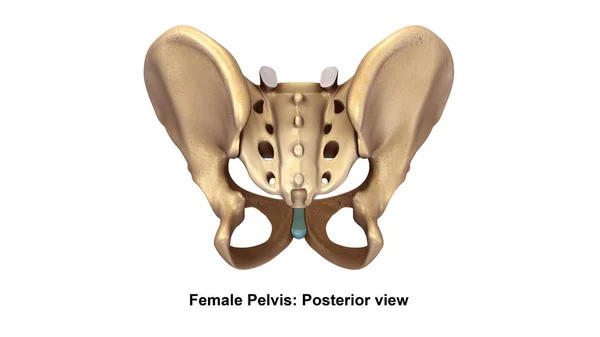
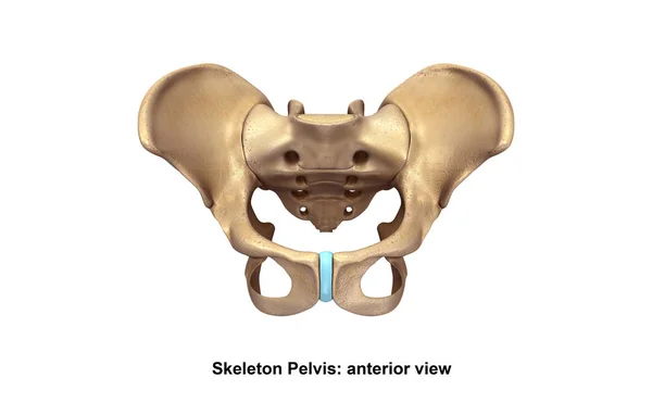
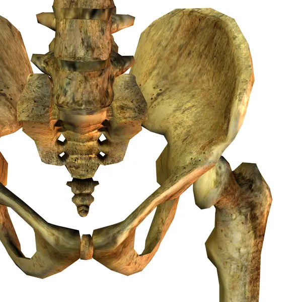
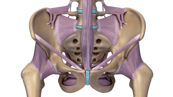
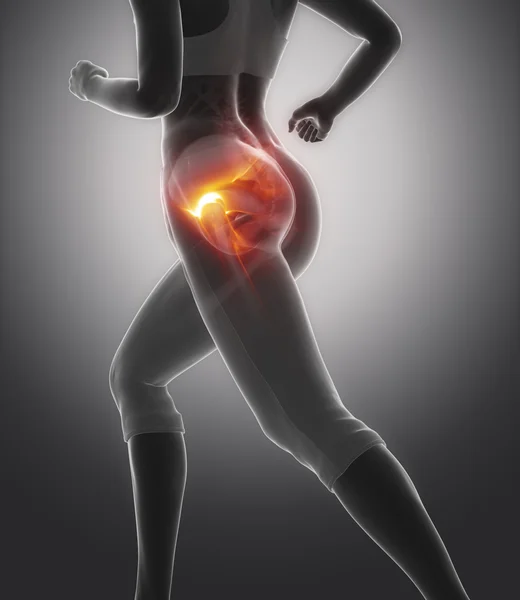
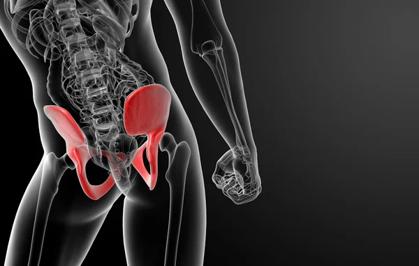
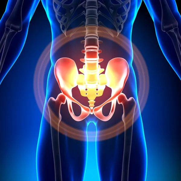
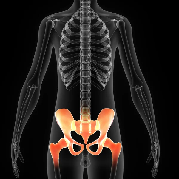
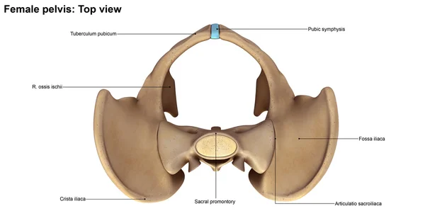
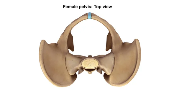
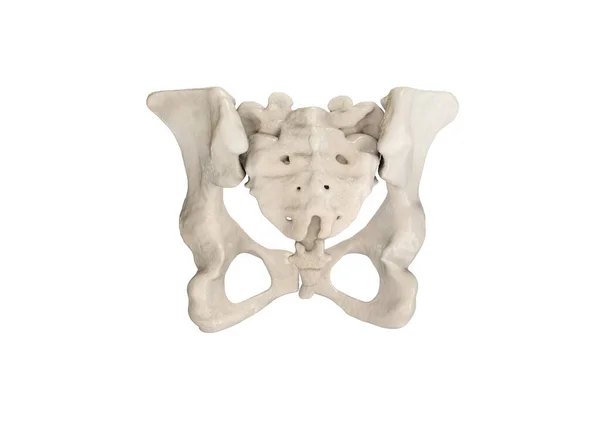
Female Hip, Sacrum, Pubis, Ischium, Ilium - Anatomy Bones
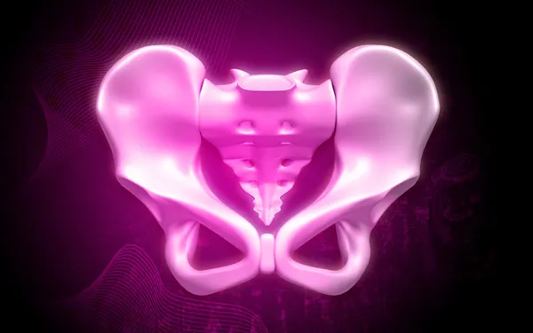
Pelvic hip Girdle anatomy
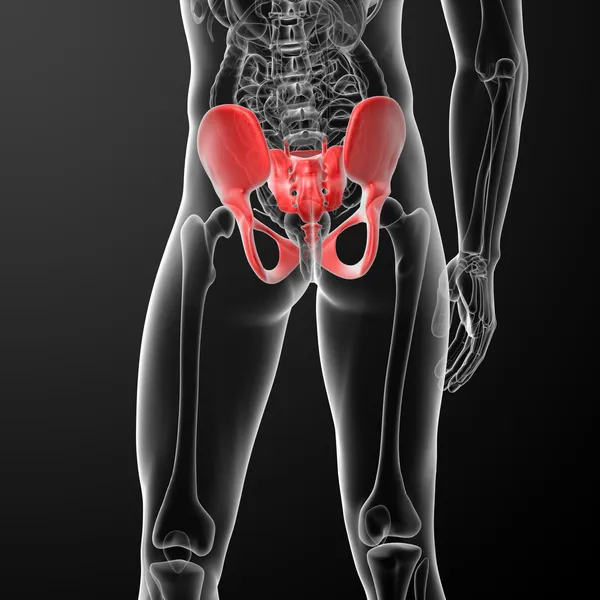
Human Pelvis Skeleton
Cropped shot of obstetrician gynecologist measuring pelvis size of pregnant woman with pelvimeter
Cropped Shot Obstetrician Gynecologist Measuring Pelvis Size Pregnant Woman Pelvimeter
X-ray of the pelvis and spinal column of a woman
Human Pelvis Skeleton
Team of doctors looking at x-ray scan
Skeleton Pelvis illustration
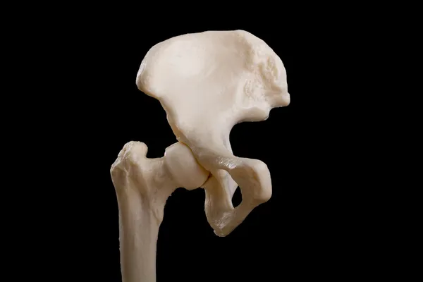
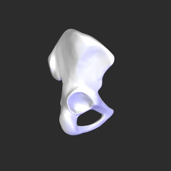
Detail left pelvic bone
Obstetrician gynecologist measuring pelvis size of pregnant woman with Beckenmesser
Obstetrician Gynecologist Measuring Pelvis Size Pregnant Woman Beckenmesser
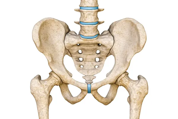
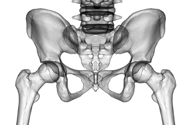
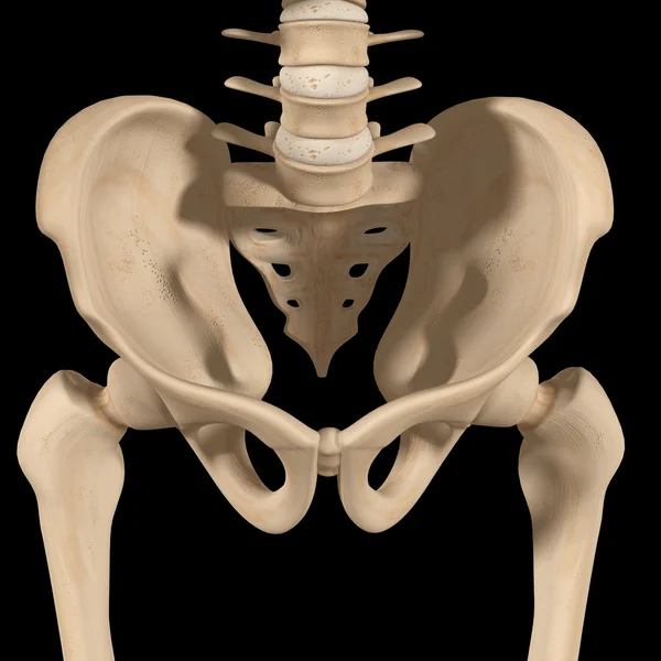
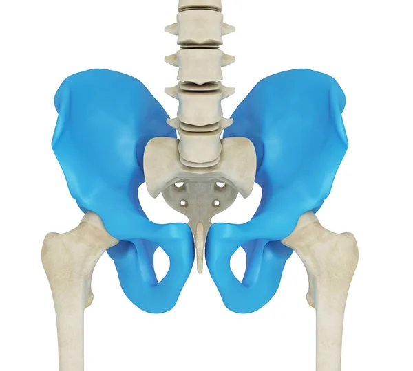
Anterior or front view of human male pelvis, sacrum, lumbar spine and femur bones isolated on white background 3D rendering illustration. Blank anatomical chart. Anatomy, medical, science, part of human skeleton concepts.
Anterior Front View Human Male Pelvis Sacrum Lumbar Spine Femur Bones Isolated White Background Rendering Illustration Blank Anatomical Chart Anatomy Medical Science Part Human Skeleton Concepts
Pregnant woman holding sample of pelvic bones in yoga studio
Pregnant Woman Holding Sample Pelvic Bones Yoga Studio
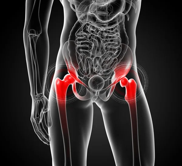
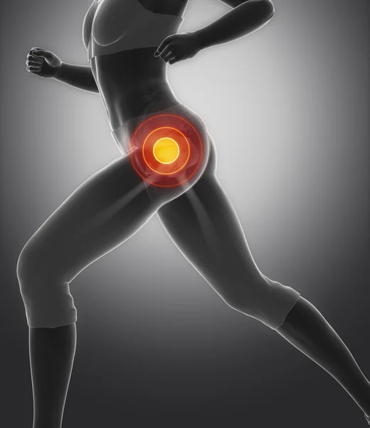
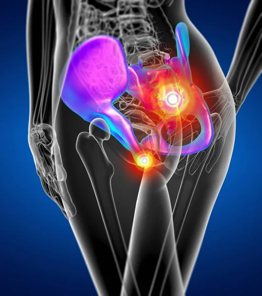
Hip injury in female body
Mature obstetrician gynecologist measuring pelvis size of pregnant woman with pelvimeter
Mature Obstetrician Gynecologist Measuring Pelvis Size Pregnant Woman Pelvimeter
Skeleton hip illustration
Doctor examining his patient pelvis
Team of doctors discussing x-ray scan
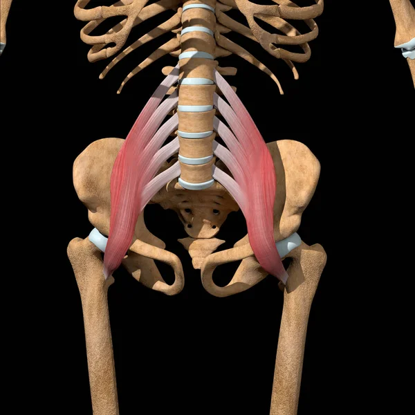
This 3d illustration shows the psoas major muscles on skeleton
Illustration Shows Psoas Major Muscles Skeleton
PELVIS bone scan
PELVIS bone scan
Team of doctors discussing x-ray scan
Medical accurate illustration : Pelvic area anatomy , 3d render
Sporty woman doing pilates
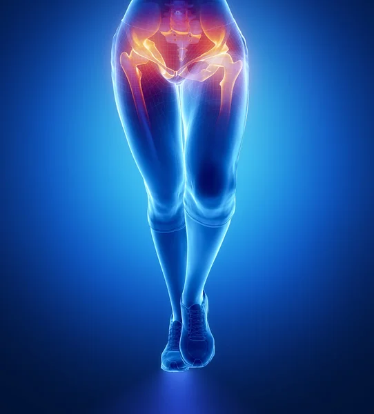
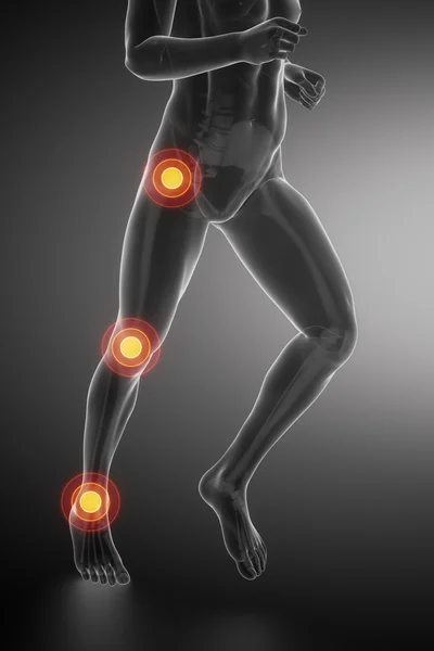
Running woman with hip injury concept
Team of doctors discussing x-ray scan
Pelvic area anatomy , 3d render
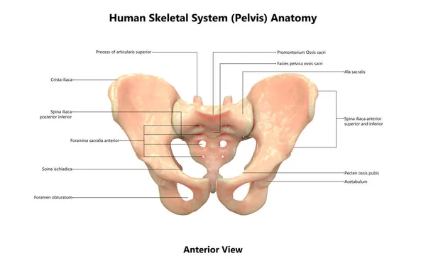
Anatomy of the pelvis bones, including the ilium, ischium, sacrum, and pubis, photorealistic 3D illustration. Front view. Perfect for educational or medical purposes.
Anatomy Pelvis Bones Including Ilium Ischium Sacrum Pubis Photorealistic Illustration Front View Perfect Educational Medical Purposes
Physiotherapist checking patients pelvis alignment
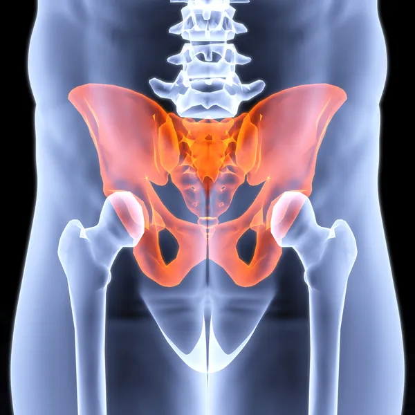
3d render pelvis under the X-rays
3D illustration of Pelvis, medical concept.
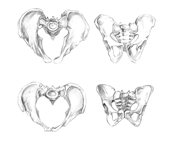
Pelvis 2 angels (male at the top and female at the bottom of drawing)
3D rendering medical illustration of the pelvis bone on blue
Rendering Medical Illustration Pelvis Bone Blue
Skeleton hip illustration
Hip Sacrum Pubis Ischium Ilium - Anatomy Bones
Pelvic hip
Pelvic hip
Doctor examining his patient pelvis
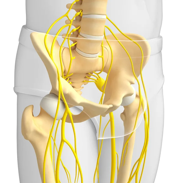
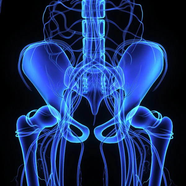
Nervous system of pelvic skeleton artwork
Hip Joint - Anatomy Bones
Female Hip, Sacrum, Pubis, Ischium, Ilium - Anatomy Bones
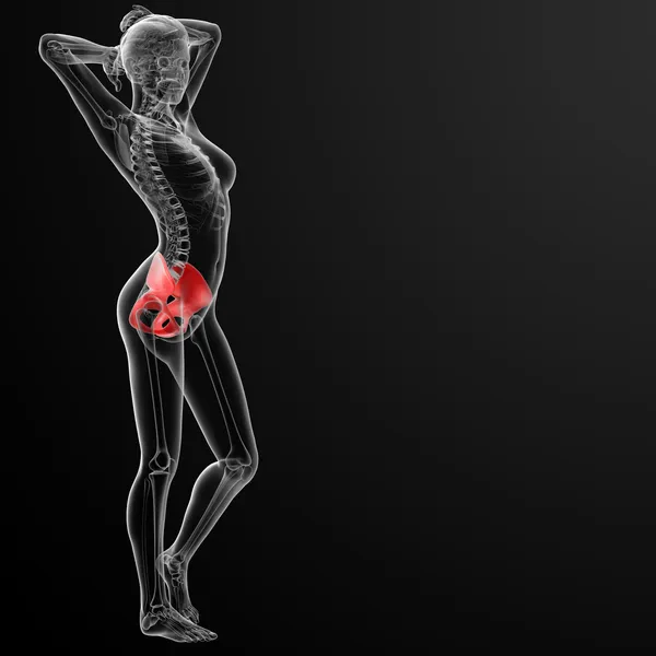
3d render female hip bone
3d illustration of the skeletal hip
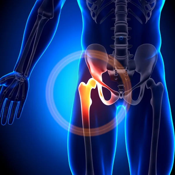
Pain in hip concept
Pain in hip concept
3D Illustration of Human Skeleton System Pelvis Anatomy
Illustration Human Skeleton System Pelvis Anatomy
Transparent digital body with shoulder pain
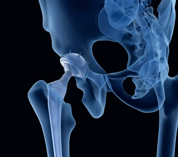
Hip replacement model
Male Osteopath Treating Female Patient With Hip Problem
Focused on hip in sports injuries
Pelvis
Pelvis
Pelvic hip bone
Pelvic hip bone
Running man with leg scan in black
Body flower and juice-1
Asian doctor writing on files
Anatomy of human hip joint and pelvis
Exercise in pregnancy
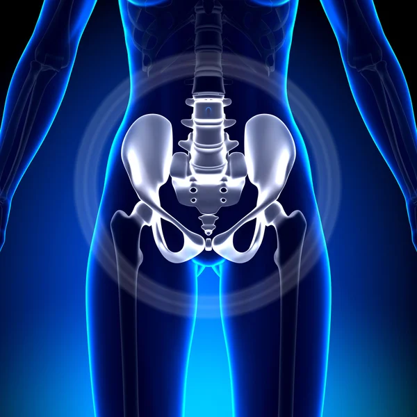
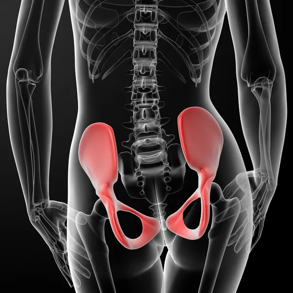
3d rendered, medical illustration of a painful hip joint -front
Bone system: hip joint (coxofemoral).
Bone System Hip Joint Coxofemoral
Pelvic hip bones
Pelvic hip bones
Human hips skeleton
Human hips skeleton
Human Pelvis Skeleton
Skeleton Pelvis illustration
Hip in sports injuries
Illustration of human pelvis
Beautiful woman body in denim jeans on white brick wall backgrou
Human pelvis
Human pelvis
Lumbar spine mobilization
Pelvis, Human skeleton, Female Pelvic Bone anatomy, hip, 3D artwork, Bones Anatomy View, black background
Pelvis Human Skeleton Female Pelvic Bone Anatomy Hip Artwork Bones Anatomy View Black Background
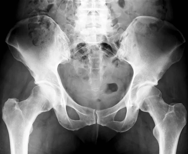
Pelvic hip x-ray
Pelvic hip x-ray
3D Illustration of Human Skeleton System Pelvis Anatomy
Illustration Human Skeleton System Pelvis Anatomy
Pelvis bone
Pelvis bone
Plastic model of a human spine
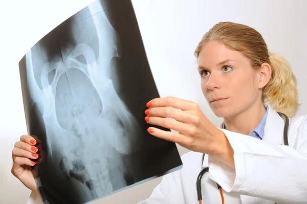
Female doctor examining an x-ray image
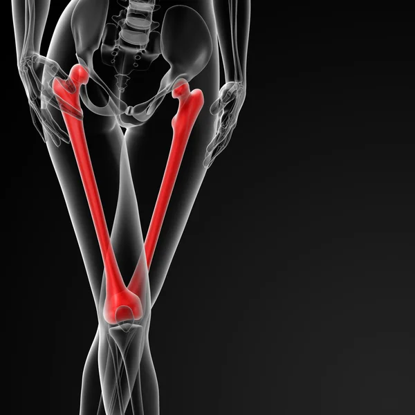
Femur bone
Femur bone
Accurate human skeletal system with adult male skeleton and body isolated on white background 3D rendering illustration. Anterior, lateral and posterior view. Anatomy, medical, osteology healthcare, science concept.
Accurate Human Skeletal System Adult Male Skeleton Body Isolated White Background Rendering Illustration Anterior Lateral Posterior View Anatomy Medical Osteology Healthcare Science Concept
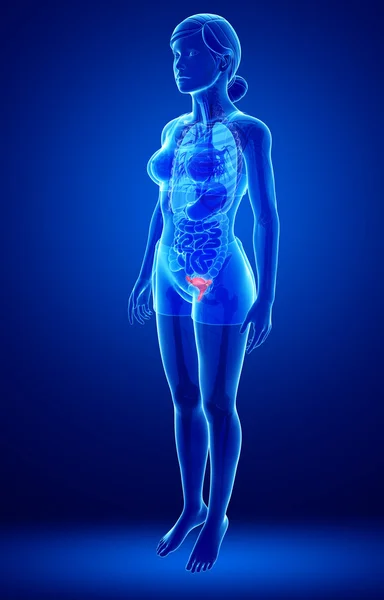
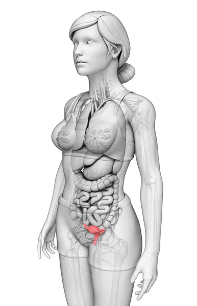
Female uterus anatomy
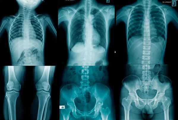
Collection human x-ray image
Horse Ligaments and Joints Tendons
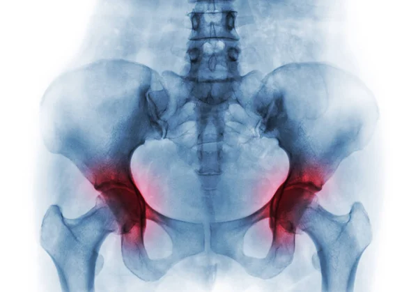
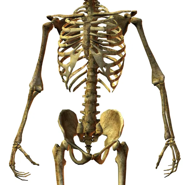
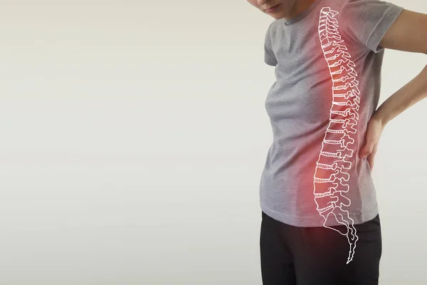
Pain in the spine, a woman with backache
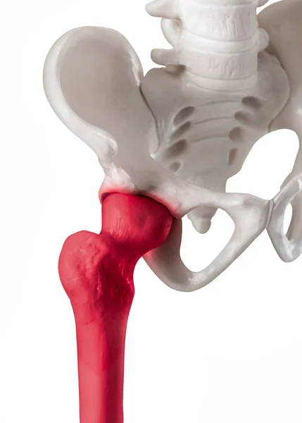
Human hip joint with red highlight on Femur or Thigh bone pain area-Healthcare-Human Anatomy and Medical concept-Isolated on white background.
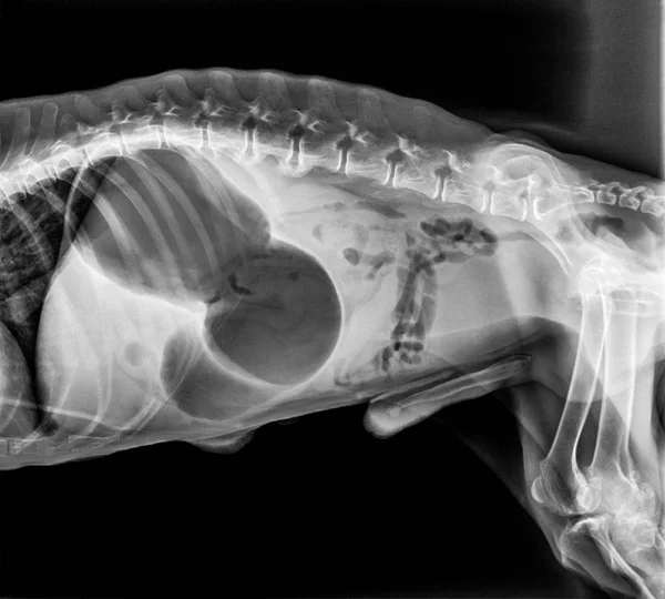
X ray of dog anterior view with Gastric dilatation volvulus GDV or stomach twists- Double bubble pattern indicates stomach torsion has occurred-Veterinary medicine and Veterinary anatomy Concept.
Ray Dog Anterior View Gastric Dilatation Volvulus Gdv Stomach Twists Double Bubble Pattern Indicates Stomach Torsion Has Occurred Veterinary Medicine Veterinary Anatomy Concept
Pelvic hip
Pelvic hip
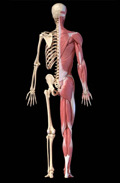
Anatomy of human male muscular and skeletal systems, back view.
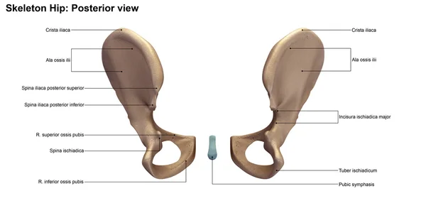
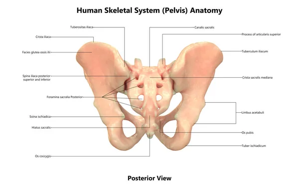
Pelvis, Human skeleton, Female Pelvic Bone anatomy, hip, 3D artwork, Bones Labeled Anatomy back View, White background
Pelvis Human Skeleton Female Pelvic Bone Anatomy Hip Artwork Bones Labeled Anatomy Back View White Background
X-ray of hip
X-ray of hip
Portrait of physiotherapist holding spine model
Orange digital skeleton
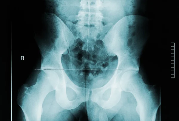
Pelvis X-Ray
Pelvis X-Ray
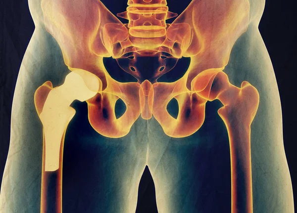
Hip replacement implant installed in the pelvis bone. X-ray view. Medically accurate 3D illustration
Hip Replacement Implant Installed Pelvis Bone Ray View Medically Accurate Illustration
Hip replacement implant installed in the pelvis bone. X-ray view. Medically accurate 3D illustration
Hip Replacement Implant Installed Pelvis Bone Ray View Medically Accurate Illustration
Hip replacement implant installed in the pelvis bone. X-ray view. Medically accurate 3D illustration
Hip Replacement Implant Installed Pelvis Bone Ray View Medically Accurate Illustration
Conceptual hip human anatomy with red hot spot inflammation articular joint pain for leg health care therapy or sport muscle concepts. 3D illustration man arthritis or bone sore osteoporosis disease
Conceptual Hip Human Anatomy Red Hot Spot Inflammation Articular Joint Pain Leg Health Care Therapy Sport Muscle Concepts Illustration Man Arthritis Bone Sore Osteoporosis Disease
3D medical illustration of the pelvis bone
Medical Illustration Pelvis Bone
Doctor hand shows the spine on a blue background.
Doctor Hand Shows Spine Blue Background
The X -ray of spine and pelvis
Ray Spine Pelvis
Film x-ray normal body of child (abdomen,buttock,thigh,knee)
Doctor with human Spine anatomy model. Spinal Cord Disorder and disease, Back pain, Lumbar, Sacral pelvis, Cervical neck, Thoracic, Coccyx, Orthopedist, chiropractic, Office Syndrome and health
Doctor Human Spine Anatomy Model Spinal Cord Disorder Disease Back Pain Lumbar Sacral Pelvis Cervical Neck Thoracic Coccyx Orthopedist Chiropractic Office Syndrome Health
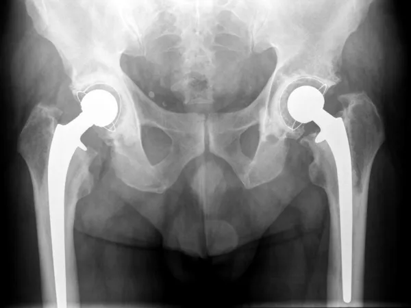
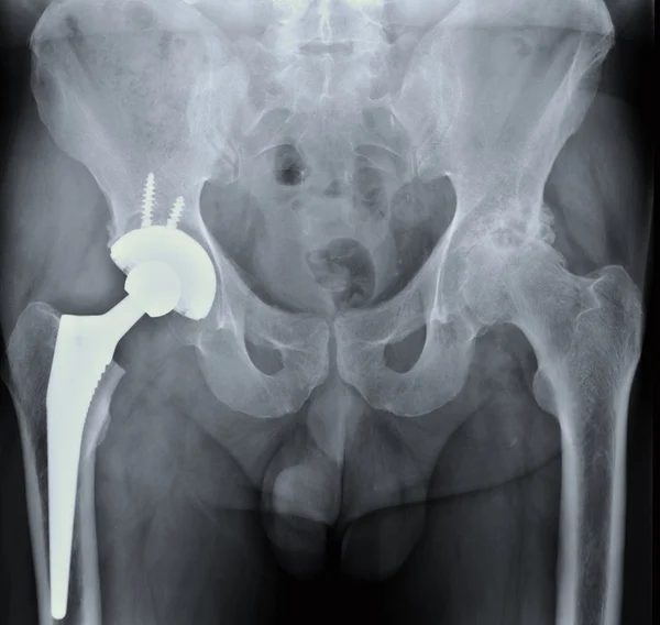
X-ray of the hip prosthesis
Human male anatomy, limbs and hip muscular and skeletal systems,
Back view x-ray image of male pelvis, femoral neck and lumbar vertebrae. Medical and human anatomy imagery.
Arthritis both hip . Film x-ray of human pelvis
Bone structure of male torso
Female uterus anatomy
Page