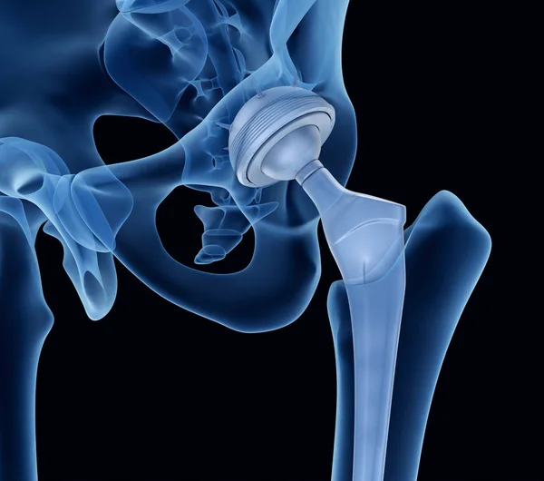
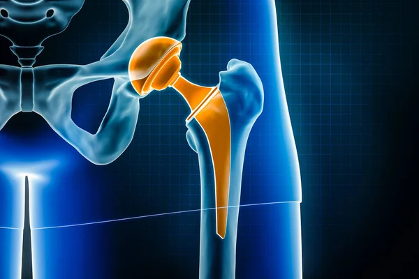
Hip replacement implant installed in the pelvis bone. Medically accurate 3D illustration
Hip Replacement Implant Installed Pelvis Bone Medically Accurate Illustration
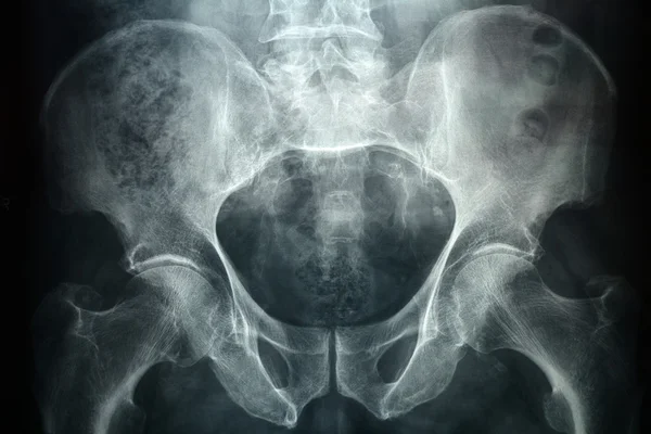
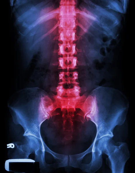
Pevic xray
Pevic xray
Portrait of physiotherapist holding
Physiotherapist explaining spine model to patient
Portrait of physiotherapist sitting with spine model
Smiling doctor showing a patient something on skeleton model
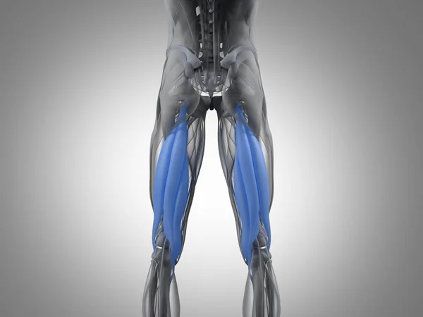
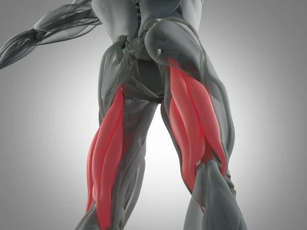
Hamstring muscle group anatomy model
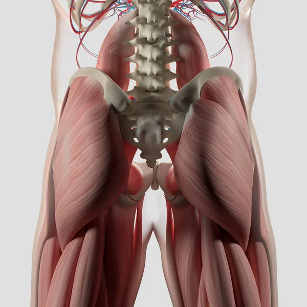
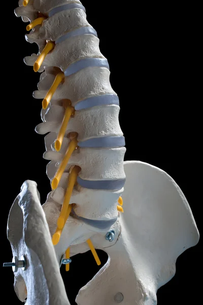
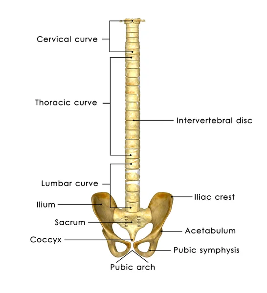
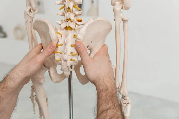
Human spine and pelvis anatomy model
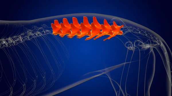
Lumbar Vertebrae Bones Dog skeleton Anatomy For Medical Concept 3D Illustration
Lumbar Vertebrae Bones Dog Skeleton Anatomy Medical Concept Illustration
Anatomy of the female genital system on white background
Anatomy Female Genital System White Background
X-ray
X-ray
Arthritis at hip joint
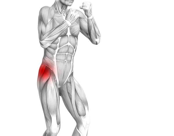
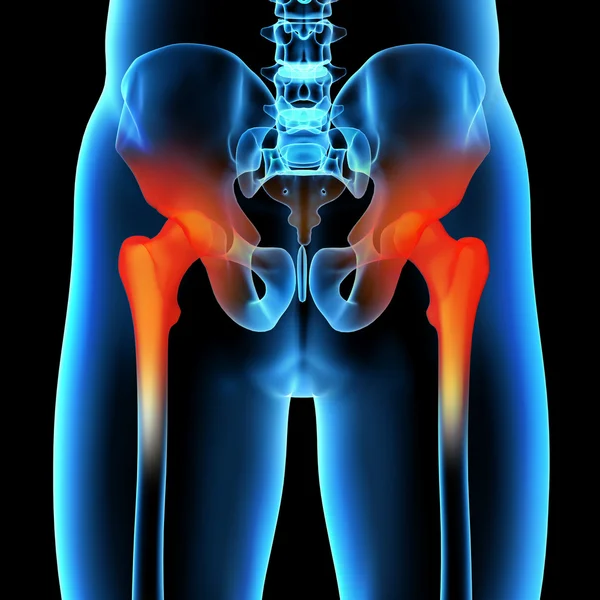
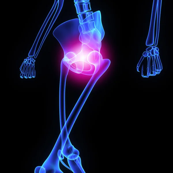
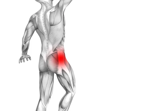
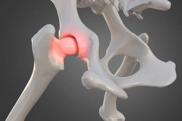
Conceptual hip human anatomy with red hot spot inflammation articular joint pain for leg health care therapy or sport muscle concepts. 3D illustration man arthritis or bone sore osteoporosis disease
Conceptual Hip Human Anatomy Red Hot Spot Inflammation Articular Joint Pain Leg Health Care Therapy Sport Muscle Concepts Illustration Man Arthritis Bone Sore Osteoporosis Disease
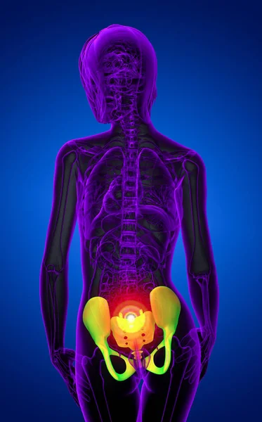
3D rendering medical illustration of the pelvis bone
Artificial human skeleton model on blue background, closeup
Artificial Human Skeleton Model Blue Background Closeup
Plastic human body model isolated on white background.
Before and after young woman side view of body. Swollen belly. Pregnancy. Diastasis recti after child birth. Fitness exercises and diet for weight loss.
Right human femur bone, posterior view, bone anatomy, black background, 3d rendering
Right Human Femur Bone Posterior View Bone Anatomy Black Background Rendering
This 3d illustration shows the obturator internus muscles on xray body
Illustration Shows Obturator Internus Muscles Xray Body
This 3d illustration shows the psoas minor muscles on skeleton
Illustration Shows Psoas Minor Muscles Skeleton
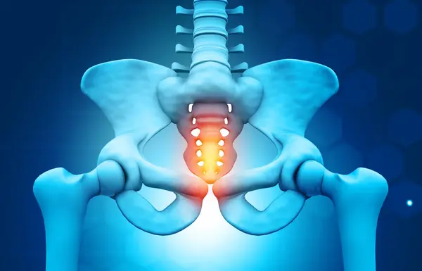
Painful sacrum
Painful sacrum
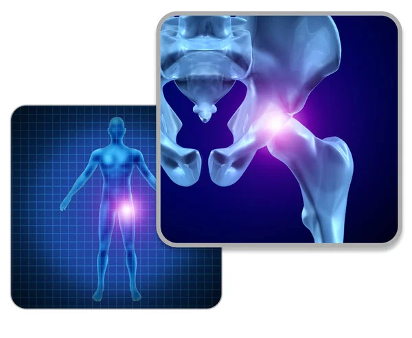
Human Hip Joint Pain
Close up. Doctor is showing vertebrae on skeleton.
Doctor model of the anatomy of hip joint in clinic. Pelvic bone pain causes, diagnosis and treatment concept
Doctor Model Anatomy Hip Joint Clinic Pelvic Bone Pain Causes Diagnosis Treatment Concept
Spondylosis
Spondylosis
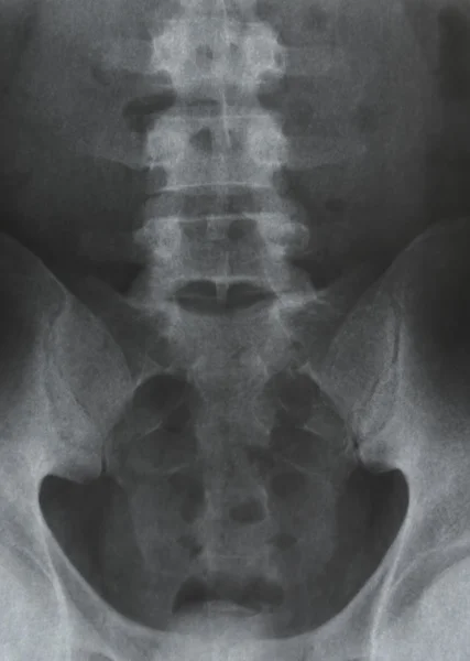
Pelvis radiograph
Pelvis radiograph
Hip x-ray of human
Hip x-ray of human
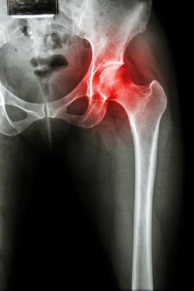
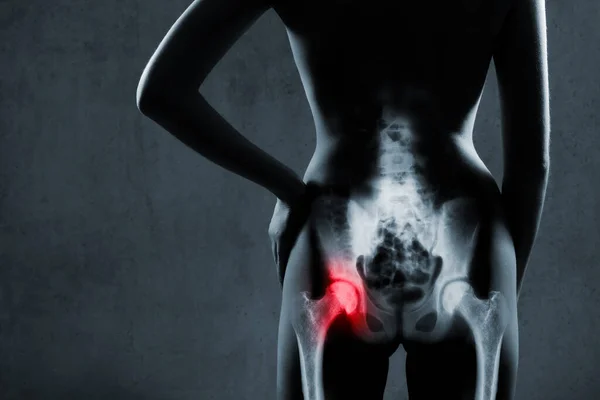
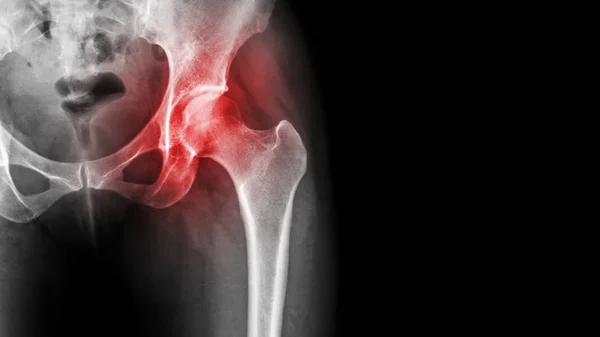
Human hip joint in x-ray on gray background. Painful hip joint is highlighted by red color. X-ray image of painful hip in woman. Osteoarthritis left hip joint at red area mark.
Human Hip Joint Ray Gray Background Painful Hip Joint Highlighted Red Color Ray Image Painful Hip Woman Osteoarthritis Left Hip Joint Red Area Mark
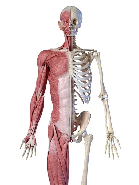
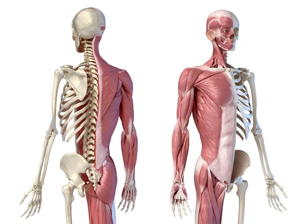
Human male anatomy, 3/4 figure muscular and skeletal systems, rear view.
Hip replacement implant installed in the pelvis bone. X-ray view. Medically accurate 3D illustration
Hip Replacement Implant Installed Pelvis Bone Ray View Medically Accurate Illustration
Skeleton hip illustration
Human cervical spine X-Ray
X-ray image of Whole Spine for diagnosis scoliosis of spine.
Ray Image Whole Spine Diagnosis Scoliosis Spine
PELVIS x--ray bones scan
Skeleton
Skeleton
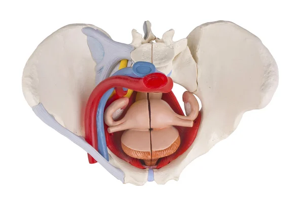
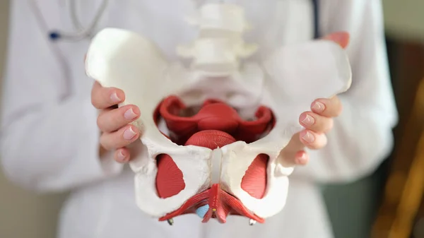
Gynecologist shows model of female pelvis with reproductive organs in hospital closeup. Women health examining and medical service. Human body
Gynecologist Shows Model Female Pelvis Reproductive Organs Hospital Closeup Women Health Examining Medical Service Human Body
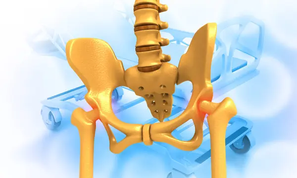
Hip joint
Hip joint
Human male anatomy, 3/4 figure muscular and skeletal systems, perspective back and front view.
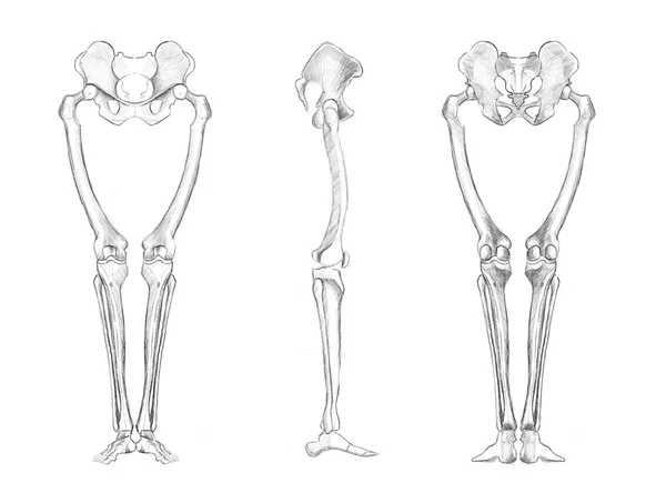
The structure of the bones of the human legs. Sketch for artists. Anatomical diagram.
Structure Bones Human Legs Sketch Artists Anatomical Diagram
Doula explains the process of childbirth on a sample of the pelvis of a pregnant woman
Doula Explains Process Childbirth Sample Pelvis Pregnant Woman
Female pelvis
Female pelvis
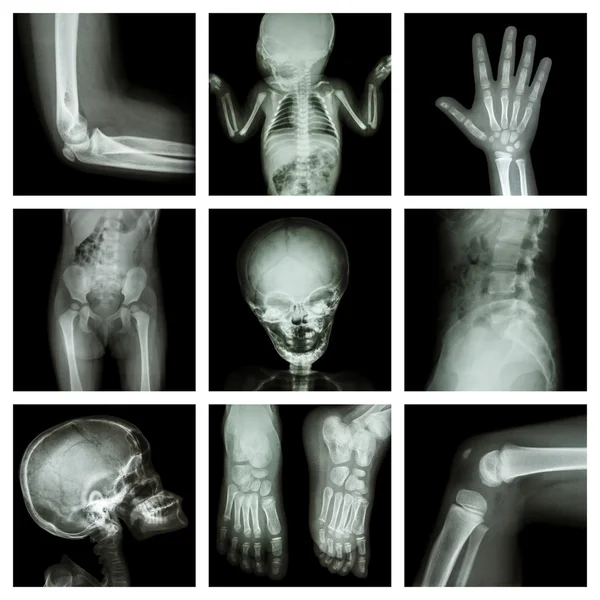
Collection X-ray part of child
Woman with human Spine anatomy model. Spinal Cord Disorder and disease, Back pain, Lumbar, Sacral pelvis, Cervical neck, Thoracic, Coccyx, Orthopedist, chiropractic, Office Syndrome and health
Woman Human Spine Anatomy Model Spinal Cord Disorder Disease Back Pain Lumbar Sacral Pelvis Cervical Neck Thoracic Coccyx Orthopedist Chiropractic Office Syndrome Health
Hip prosthesis x-ray 3D rendering illustration. Total hip joint replacement surgery or arthroplasty, medical and healthcare, arthritis, pathology, science, osteology, orthopedics concepts.
Hip Prosthesis Ray Rendering Illustration Total Hip Joint Replacement Surgery Arthroplasty Medical Healthcare Arthritis Pathology Science Osteology Orthopedics Concepts
Osteoporosismedical illustration design
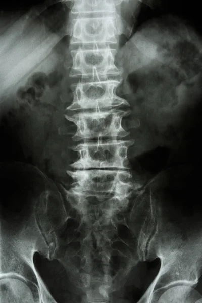
X-ray lumbo-sacral spine and pelvis and inflammation at spine
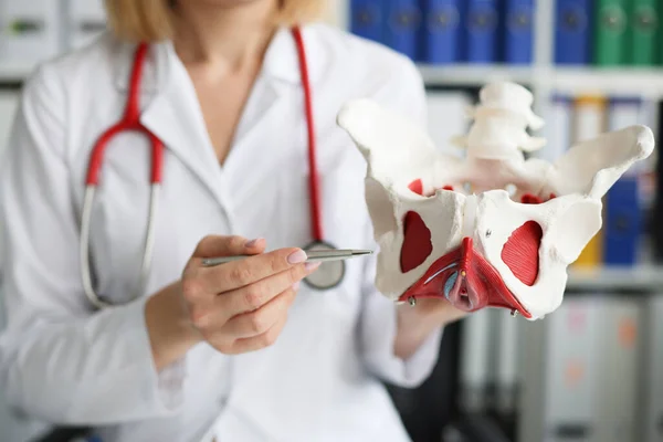
Gynecologist doctor holds model of bones of pelvic floor
Female doctor holding spine model
Human pelvic bone. Vector illustration
Human Pelvic Bone Vector Illustration
Human Skeleton bones anatomy structure of the human body isolated - 3d render
Free
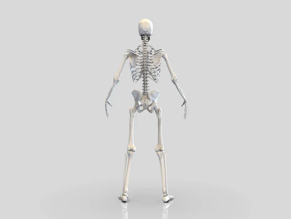
Human Skeleton Bones Anatomy Structure Human Body Isolated Render
Hamstring muscle group anatomy model
Female gynecologist showing model of female pelvis with muscles close-up.
Female Gynecologist Showing Model Female Pelvis Muscles Close
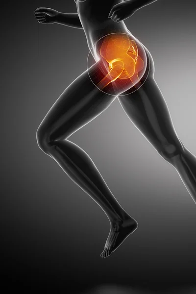
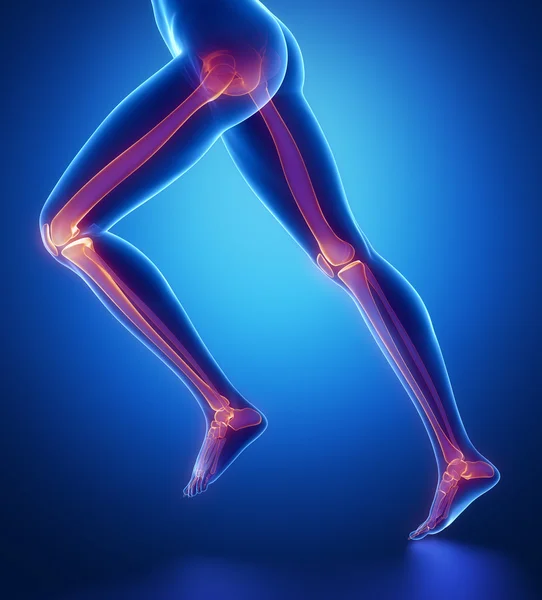
Running woman with hip anatomy
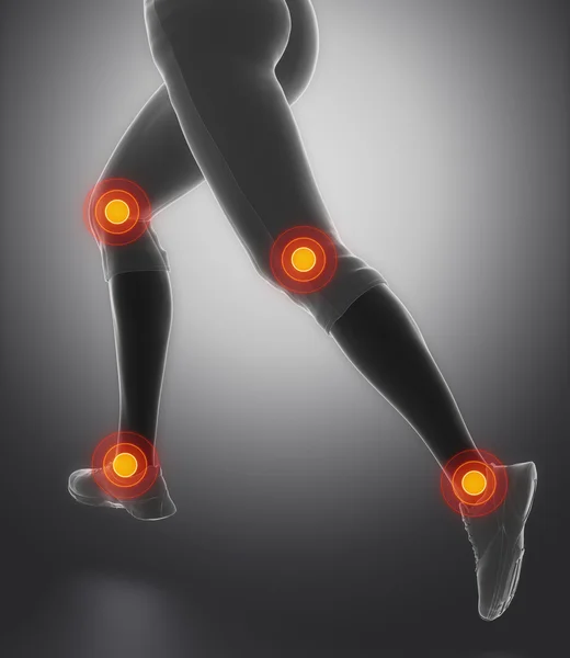
Leg most injured regoins in sport
Coccyx and sacrum of the human spine with nerve
Coccyx Sacrum Human Spine Nerve
Word writing text Pelvic Pain. Business concept for Pain perceived in the area of the lower part of the abdomen Mini blue alarm clock stand tilted above buffer wire in front of notepaper.
Human hip pain and pelvis anatomy. 3d illustration
Human Hip Pain Pelvis Anatomy Illustration
Human pelvis skeleton on medical background. 3d illustration
Human Pelvis Skeleton Medical Background Illustration
Human spinal column
Human spinal column
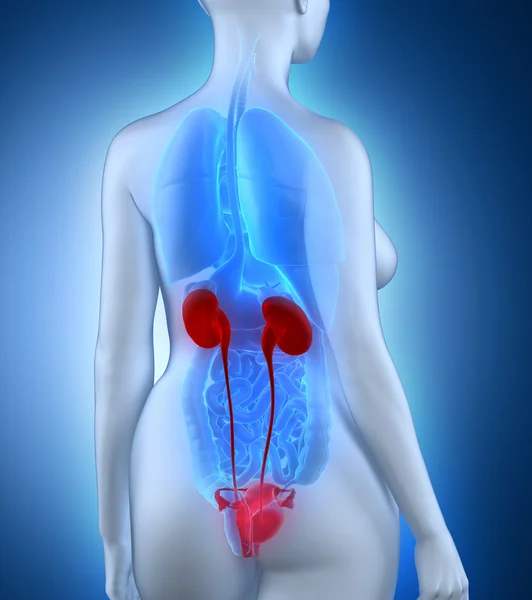
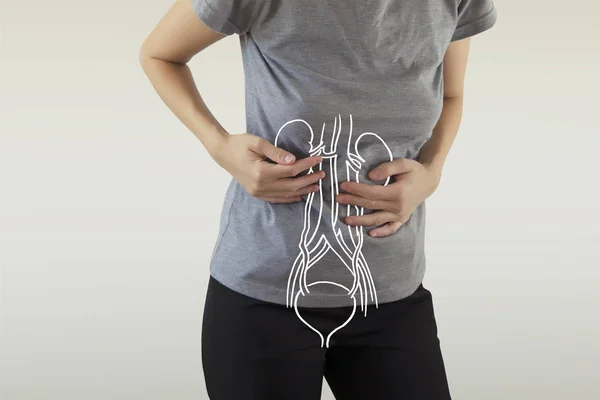
Woman urogenital anatomy
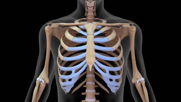
Human rib cage in human skeletal system 3d rendered
Human Rib Cage Human Skeletal System Rendered
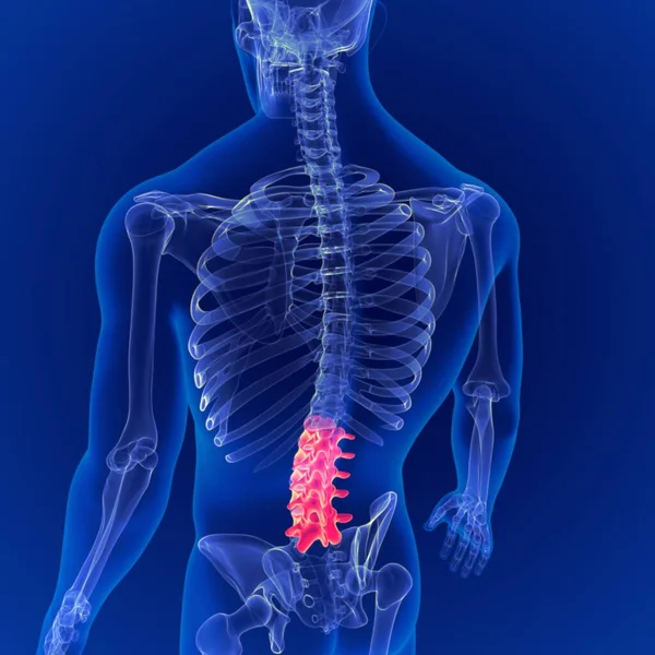
Human Skeleton Vertebral Column Lumbar Vertebrae Anatomy 3D Illustration
Human Skeleton Vertebral Column Lumbar Vertebrae Anatomy Illustration
Conceptual hip human anatomy with red hot spot inflammation articular joint pain for leg health care therapy or sport muscle concepts. 3D illustration man arthritis or bone sore osteoporosis disease
Conceptual Hip Human Anatomy Red Hot Spot Inflammation Articular Joint Pain Leg Health Care Therapy Sport Muscle Concepts Illustration Man Arthritis Bone Sore Osteoporosis Disease
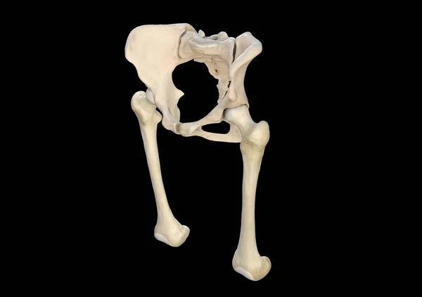
Bones, pelvis, femur, black background, 3d rendering
Bones Pelvis Femur Black Background Rendering
Arthritis at hip joint . Film x-ray show inflamed of hip joint and blank area at right side . Avascular necrosis concept
Human male anatomy, 3/4 figure muscular and skeletal systems, pperspective back and front view.
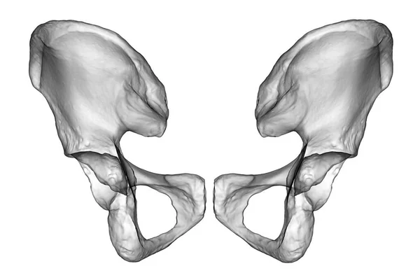
Anatomy of the coxal bones, showcasing their intricate details and features, 3D illustration. Front view. Perfect for educational or medical purposes
Anatomy Coxal Bones Showcasing Intricate Details Features Illustration Front View Perfect Educational Medical Purposes
Vintage Illustration of Human skeleton.
Vintage Illustration Human Skeleton
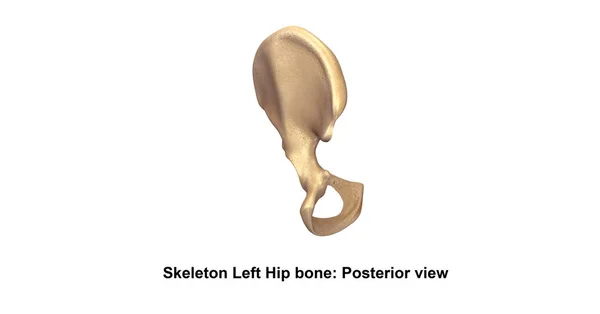
Human Left hip bone
Human Left hip bone
Shoulder pain on skeleton
Smiling physiotherapist looking at spine model
Content doctor holding skeleton and talking to patient
This 3d illustration shows the gluteus medius muscles anatomical position on xray body
Illustration Shows Gluteus Medius Muscles Anatomical Position Xray Body
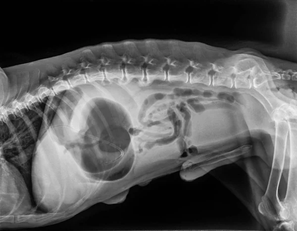
X ray of dog lateral view with Gastric dilatation volvulus GDV or stomach twists- Double bubble pattern indicates stomach torsion has occurred-Veterinary medicine and Veterinary anatomy Concept.
Ray Dog Lateral View Gastric Dilatation Volvulus Gdv Stomach Twists Double Bubble Pattern Indicates Stomach Torsion Has Occurred Veterinary Medicine Veterinary Anatomy Concept
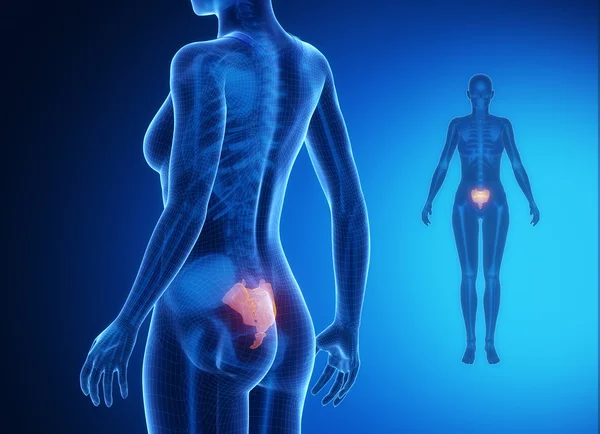
Female body with SACRUM bone scan
Film x-ray pelvis and arthritis both hip
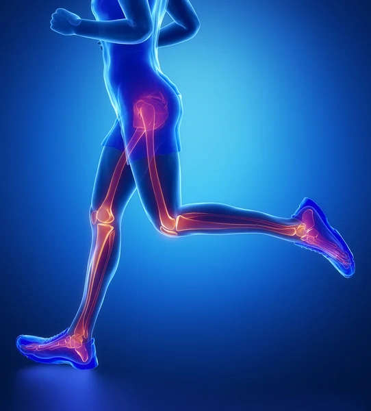
Running man with leg scan
3D medical illustration of the pelvis bone
Medical Illustration Pelvis Bone
3D Illustration Human Skeleton Vertebral Column Lumbar Vertebrae Anatomy
Illustration Human Skeleton Vertebral Column Lumbar Vertebrae Anatomy
Human male anatomy, 3/4 figure muscular and skeletal systems, front view.
Backbone with hips
Backbone with hips
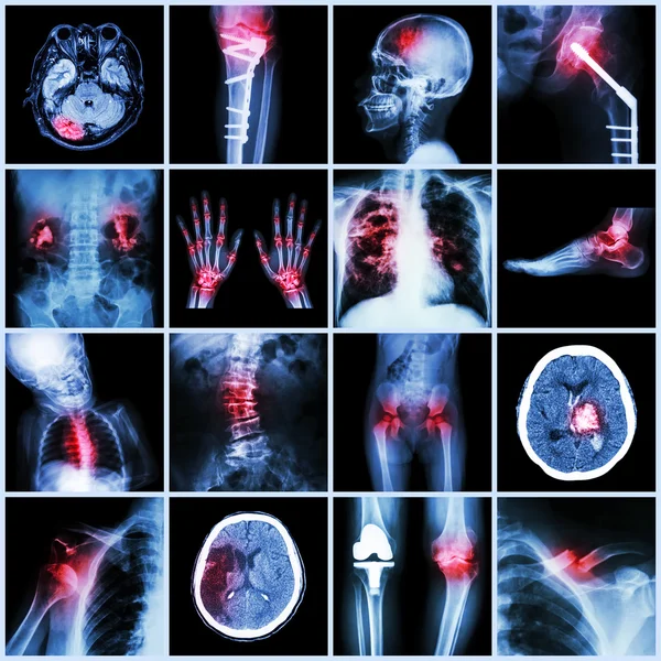
Set of X-ray multiple part of human,Multiple disease,orthopedic,surgery (Stroke,Bone fracture,Orthopedic operation,Kidney stone,Arthritis,Gout,Pulmonary tuberculosis,Heart disease,Scoliosis,etc)
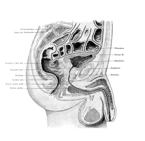
Midsection of the male pelvis in the old book Atlas der Anatomie by Fischer, 1894, Jena
Midsection Male Pelvis Old Book Atlas Der Anatomie Fischer 1894 Jena
Hips pelvis xray scan test result.
Hips Pelvis Xray Scan Test Result
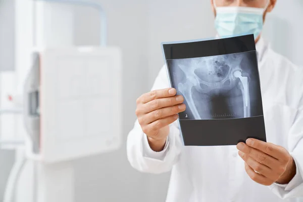
X-ray of the hip prosthesis
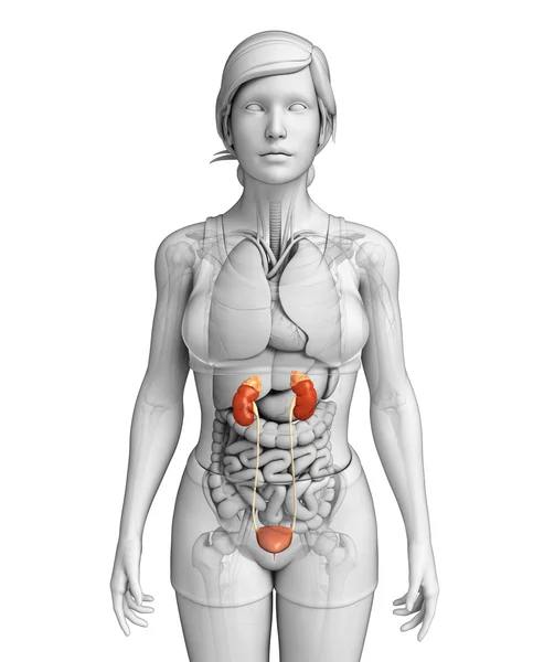
Female urinary system
Elderly woman having back body ache for concepts of Back Muscle Strain, Piriformis Syndrome, Low Back Pain, Lumbar Disc Degeneration, Herniated, Osteoporosis, Spinal Compression and Health care
Elderly Woman Having Back Body Ache Concepts Back Muscle Strain Piriformis Syndrome Low Back Pain Lumbar Disc Degeneration Herniated Osteoporosis Spinal Compression Health Care
Human skeleton anatomy Radius Bone 3D Rendering For Medical Concept
Human Skeleton Anatomy Radius Bone Rendering Medical Concept
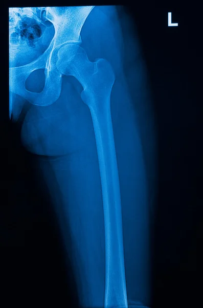
FEMUR x--ray bones scan
Doctor man pointing on pelvis of human skeleton anatomical model. Physiotherapist explaining joints model. Chiropractor or osteopath points to the skeleton of human body. Bones anatomy close up
Human skeleton anatomy Radius Bone 3D Rendering For Medical Concept
Human Skeleton Anatomy Radius Bone Rendering Medical Concept
Focused on leg bones anatomy
Female uterus anatomy
This 3d illustration shows the superior and inferior gemellus muscles on xray body
Illustration Shows Superior Inferior Gemellus Muscles Xray Body
Canine dysplasia, dog bone with visible hip joint and femur affected by inflammation due to dysplasia, 3d illustration
Canine Dysplasia Dog Bone Visible Hip Joint Femur Affected Inflammation Due Dysplasia Illustration
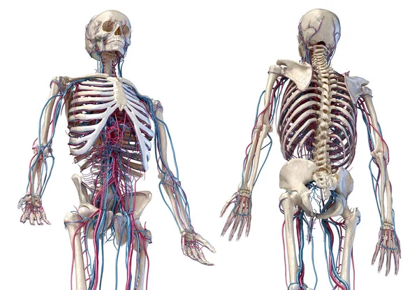
Human skeleton with veins and arteries. 3/4 upper part, Front and back views.
X-rays
X-rays
Scan with image of hip bone in doctors hands.
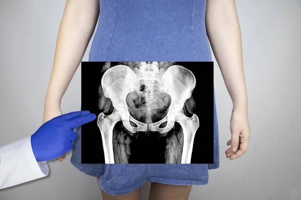
X-ray of the pelvic bones of a woman. Radiologist examines X-ray examination. A picture of the hip joint is superimposed on the patient's body.
Ray Pelvic Bones Woman Radiologist Examines Ray Examination Picture Hip Joint Superimposed Patient Body
Human Female Kidney Anatomy highlighted white
Page