Gelatinase is a molecular chemical formula. Enzyme of the stomach. Infographics. Vector illustration on isolated background
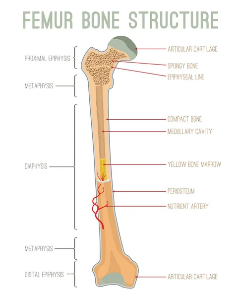
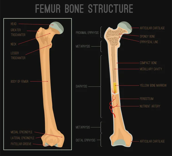
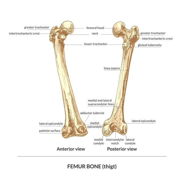
Femur Bone Structure
Bone Structure. Vector scheme
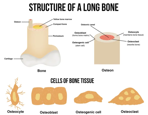
Structure of a long bone
Skull drawing in a vintage retro woodcut etched or engraved style
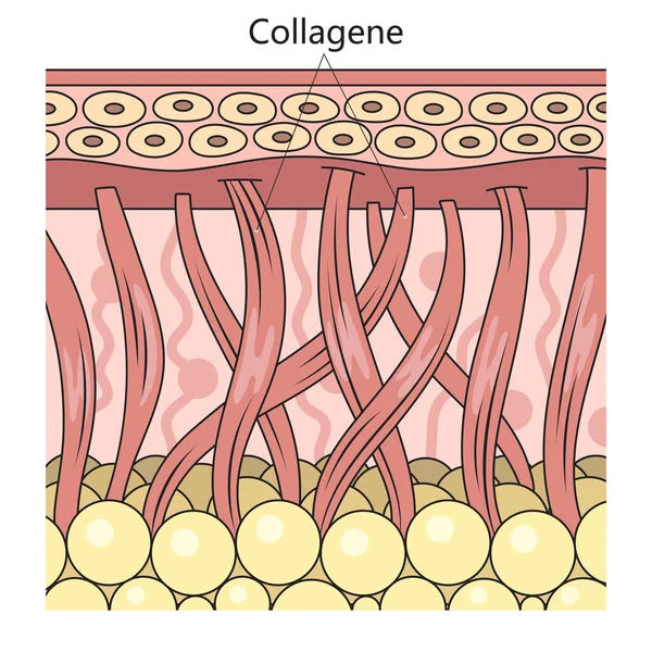
Collagen protein in skin structure diagram schematic vector illustration. Medical science educational illustration
Collagen Protein Skin Structure Diagram Schematic Vector Illustration Medical Science Educational Illustration
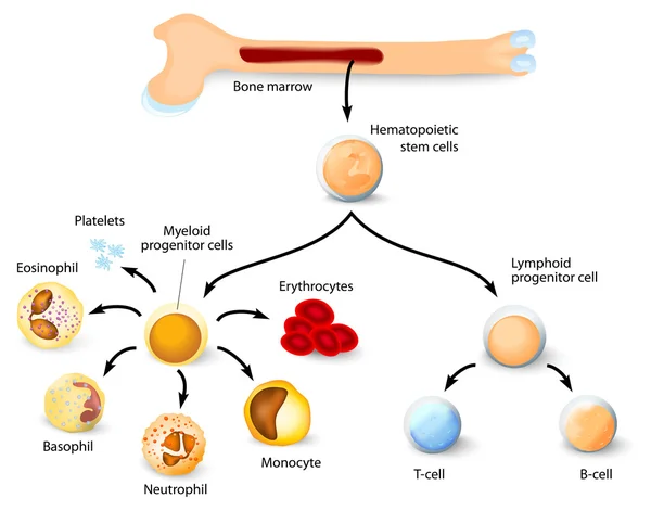
Human hematopoiesis blood cell formation from bone marrow, hematopoietic stem cells differentiation structure diagram hand drawn schematic vector illustration. Medical science educational illustration
Human Hematopoiesis Blood Cell Formation Bone Marrow Hematopoietic Stem Cells Differentiation Structure Diagram Hand Drawn Schematic Vector Illustration Medical Science Educational Illustration
Vestigial structures brief sketch shows evolution by comparing coccyx, whale pelvic bone, and appendix, illustrating reduced ancestral functions across species. Outline diagram
Vestigial Structures Brief Sketch Shows Evolution Comparing Coccyx Whale Pelvic Bone Appendix Illustrating Reduced Ancestral Functions Species Outline Diagram
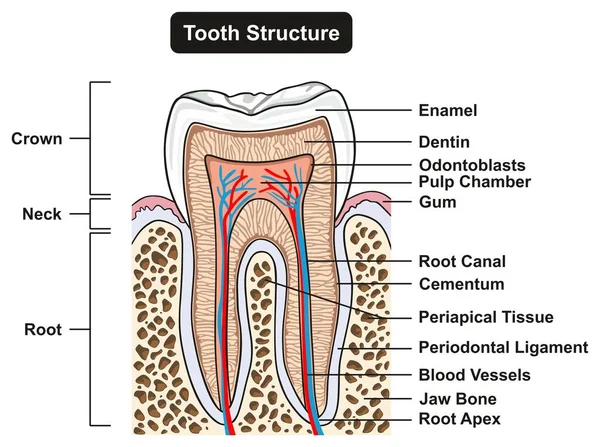
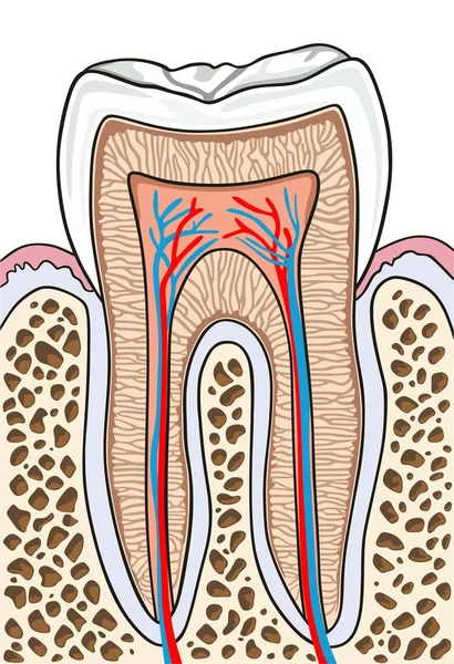
Labeled Tooth Cross Section Anatomy with all parts including crown neck enamel dentin pulp cavity gums root canal cement bone and blood supply for medical science education and dental health care
Labeled Tooth Cross Section Anatomy All Parts Including Crown Neck Enamel Dentin Pulp Cavity Gums Root Canal Cement Bone Blood Supply Medical Science Education Dental Health Care
Biological Hierarchy Color Flowchart
Bone structure set. Biology educational poster design. Scheme of human bone anatomy. Flat vector illustration with detailed information isolated on white background.
Bone Structure Set Biology Educational Poster Design Scheme Human Bone Anatomy Flat Vector Illustration Detailed Information Isolated White Background
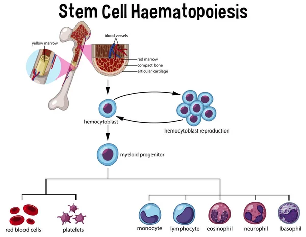
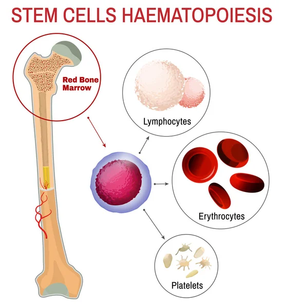
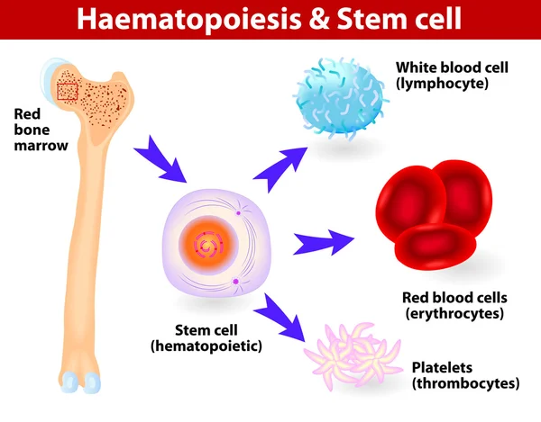
Stem Cell Haematopoiesis Diagram illustration
Stem Cell Haematopoiesis Diagram Illustration
Bone structure. Biology educational poster design. Scheme of human bone anatomy. Flat vector illustration with detailed information isolated on white background.
Bone Structure Biology Educational Poster Design Scheme Human Bone Anatomy Flat Vector Illustration Detailed Information Isolated White Background
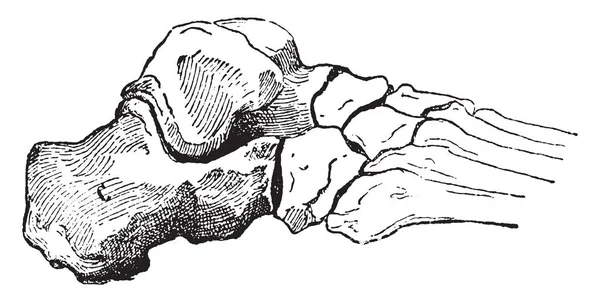
Calcaneus or Heel Bone, vintage engraved illustration. Dictionary of words and things - Larive and Fleury - 1895
Calcaneus Heel Bone Vintage Engraved Illustration Dictionary Words Things Larive Fleury 1895
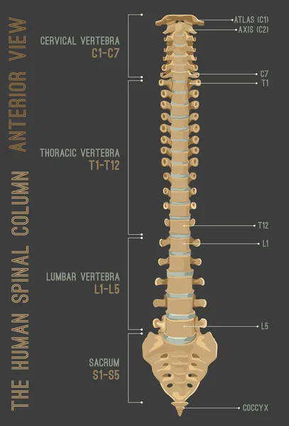
Human Spine Column
Human Spine Column
Bone remodeling from resorption to new bone formation. Close-up of Trabecular bone matrix, Osteoblasts, Osteoclasts and Osteocytes. Vector illustration
Bone Remodeling Resorption New Bone Formation Close Trabecular Bone Matrix Osteoblasts Osteoclasts Osteocytes Vector Illustration
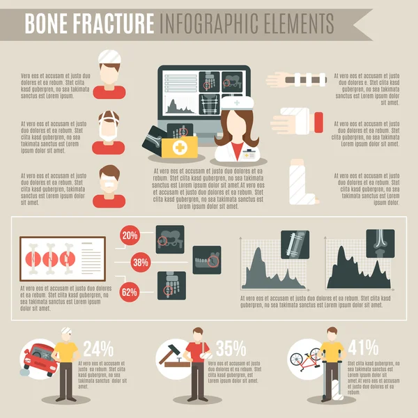
Fracture Bone Infographics
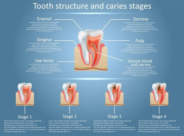
Human tooth structure vector diagram and caries stages. Dental anatomy and tooth decay or cavities development concept. Training medical anatomical poster.
Human Tooth Structure Vector Diagram Caries Stages Dental Anatomy Tooth Decay Cavities Development Concept Training Medical Anatomical Poster
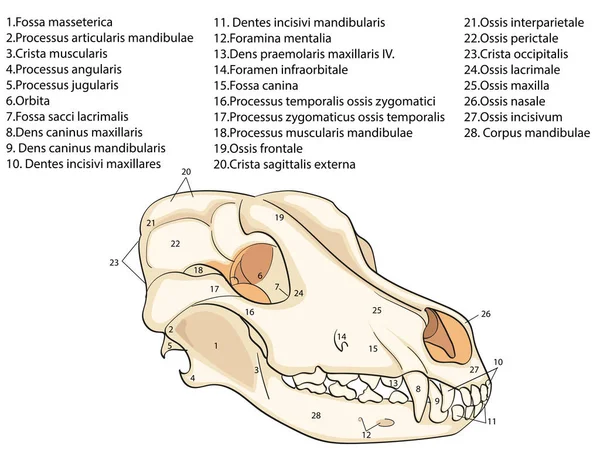
The skull of a dog. Structure of the bones of the head, anatomical design. In Latin
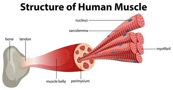
A Structure of Human Muscle illustration
Structure Human Muscle Illustration
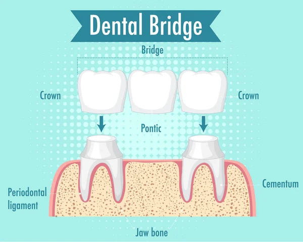
Infographic of human in dental bridge on white background illustration
Infographic Human Dental Bridge White Background Illustration
Illustration of biology and medical, Anatomy of bone, Bone is living tissue that makes up the body's skeleton, Compact tissue, Cancellous tissue
Illustration Biology Medical Anatomy Bone Bone Living Tissue Makes Body Skeleton Compact Tissue Cancellous Tissue
Illustration of the human right scapula bone with the name and d
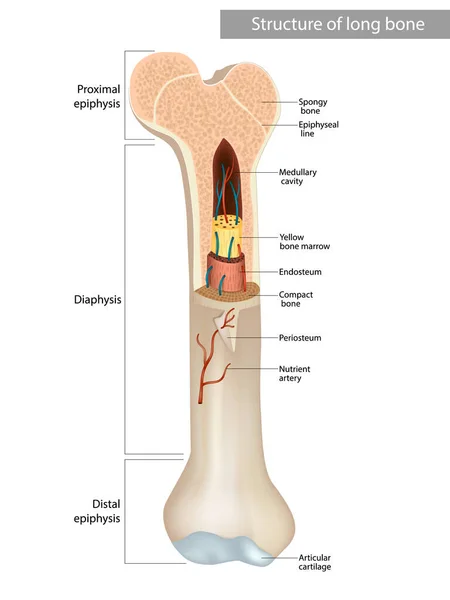
Structure and components of long bone. Proximal epiphysis, Distal epiphysis, Diaphysis. Compact bone and an inner medullary cavity containing bone marrow.
Structure Components Long Bone Proximal Epiphysis Distal Epiphysis Diaphysis Compact Bone Inner Medullary Cavity Containing Bone Marrow
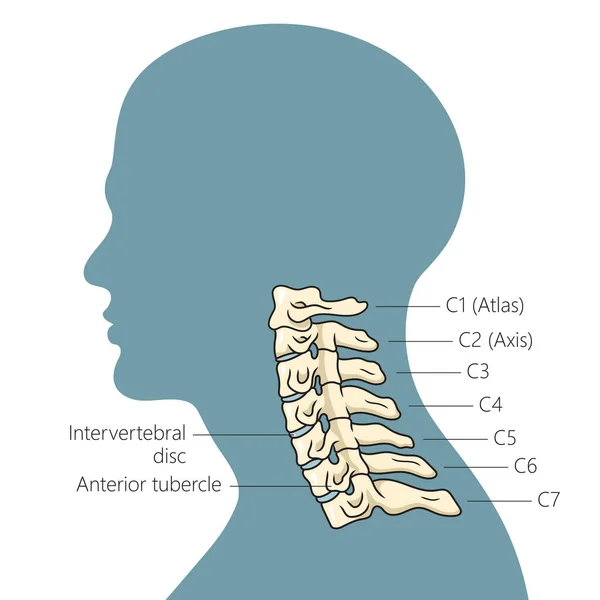
Structure of the human cervical spine schematic vector illustration. Medical science educational illustration
Structure Human Cervical Spine Schematic Vector Illustration Medical Science Educational Illustration
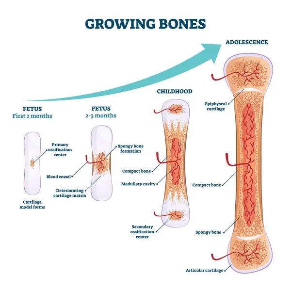
Growing bones vector illustration. Educational fetus and adolescence stages
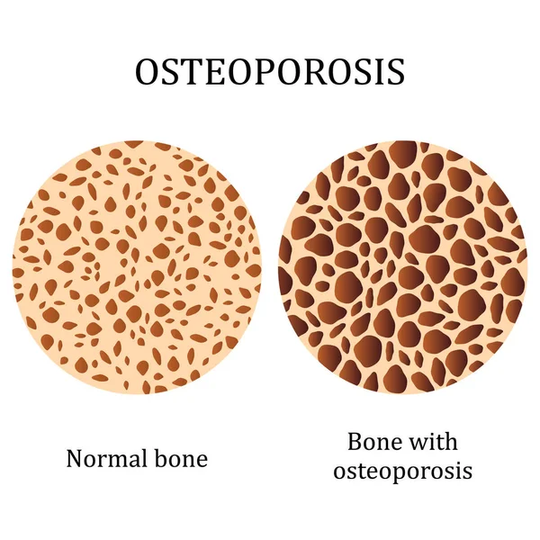
Healthy bone and bone with osteoporosis.
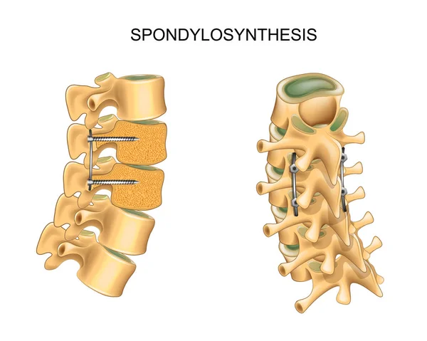
Spondylosynthesis. vertebral osteosynthesis
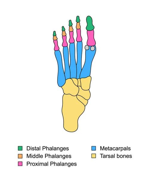
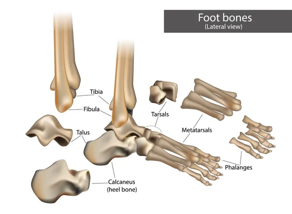
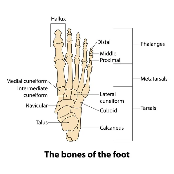
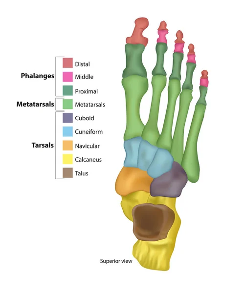
Foot bones anatomy with descriptions. Colored leg base parts structure. Educational diagram with internal organ illustration. Distal, proximal and middle phalanges, metacarpal, tarsal bones foot parts
Foot Bones Anatomy Descriptions Colored Leg Base Parts Structure Educational Diagram Internal Organ Illustration Distal Proximal Middle Phalanges Metacarpal Tarsal Bones Foot Parts
Bone structure medical educational vector
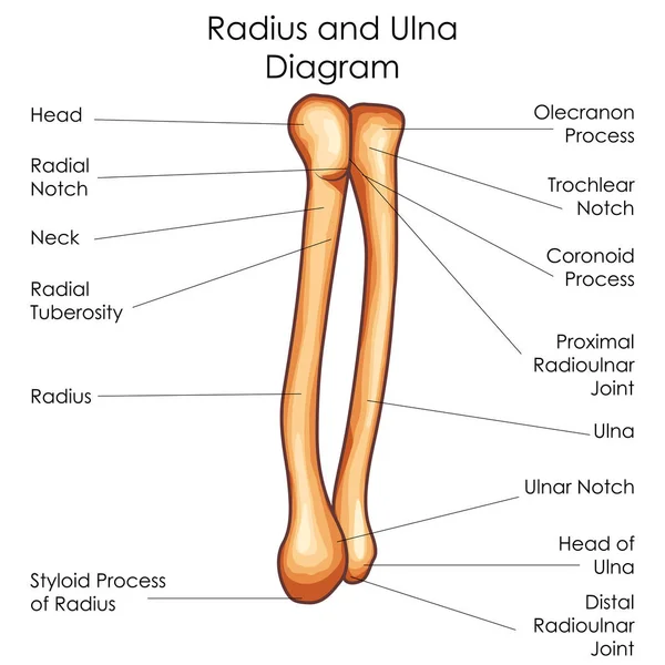
Medical Education Chart of Biology for Radius and Ulna Diagram
Vector dental anatomy and tooth structure diagram
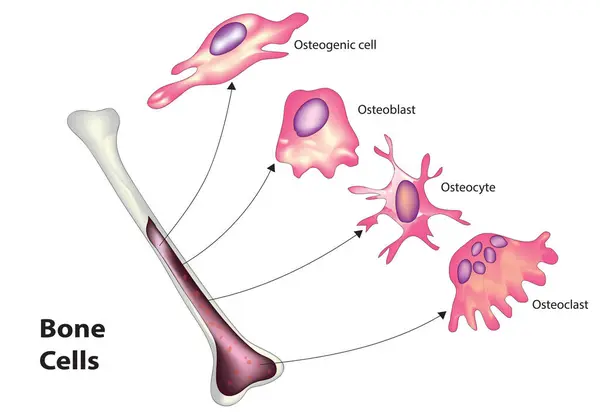
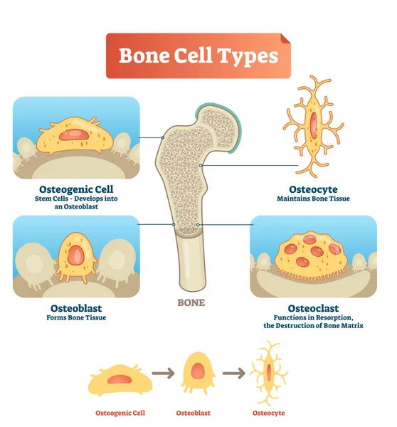
Bone cell classification (Types of bone cell)
Bone Cell Classification Types Bone Cell
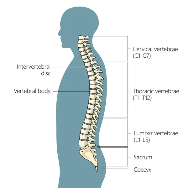
The structure of the spine. Side view. The intervertebral discs. Infographics. Vector illustration on isolated background
Fifth metatarsal or foot little finger fracture after injury outline diagram. Labeled educational scheme with feet trauma after twisting motion vector illustration. Anatomical skeletal bone zones.
Fifth Metatarsal Foot Little Finger Fracture Injury Outline Diagram Labeled Educational Scheme Feet Trauma Twisting Motion Vector Illustration Anatomical Skeletal Bone Zones
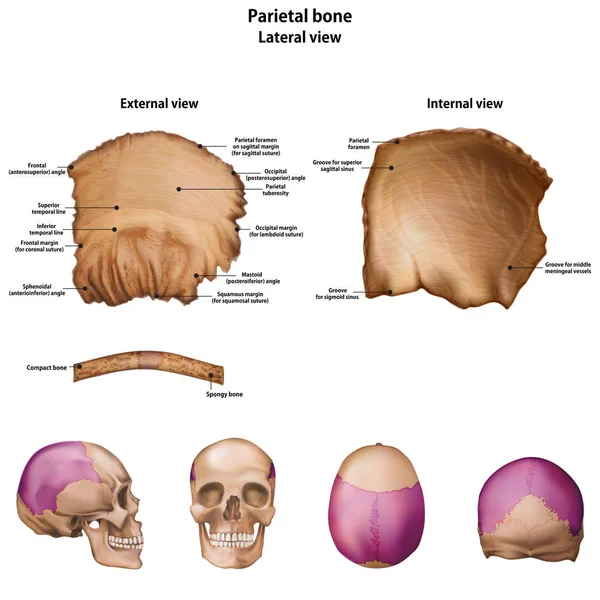
Parietal bone. With the name and description of all sites.
Parietal Bone Name Description All Sites
Anatomy Bones of the Feet. Superior view
Vector illustration bone cell types diagram. Scheme of osteogenic cell, osteoblast, osteocyte. Medical visualization of stem cells, bone tissue and resorption.
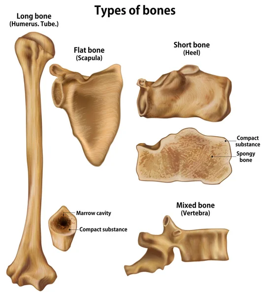
Types of bones.Long(Humerus).Shirt(Heel).Flat(Scapula).Mixed(Vertebra)
Types Bones Long Humerus Shirt Heel Flat Scapula Mixed Vertebra
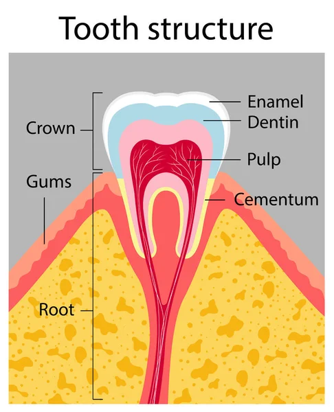
Tooth structure. Medical poster
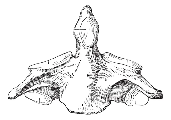
Fig. 136. Axis (second cervical vertebra), vintage engraving.
Human spine structure vertebral column diagram schematic vector illustration. Medical science educational illustration
Human Spine Structure Vertebral Column Diagram Schematic Vector Illustration Medical Science Educational Illustration
Vitamin D3 and K2 for Bone Health: A Dynamic Vector Illustration of Vital Nutrients.
Vitamin Bone Health Dynamic Vector Illustration Vital Nutrients
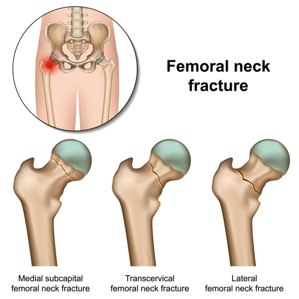
Femoral neck fractures medical vector illustration on white background
Femoral Neck Fractures Medical Vector Illustration White Background
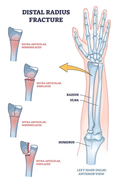
Distal radius fracture and broken arm bone types anatomy outline diagram. Labeled educational scheme with extra articular nondisplaced and displaced radius and ulna comparison vector illustration.
Distal Radius Fracture Broken Arm Bone Types Anatomy Outline Diagram Labeled Educational Scheme Extra Articular Nondisplaced Displaced Radius Ulna Comparison Vector Illustration
Hip joint anatomy. Ball-and-socket joint. Vector poster
Hip Joint Anatomy Ball Socket Joint Vector Poster
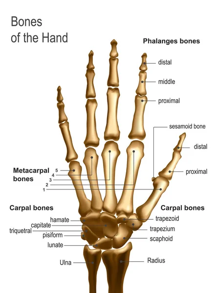
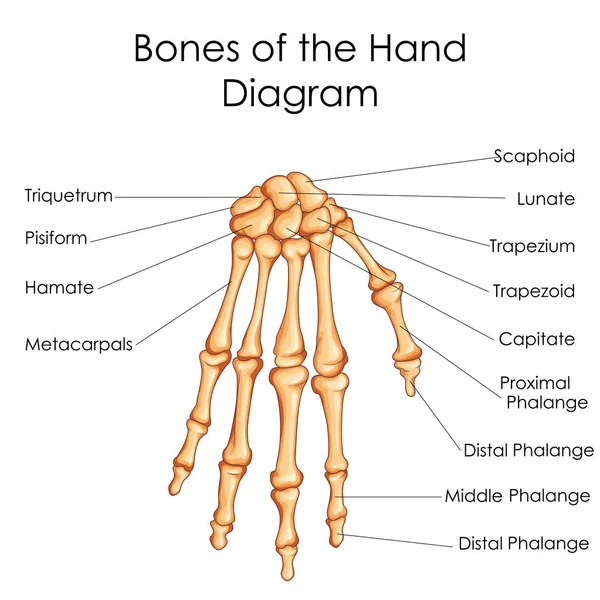
Frontal view image of bones the of hand with anotations.
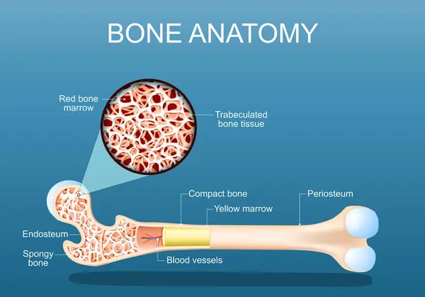
Bone anatomy. Structure of a femur. Close-up of a cross section of Spongy Trabeculated bone tissue with Red bone marrow. vector illustration
Bone Anatomy Structure Femur Close Cross Section Spongy Trabeculated Bone Tissue Red Bone Marrow Vector Illustration
Bone remodeling. Close-up of a Osteoclast resorbs bone, and Osteoblasts synthesized bone tissue. Vector illustration
Bone Remodeling Close Osteoclast Resorbs Bone Osteoblasts Synthesized Bone Tissue Vector Illustration
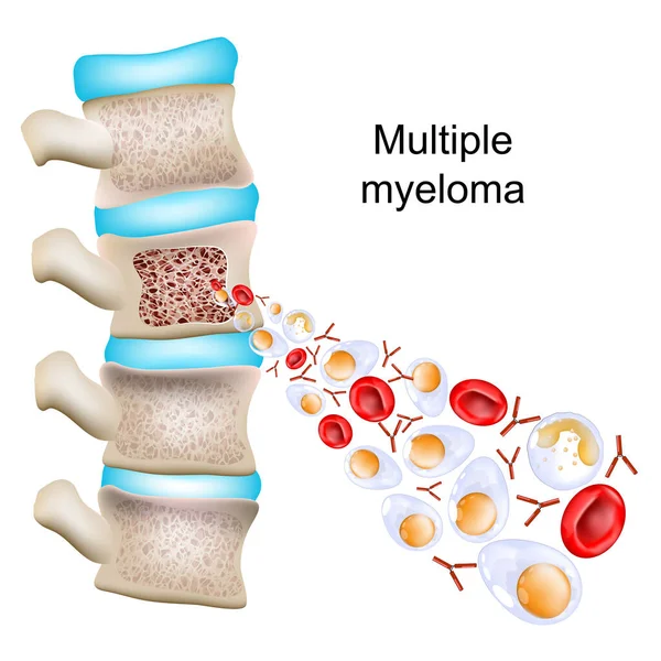
Multiple myeloma. Part of a Vertebral column with cross section of a vertebra with bone structure and bone marrow. Close-up of Abnormal plasma cells, red blood cells, and antibodies. Vector illustration
Multiple Myeloma Part Vertebral Column Cross Section Vertebra Bone Structure Bone Marrow Close Abnormal Plasma Cells Red Blood Cells Antibodies Vector Illustration
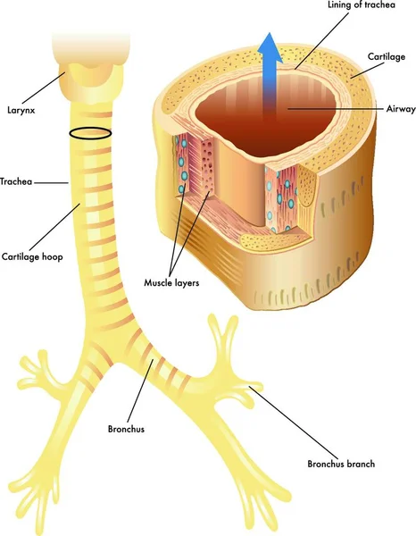
Vector medical illustration of the anatomy of the trachea
Vector Medical Illustration Anatomy Trachea
Femur Bone Structure
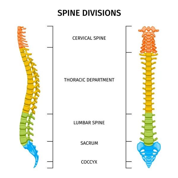
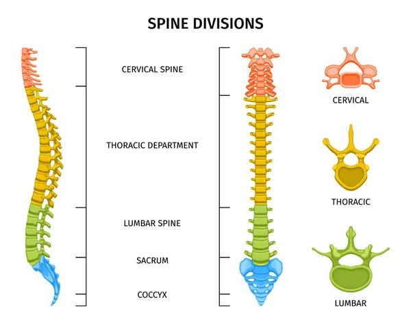
Spine structure anatomy composition with spine divisions chart editable text captions and colored images of spine vector illustration
Spine Structure Anatomy Composition Spine Divisions Chart Editable Text Captions Colored Images Spine Vector Illustration
Ankle joint fracture Type B medical vector illustration on white background
Ankle Joint Fracture Type Medical Vector Illustration White Background
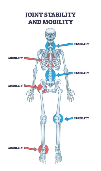
Joint stability or body mobility skeletal bone division outline diagram. Labeled educational anatomical scheme with skeleton functions as spinal, chest, hip, knee or ankle purpose vector illustration
Joint Stability Body Mobility Skeletal Bone Division Outline Diagram Labeled Educational Anatomical Scheme Skeleton Functions Spinal Chest Hip Knee Ankle Purpose Vector Illustration
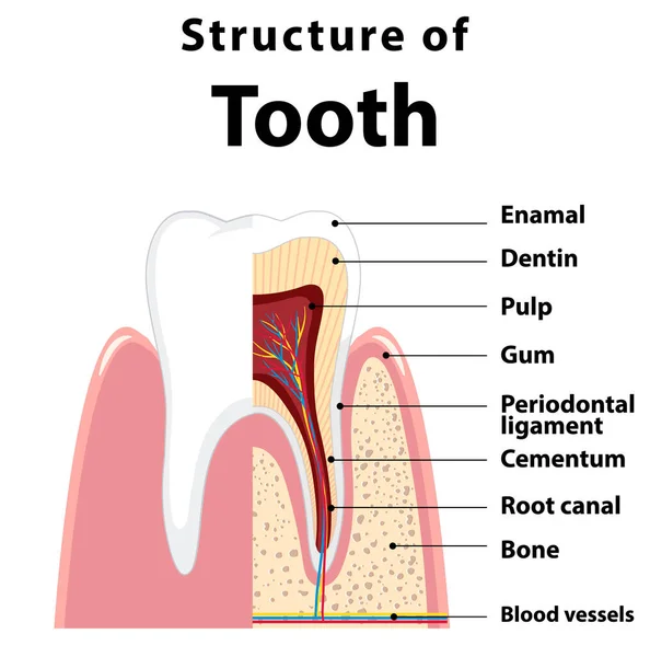
Infographic of human in structure of tooth illustration
Infographic Human Structure Tooth Illustration
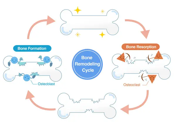
Mechanisms of bone resorption and bone formation by osteoclasts and osteoblasts. Bone Remodeling Cycle
Mechanisms Bone Resorption Bone Formation Osteoclasts Osteoblasts Bone Remodeling Cycle
Scoliosis flat vector illustration
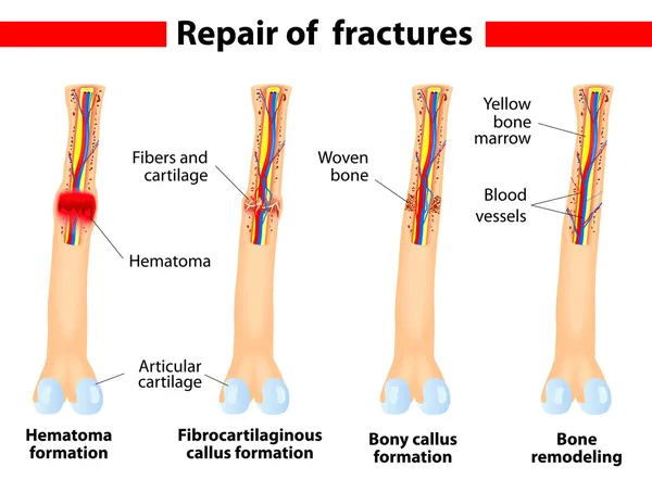
Bone fracture healing process
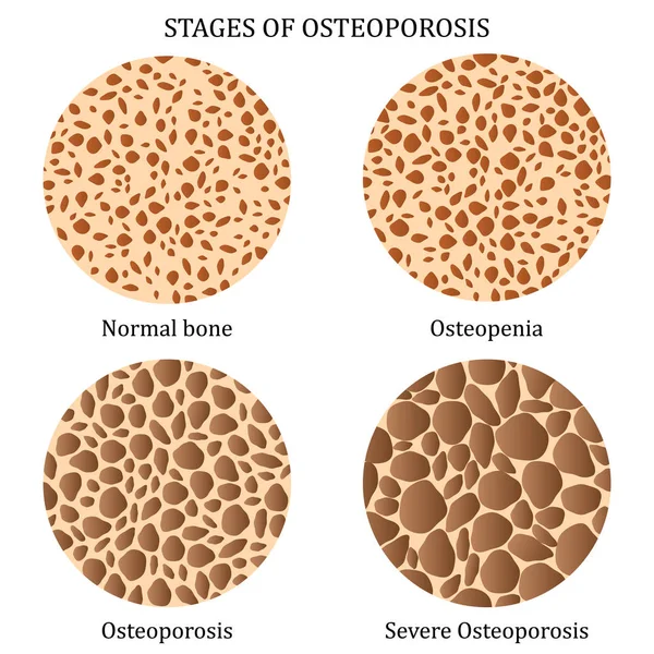
Stages of osteoporosis.
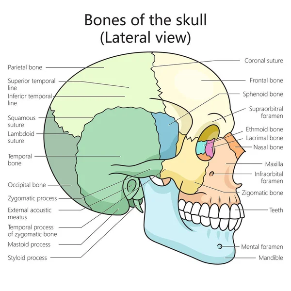
Human skull bones structure lateral view diagram schematic vector illustration. Medical science educational illustration
Human Skull Bones Structure Lateral View Diagram Schematic Vector Illustration Medical Science Educational Illustration
Hematopoietic stem cell
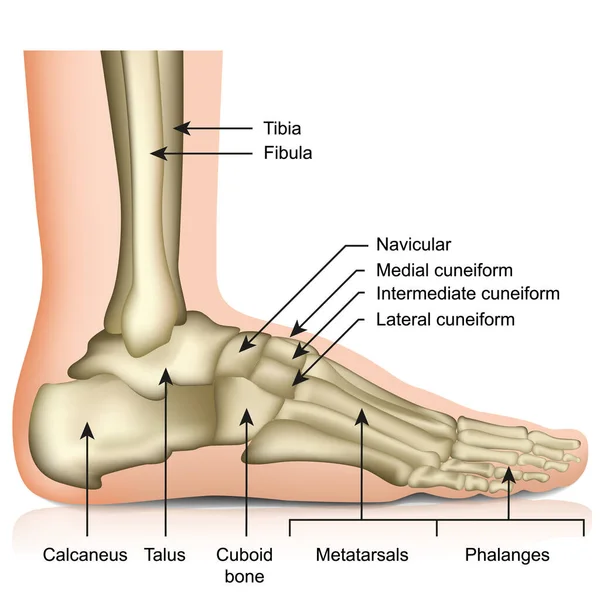
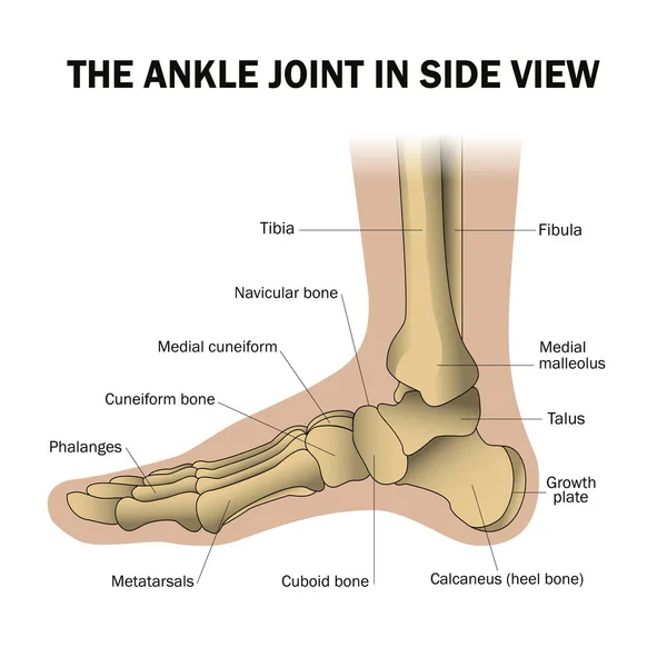
Bones of the foot and ankle joint medical vector illustration isolated on white background eps 10 infographic
Bones Foot Ankle Joint Medical Vector Illustration Isolated White Background Eps Infographic
Types of bone cells for Bone formation, resorption and remodeling. Osteocyte, lining cells, osteoblast, osteoclast. Osteogenesis. Isometric flat vector Illustration
Types Bone Cells Bone Formation Resorption Remodeling Osteocyte Lining Cells Osteoblast Osteoclast Osteogenesis Isometric Flat Vector Illustration
Blood stem cells haematopoiesis
Infographic of human in dental bridge illustration
Infographic Human Dental Bridge Illustration
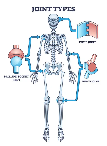
Joint types with fixed, hinge or ball and socket connections outline diagram. Labeled educational medical scheme with skeletal system and bone support vector illustration. Simple anatomical examples.
Joint Types Fixed Hinge Ball Socket Connections Outline Diagram Labeled Educational Medical Scheme Skeletal System Bone Support Vector Illustration Simple Anatomical Examples
The skull of a dog. Structure of the bones of the head, anatomical design. In Latin
Tooth Cross Section Anatomy with all parts including crown neck enamel dentin pulp cavity gums root canal cement bone and blood supply for medical science education and dental health care
Tooth Cross Section Anatomy All Parts Including Crown Neck Enamel Dentin Pulp Cavity Gums Root Canal Cement Bone Blood Supply Medical Science Education Dental Health Care
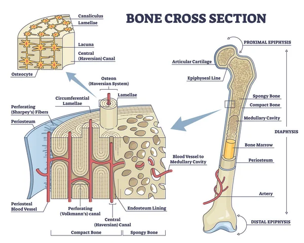
Bone cross section and isolated anatomical detailed structure outline diagram
Anatomical structure of the human Foot bones.
Mandibular fractures, including body, angle, ramus, parasymphyseal, condylar neck, and coronoid fractures diagram hand drawn schematic vector illustration. Medical science educational illustration
Mandibular Fractures Including Body Angle Ramus Parasymphyseal Condylar Neck Coronoid Fractures Diagram Hand Drawn Schematic Vector Illustration Medical Science Educational Illustration
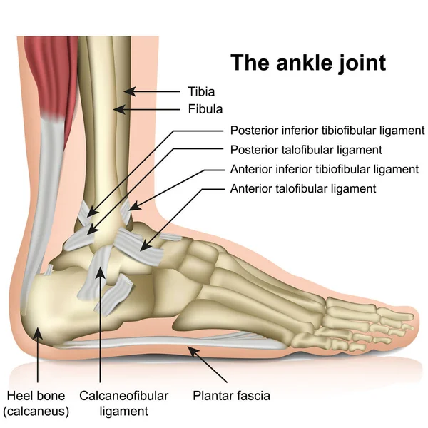
The ankle joint, tendons of the ankle joint foot anatomy vector illustration eps 10 infographic
Ankle Joint Tendons Ankle Joint Foot Anatomy Vector Illustration Eps Infographic
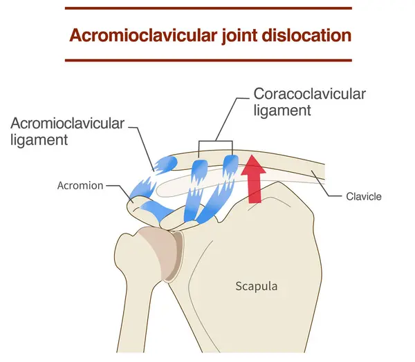
Dislocation of the acromioclavicular joint and ligaments and its mechanism
Dislocation Acromioclavicular Joint Ligaments Its Mechanism
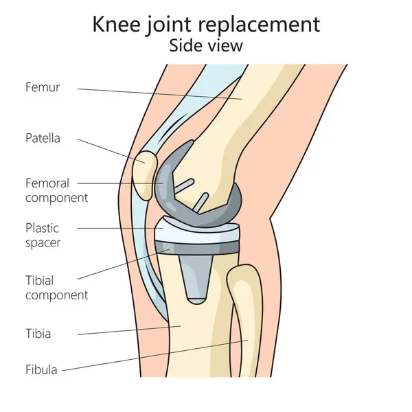
Knee joint replacement structure diagram hand drawn schematic vector illustration. Medical science educational illustration
Knee Joint Replacement Structure Diagram Hand Drawn Schematic Vector Illustration Medical Science Educational Illustration
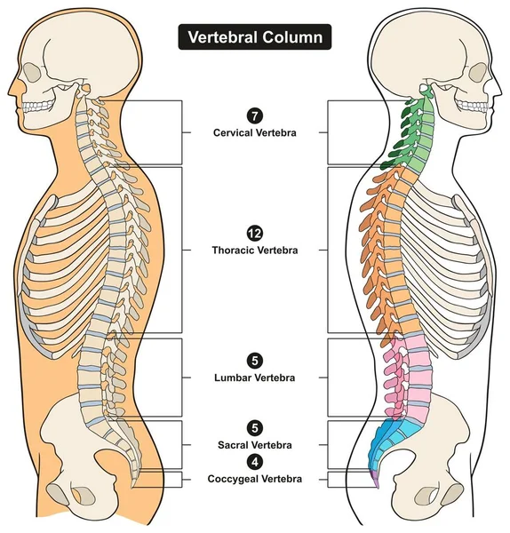
Vertebral Column of Human Body Anatomy infograpic diagram including all vertebra cervical thoracic lumbar sacral and coccygeal for medical science education and healthcare
Vertebral Column Human Body Anatomy Infograpic Diagram Including All Vertebra Cervical Thoracic Lumbar Sacral Coccygeal Medical Science Education Healthcare
Illustration foot bone. the bones of the foot
Illustration Foot Bone Bones Foot
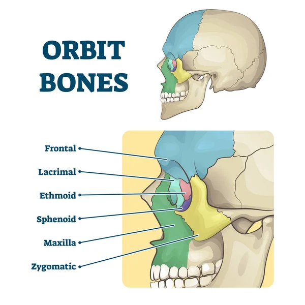
Orbit bones labeled educational skeletal division scheme vector illustration
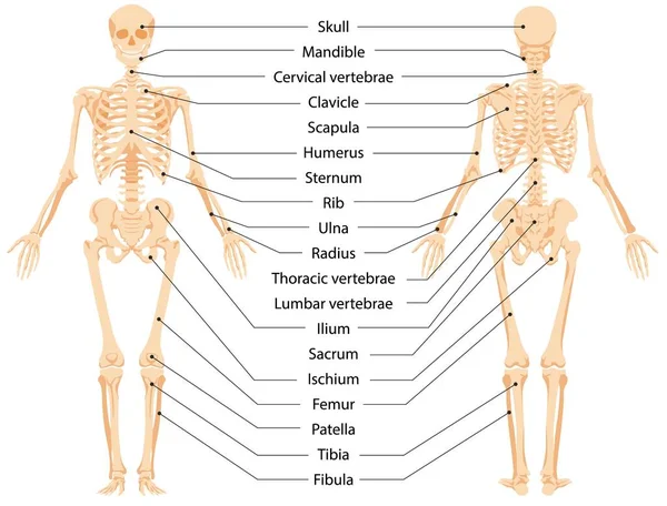
Human anatomical skeleton infographic front view and back view vector graphic illustration
Spine structure anatomy composition with colored view of spine divisions with editable text captions and vertebrae vector illustration
Spine Structure Anatomy Composition Colored View Spine Divisions Editable Text Captions Vertebrae Vector Illustration
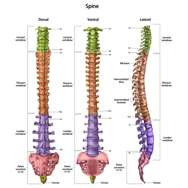
The human spine (vertebral column) with the name and description of all sites. Dorsal, lateral, ventral sides. Human anatomy. Vector illustration isolated on white background.
Stem cells & Hematopoiesis
Ligaments and joints of the cervical vertebrae and the occipital bone. Back view. Vector illustration
Ligaments Joints Cervical Vertebrae Occipital Bone Back View Vector Illustration
Medical Education Chart of Biology for Bones of Hand Diagram
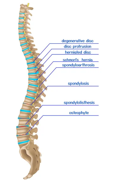
Damaged spine. Infographics of various back diseases. Vector illustration
Damaged Spine Infographics Various Back Diseases Vector Illustration
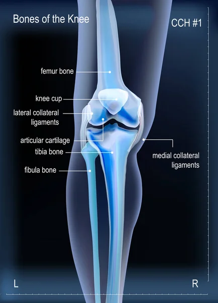
X ray bones the of knee
Human foot structure, anatomical medical teaching poster, bones of the heel, phalanges, joint, ankle. Healthcare mockup, vector illustration.
Human Foot Structure Anatomical Medical Teaching Poster Bones Heel Phalanges Joint Ankle Healthcare Mockup Vector Illustration
Structure of a Long bone. Humerus. Tube.
Structure Long Bone Humerus Tube
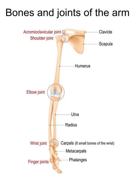
Human arm anatomy. Bones and joints of the arm, forearm, and hand. Upper limb of Skeleton front view. Vector illustration
Human Arm Anatomy Bones Joints Arm Forearm Hand Upper Limb Skeleton Front View Vector Illustration
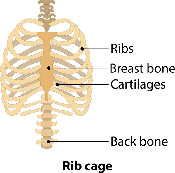
Hand drawn line art anatomically human ribcage. White background vector illustration. Anatomy of human rib. Illustration from vector about science and medical.
Hand Drawn Line Art Anatomically Human Ribcage White Background Vector Illustration Anatomy Human Rib Illustration Vector Science Medical
Jaw bone loss and Deterioration.
Human vertebral column in anterior posterior view, with spine parts labeled. Vector flat realistic concept illustration
Sternum or breastbone. Structure
414_bone, human thigh (thigh), back view, front view bone, human thigh (thigh), back view, front view, natural detailed hand drawing, graphics,
414_Bone Human Thigh Thigh Back View Front View Bone Human Thigh Thigh Back View Front View Natural Detailed Hand Drawing Graphics
MLC or medial collateral ligament anatomical location in knee outline diagram. Labeled educational leg skeletal system with bones and ligaments vector illustration. ACL, PCL and LCL medical study.
Mlc Medial Collateral Ligament Anatomical Location Knee Outline Diagram Labeled Educational Leg Skeletal System Bones Ligaments Vector Illustration Acl Pcl Lcl Medical Study
Human Spine Column
Human Spine Column
Bones of the Foot .Tarsals or tarsus, Metatarsals, Phalanges
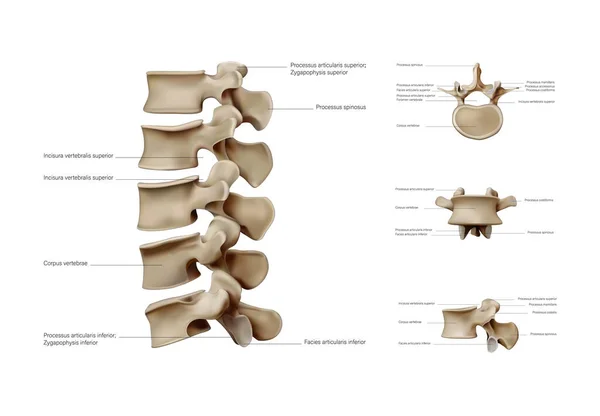
Anatomy of the vertebral column. Vertebra
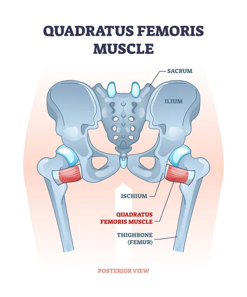
Quadratus femoris muscle as hip and groin rotator joint outline diagram
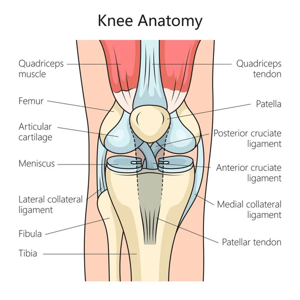
Anatomy of the human knee joint structure diagram schematic vector illustration. Medical science educational illustration
Anatomy Human Knee Joint Structure Diagram Schematic Vector Illustration Medical Science Educational Illustration
Page