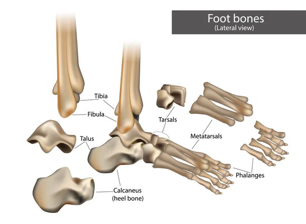
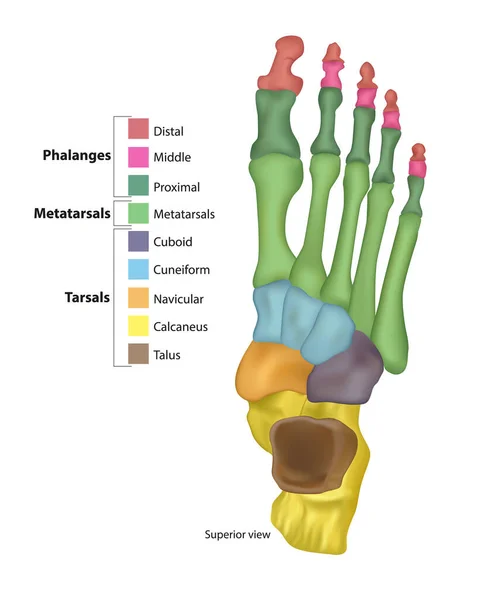
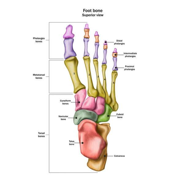
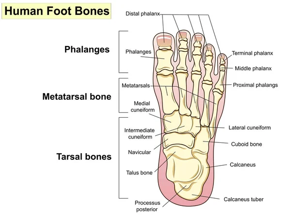
Anatomical structure of the human Foot bones.
Mandibular fractures, including body, angle, ramus, parasymphyseal, condylar neck, and coronoid fractures diagram hand drawn schematic vector illustration. Medical science educational illustration
Mandibular Fractures Including Body Angle Ramus Parasymphyseal Condylar Neck Coronoid Fractures Diagram Hand Drawn Schematic Vector Illustration Medical Science Educational Illustration
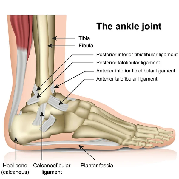
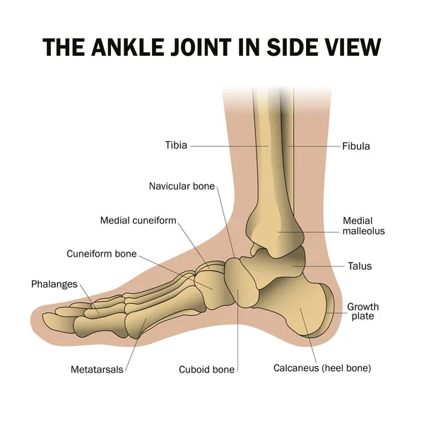
The ankle joint, tendons of the ankle joint foot anatomy vector illustration eps 10 infographic
Ankle Joint Tendons Ankle Joint Foot Anatomy Vector Illustration Eps Infographic
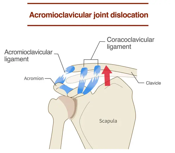
Dislocation of the acromioclavicular joint and ligaments and its mechanism
Dislocation Acromioclavicular Joint Ligaments Its Mechanism
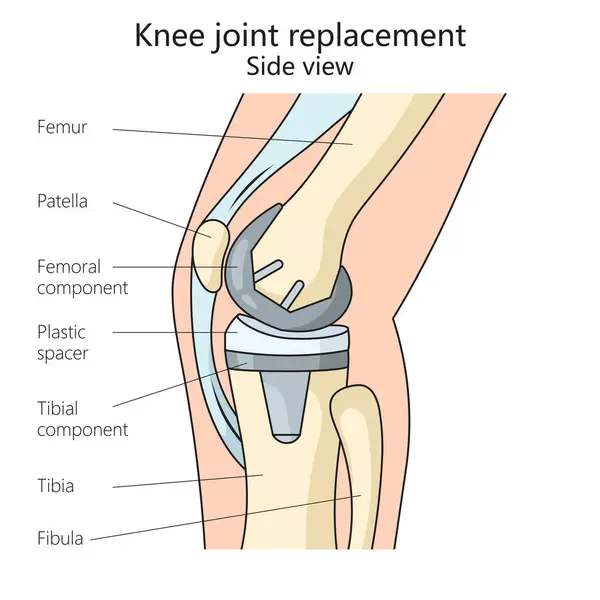
Knee joint replacement structure diagram hand drawn schematic vector illustration. Medical science educational illustration
Knee Joint Replacement Structure Diagram Hand Drawn Schematic Vector Illustration Medical Science Educational Illustration
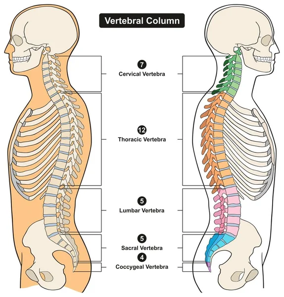
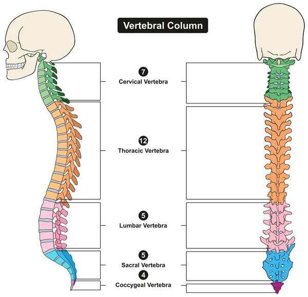
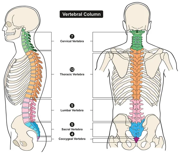
Vertebral Column of Human Body Anatomy infograpic diagram including all vertebra cervical thoracic lumbar sacral and coccygeal for medical science education and healthcare
Vertebral Column Human Body Anatomy Infograpic Diagram Including All Vertebra Cervical Thoracic Lumbar Sacral Coccygeal Medical Science Education Healthcare
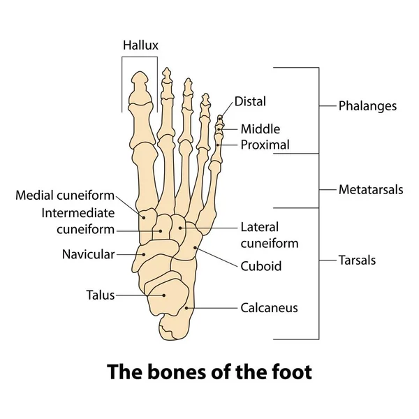
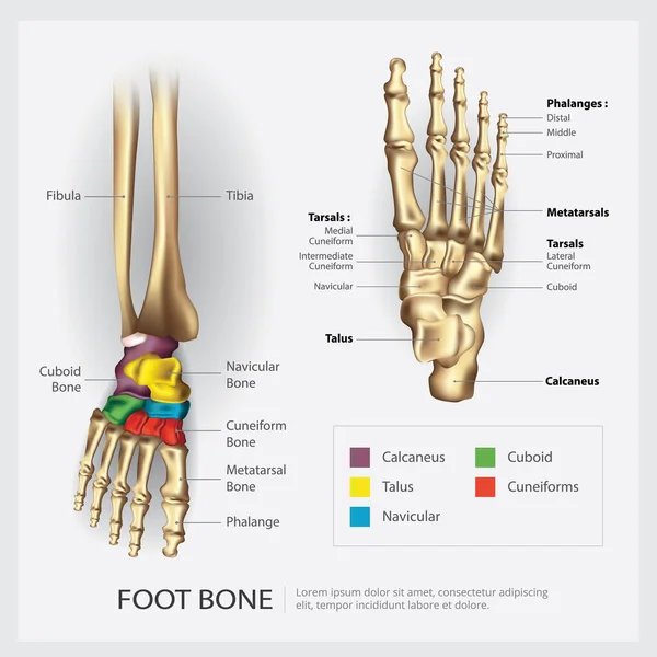
Illustration foot bone. the bones of the foot
Illustration Foot Bone Bones Foot
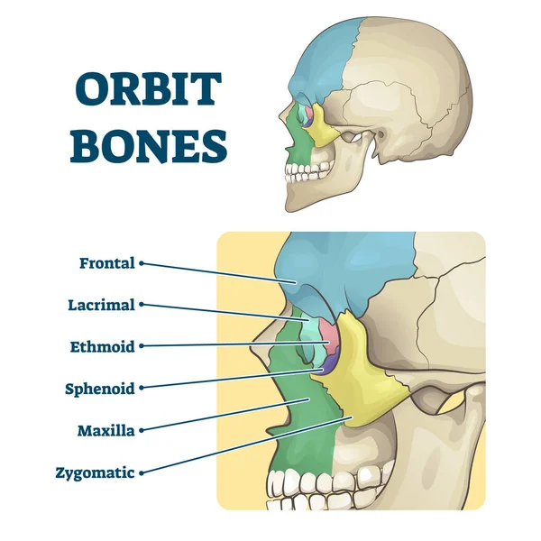
Orbit bones labeled educational skeletal division scheme vector illustration
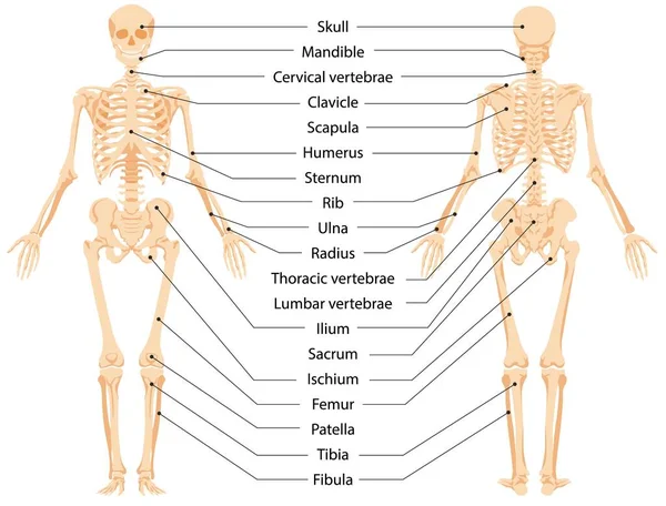
Human anatomical skeleton infographic front view and back view vector graphic illustration
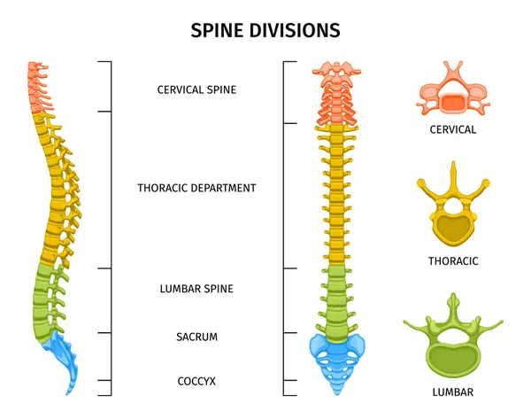
Spine structure anatomy composition with colored view of spine divisions with editable text captions and vertebrae vector illustration
Spine Structure Anatomy Composition Colored View Spine Divisions Editable Text Captions Vertebrae Vector Illustration
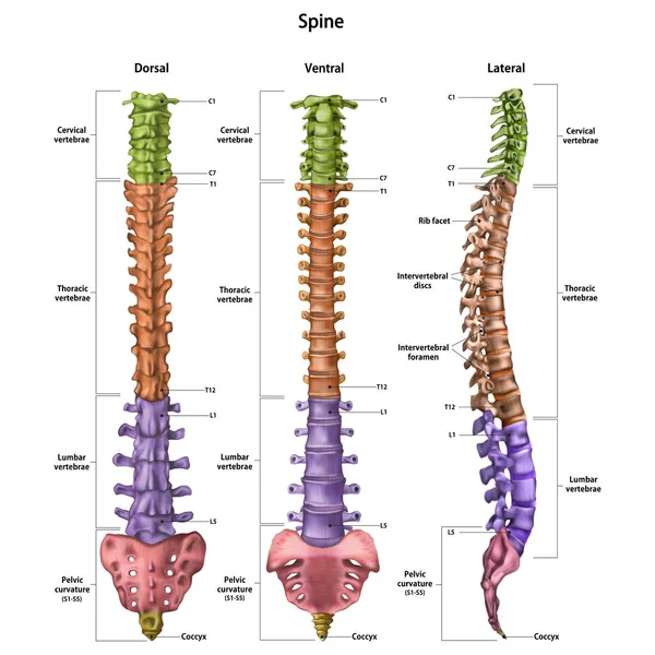
The human spine (vertebral column) with the name and description of all sites. Dorsal, lateral, ventral sides. Human anatomy. Vector illustration isolated on white background.
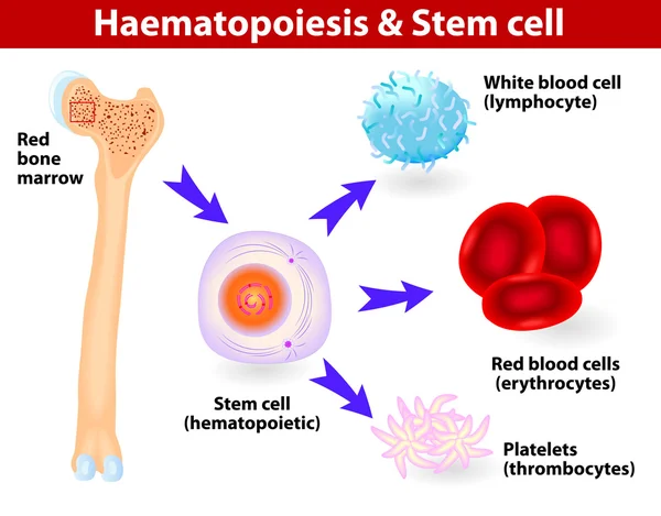
Stem cells & Hematopoiesis
Ligaments and joints of the cervical vertebrae and the occipital bone. Back view. Vector illustration
Ligaments Joints Cervical Vertebrae Occipital Bone Back View Vector Illustration
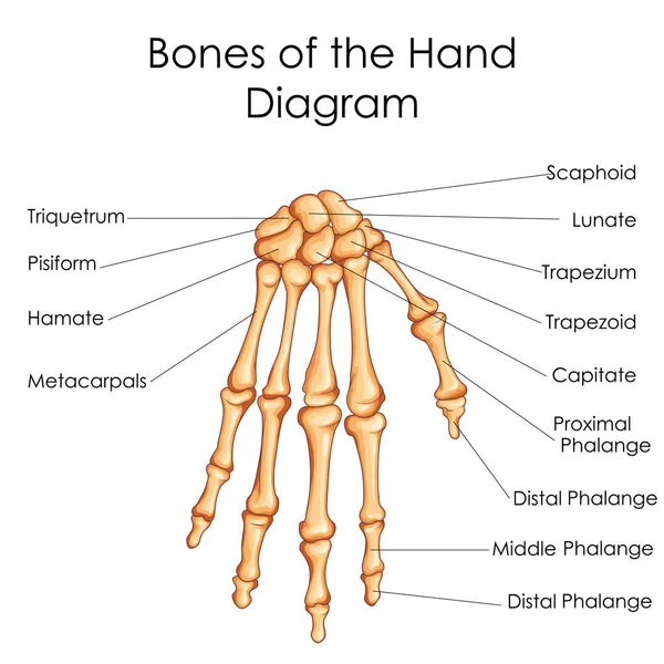
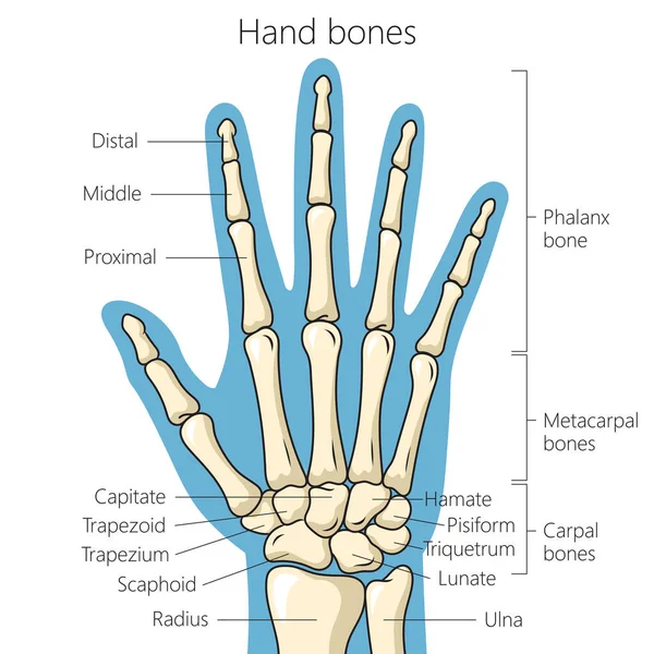
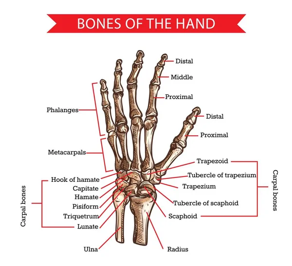
Medical Education Chart of Biology for Bones of Hand Diagram
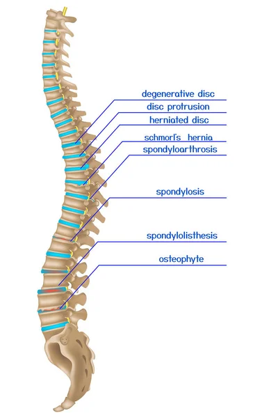
Damaged spine. Infographics of various back diseases. Vector illustration
Damaged Spine Infographics Various Back Diseases Vector Illustration
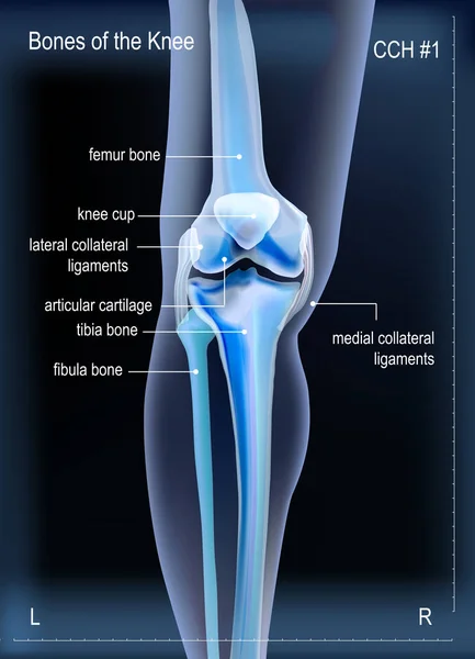
X ray bones the of knee
Human foot structure, anatomical medical teaching poster, bones of the heel, phalanges, joint, ankle. Healthcare mockup, vector illustration.
Human Foot Structure Anatomical Medical Teaching Poster Bones Heel Phalanges Joint Ankle Healthcare Mockup Vector Illustration
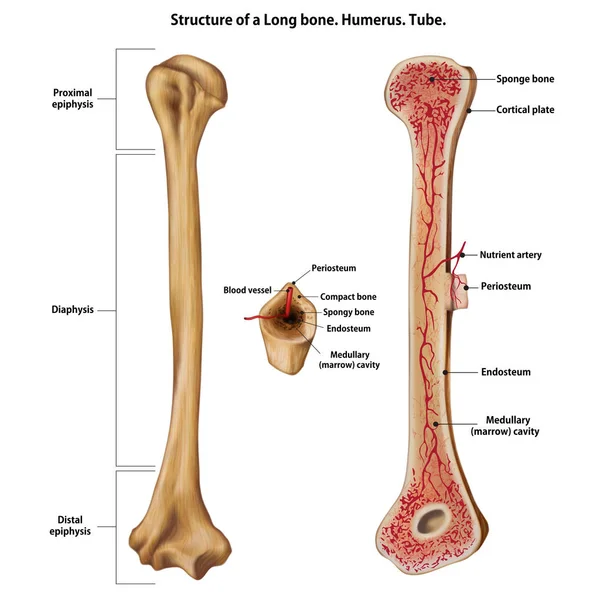
Structure of a Long bone. Humerus. Tube.
Structure Long Bone Humerus Tube
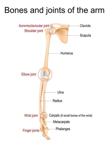
Human arm anatomy. Bones and joints of the arm, forearm, and hand. Upper limb of Skeleton front view. Vector illustration
Human Arm Anatomy Bones Joints Arm Forearm Hand Upper Limb Skeleton Front View Vector Illustration
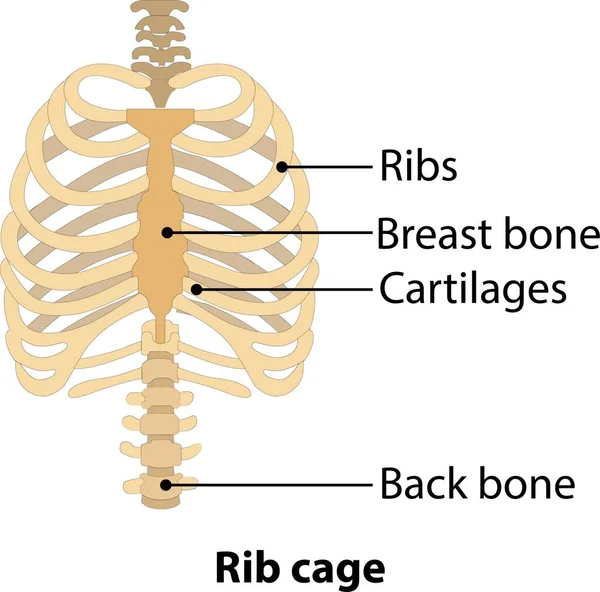
Hand drawn line art anatomically human ribcage. White background vector illustration. Anatomy of human rib. Illustration from vector about science and medical.
Hand Drawn Line Art Anatomically Human Ribcage White Background Vector Illustration Anatomy Human Rib Illustration Vector Science Medical
Jaw bone loss and Deterioration.
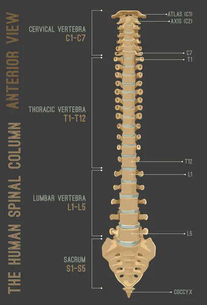
Human vertebral column in anterior posterior view, with spine parts labeled. Vector flat realistic concept illustration
Sternum or breastbone. Structure
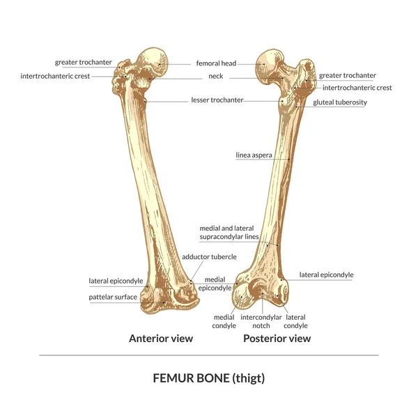
414_bone, human thigh (thigh), back view, front view bone, human thigh (thigh), back view, front view, natural detailed hand drawing, graphics,
414_Bone Human Thigh Thigh Back View Front View Bone Human Thigh Thigh Back View Front View Natural Detailed Hand Drawing Graphics
MLC or medial collateral ligament anatomical location in knee outline diagram. Labeled educational leg skeletal system with bones and ligaments vector illustration. ACL, PCL and LCL medical study.
Mlc Medial Collateral Ligament Anatomical Location Knee Outline Diagram Labeled Educational Leg Skeletal System Bones Ligaments Vector Illustration Acl Pcl Lcl Medical Study
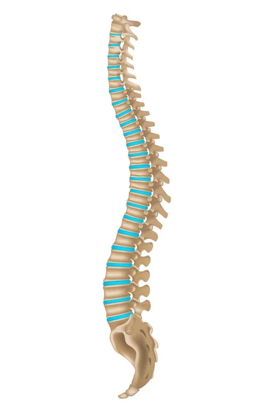
Human Spine Column
Human Spine Column
Bones of the Foot .Tarsals or tarsus, Metatarsals, Phalanges
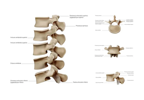
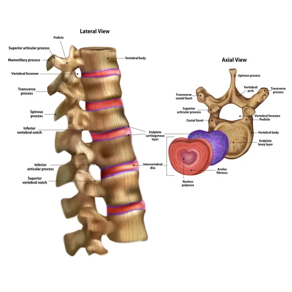
Anatomy of the vertebral column. Vertebra
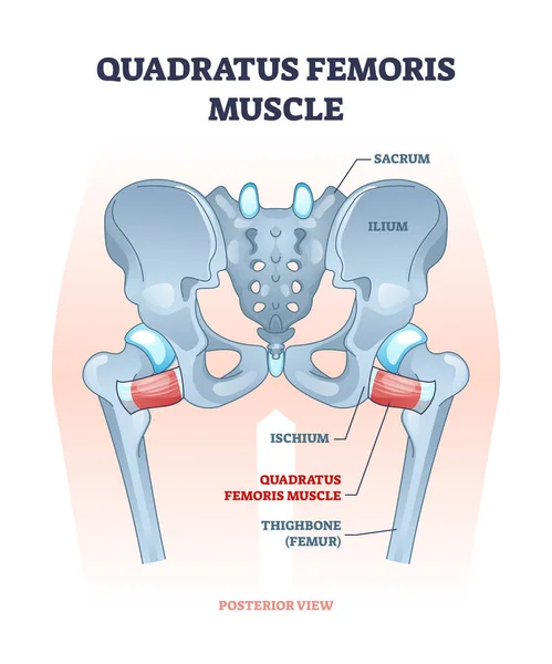
Quadratus femoris muscle as hip and groin rotator joint outline diagram
Anatomy of the human knee joint structure diagram schematic vector illustration. Medical science educational illustration
Anatomy Human Knee Joint Structure Diagram Schematic Vector Illustration Medical Science Educational Illustration
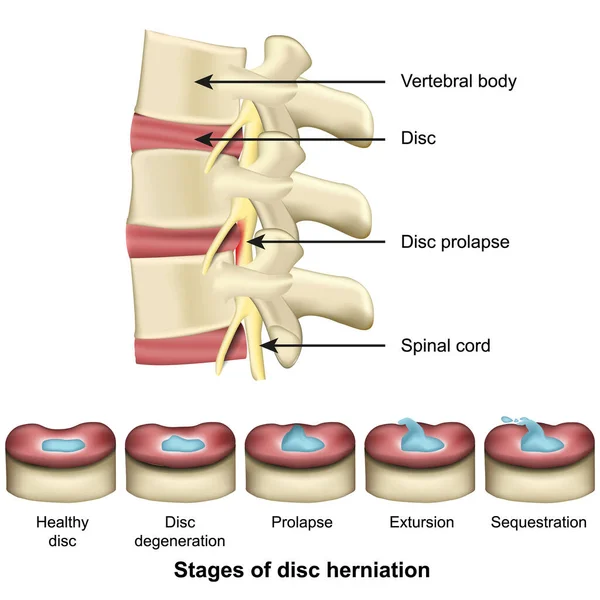
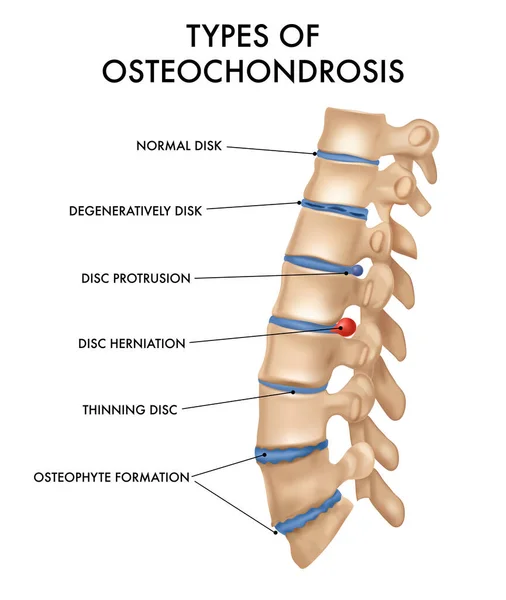
Stages of disc herniation spine and disc anatomy 3d medical vector illustration eps 10
Stages Disc Herniation Spine Disc Anatomy Medical Vector Illustration Eps
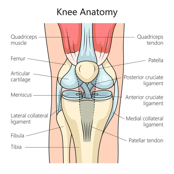
Human Knee joint anatomy. Knee Tendons, anatomical diagram
Human Knee Joint Anatomy Knee Tendons Anatomical Diagram
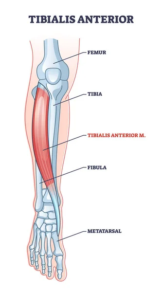
Tibialis anterior muscle with human leg skeletal structure outline diagram
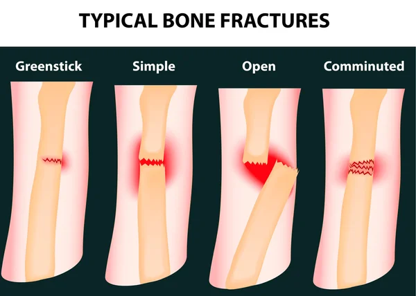
Typical bone fractures
Vertebral Column of Human Body Anatomy infograpic diagram including all vertebra cervical thoracic lumbar sacral and coccygeal for medical science education and healthcare
Vertebral Column Human Body Anatomy Infograpic Diagram Including All Vertebra Cervical Thoracic Lumbar Sacral Coccygeal Medical Science Education Healthcare
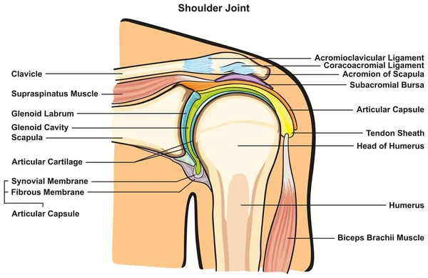
Shoulder Joint of Human Body Anatomy infographic diagram with all parts including bones ligaments muscles bursa cavity capsule cartilage membrane for medical science education and health care
Shoulder Joint Human Body Anatomy Infographic Diagram All Parts Including Bones Ligaments Muscles Bursa Cavity Capsule Cartilage Membrane Medical Science Education Health Care
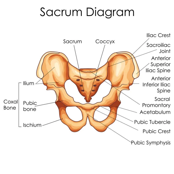
Medical Education Chart of Biology for Sacrum Diagram
Knee arthrosis. Healthy knee structure. Bone exposure, healthy knee structure comparison. Medical poster with foot bones. Isolated vector illustration.
Knee Arthrosis Healthy Knee Structure Bone Exposure Healthy Knee Structure Comparison Medical Poster Foot Bones Isolated Vector Illustration
The anatomical structure of the tooth. Infographics. Vector illustration on isolated background
Bones of the human foot with the name and description of all sit
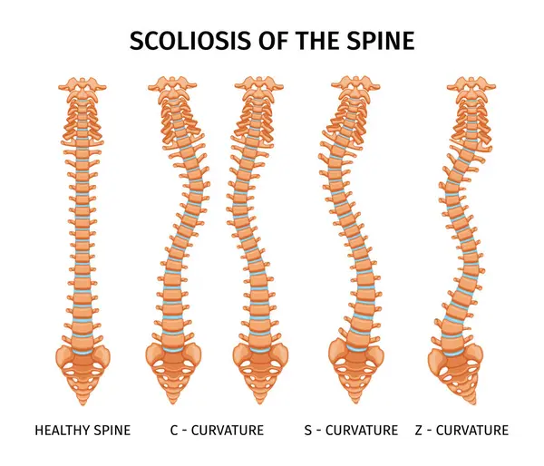
Types of scoliosis of spine infographics with set of isolated spine curvature images on blank background vector illustration
Types Scoliosis Spine Infographics Set Isolated Spine Curvature Images Blank Background Vector Illustration
Human skull bones structure diagram schematic vector illustration. Medical science educational illustration
Human Skull Bones Structure Diagram Schematic Vector Illustration Medical Science Educational Illustration
Human vertebral column in anterior posterior view, Intervertebral disc with spine parts labeled. Vector flat realistic
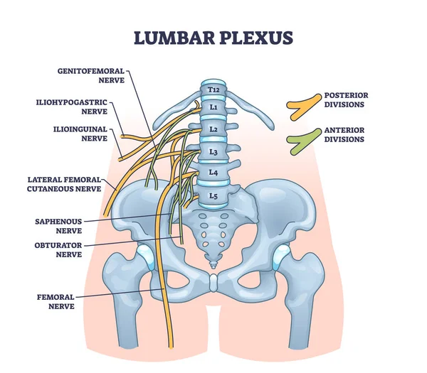
Lumbar plexus as anatomical web of lumbosacral nerves outline diagram. Labeled educational scheme with medical location of genitofemoral, iliohypogastric and ilioinguinal nerves vector illustration.
Lumbar Plexus Anatomical Web Lumbosacral Nerves Outline Diagram Labeled Educational Scheme Medical Location Genitofemoral Iliohypogastric Ilioinguinal Nerves Vector Illustration
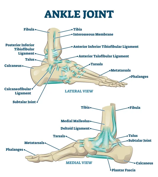
Ankle joint vector illustration. Labeled educational leg structure scheme.
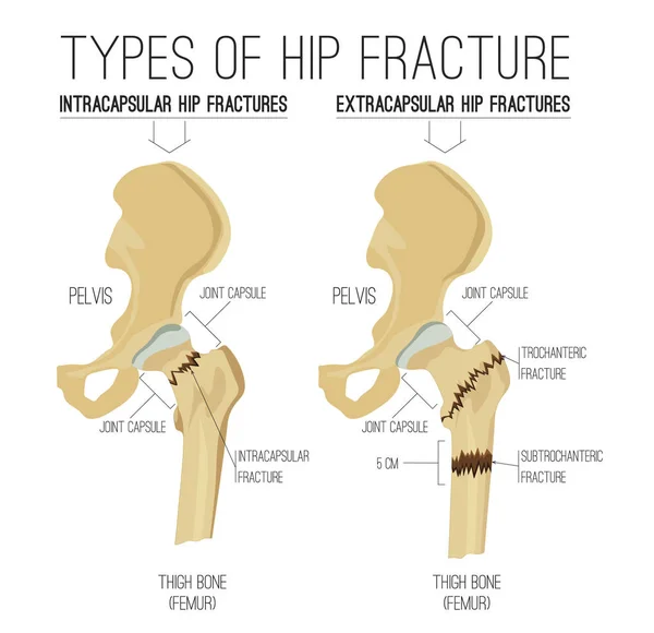
Types of hip fracture
Skeleton. Intervertebral disc with the name and description of all sites.
Skeleton Intervertebral Disc Name Description All Sites
Bones of the hand anatomy schematic diagram vector illustration. Medical science educational illustration
Bones Hand Anatomy Schematic Diagram Vector Illustration Medical Science Educational Illustration
The structure of the ulna bone with the name and description of
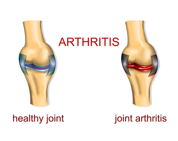
JOINT ARTHRITIS
JOINT ARTHRITIS
Spinal disc herniation vector illustration
Spinal Disc Herniation Vector Illustration
Myeloma pathology illustration. Illustration of a bone marrow with cancer producing M-protein
Myeloma Pathology Illustration Illustration Bone Marrow Cancer Producing Protein
Human spine anatomy in two projections. Intervertebral discs. 3d rendering. Vector illustration.
Human Spine Anatomy Two Projections Intervertebral Discs Rendering Vector Illustration
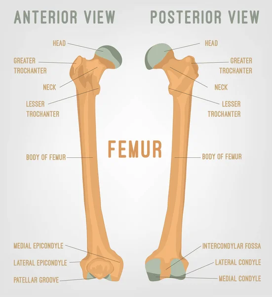
Human femur bones
Human femur bones
Anatomy of human knee vector sketch of leg bones and joint, medicine design. Side and front view of knee bones, hand drawn femur, patella, tibia and fibula, tibial plateau and lateral condyle
Anatomy Human Knee Vector Sketch Leg Bones Joint Medicine Design Side Front View Knee Bones Hand Drawn Femur Patella Tibia Fibula Tibial Plateau Lateral Condyle
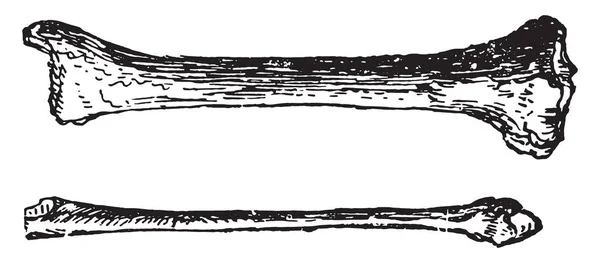
Ulna and radius, vintage engraved illustration
Ulna Radius Vintage Engraved Illustration
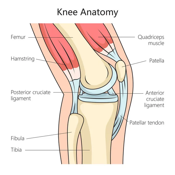
Anatomy of the human knee joint side view structure diagram schematic vector illustration. Medical science educational illustration
Anatomy Human Knee Joint Side View Structure Diagram Schematic Vector Illustration Medical Science Educational Illustration
Spine Hernia Realistic Chart
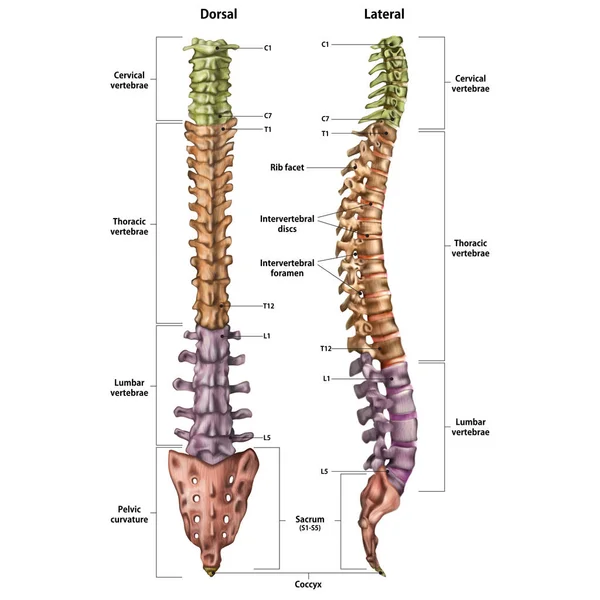
Illustration of the human spine with the name and description of all sites. Lateral and spinal views.
Illustration Human Spine Name Description All Sites Lateral Spinal Views
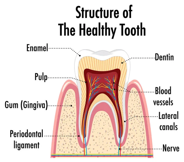
Infographic of human in structure of the healthy tooth illustration
Infographic Human Structure Healthy Tooth Illustration
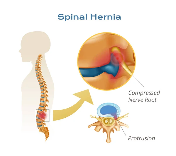
Vertebrae spinal cord anatomy infographics with body silhouette with spine and compressed nerve root protrusion views vector illustration
Vertebrae Spinal Cord Anatomy Infographics Body Silhouette Spine Compressed Nerve Root Protrusion Views Vector Illustration
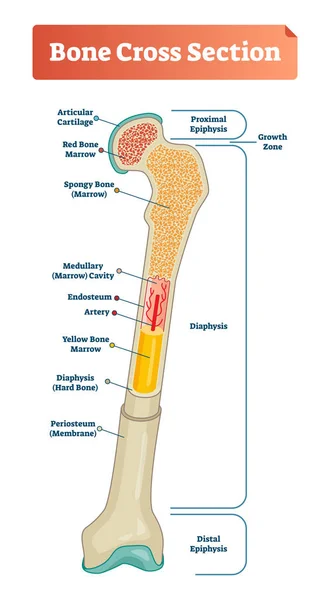
Vector illustration scheme of bone cross section. Diagram with articular cartilage, marrow, spongy bone, medullary cavity, endosteum, diaphysis, and periosteum.
Types of hip fracture. Femoral neck fracture. Medical vector illustration
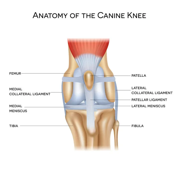
Anatomy of the canine (dog's) knee joint colorful design, healthy joint info poster illustration
Anatomy Canine Dog Knee Joint Colorful Design Healthy Joint Info Poster Illustration
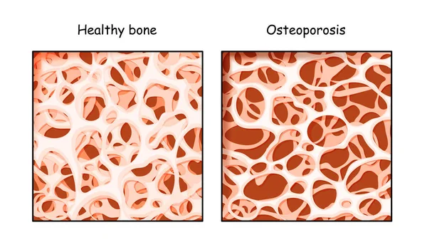
Healthy bone and Osteoporosis in comparison isolated on a white background. disease of bones that leads to an increased risk of fracture. low density calcium. Vector illustration for medical, educational or scientific graphic design.
Healthy Bone Osteoporosis Comparison Isolated White Background Disease Bones Leads Increased Risk Fracture Low Density Calcium Vector Illustration Medical Educational Scientific Graphic Design
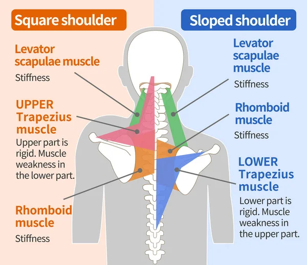
Muscular structure of the square shoulder and sloped shoulder
Muscular Structure Square Shoulder Sloped Shoulder
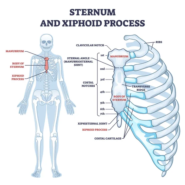
Sternum and xiphoid process with breastbone bone structure outline diagram
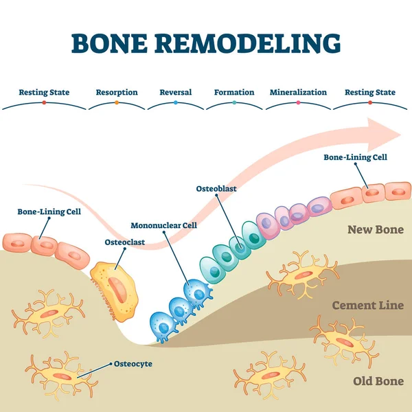
Bone remodeling process educational explanation with labeled structure scheme
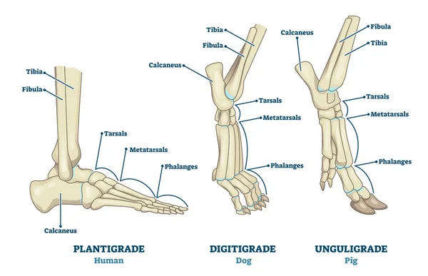
Plantigrade, Digitigrade and Unguligrade comparison vector illustration. Educational labeled structure scheme with human, dog and pig legs collection. Bone skeleton parts with location explanation.
Plantigrade Digitigrade Unguligrade Comparison Vector Illustration Educational Labeled Structure Scheme Human Dog Pig Legs Collection Bone Skeleton Parts Location Explanation
Anatomy. Human foot bon Anatomy. Human foot bones. The main parts that make up the foot are shown. For basic medical education. Signatures and text. Also for clinics set the textes.
Anatomy Human Foot Bon Anatomy Human Foot Bones Main Parts Make Foot Shown Basic Medical Education Signatures Text Also Clinics Set Textes
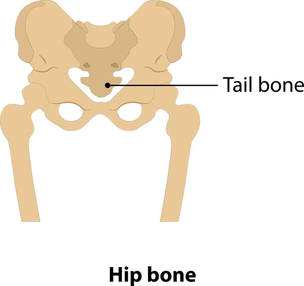
Human hip bone and tail bone. vector. on white background
Human Hip Bone Tail Bone Vector White Background
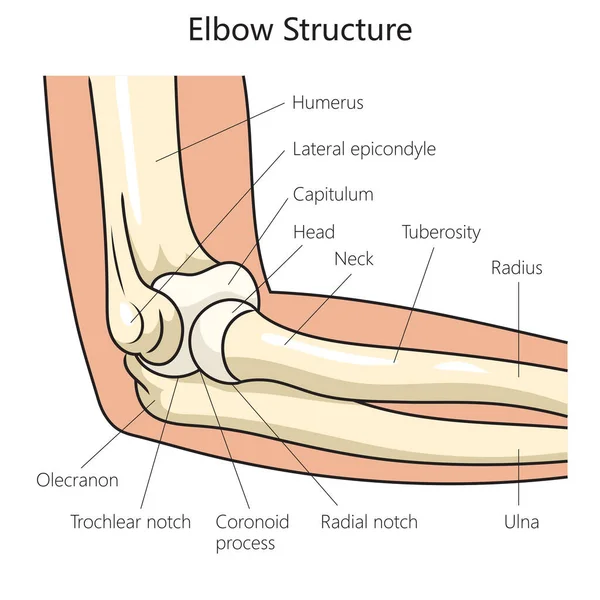
Anatomy structure of the human elbow diagram schematic vector illustration. Medical science educational illustration
Anatomy Structure Human Elbow Diagram Schematic Vector Illustration Medical Science Educational Illustration
Hand and wrist bones vector sketch of human anatomy and medicine design. Hand drawn arm of skeleton with radius, ulna, finger phalanges and palm metacarpals, trapezoid, scaphoid and carpal bones
Hand Wrist Bones Vector Sketch Human Anatomy Medicine Design Hand Drawn Arm Skeleton Radius Ulna Finger Phalanges Palm Metacarpals Trapezoid Scaphoid Carpal Bones
Bone remodeling process. Osteoblast, osteoclast, and osteocyte.
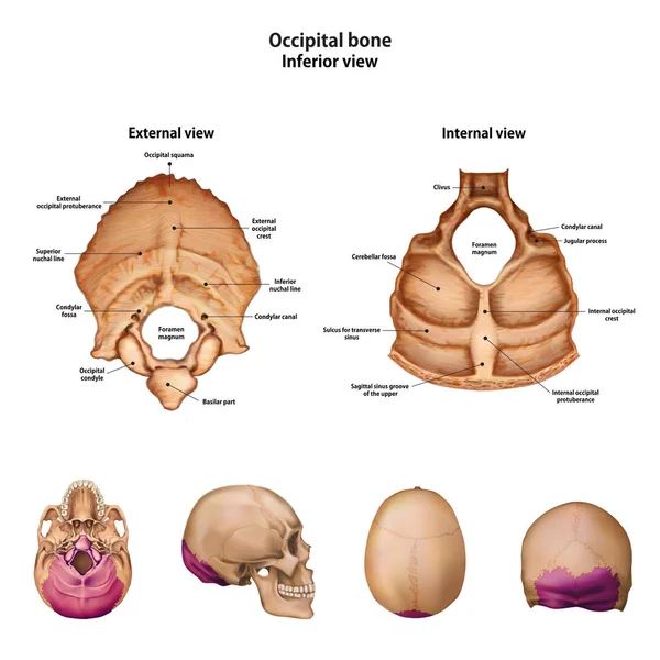
Occipital bone. With the name and description of all sites.
Occipital Bone Name Description All Sites
Structure of the humerus bone with the name and description of a
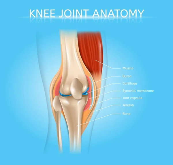
Human Knee Joint Anatomy Realistic Vector Scheme
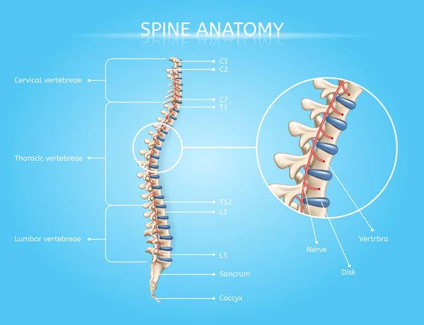
Human Spine Anatomy Vector Medical Infographic
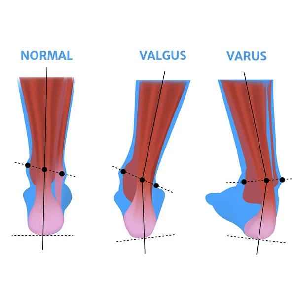
Valgus deformity of the foot. Orthopedic pathology. Abnormal deformity of joints. Curvature of the feet. Vector illustration.
Valgus Deformity Foot Orthopedic Pathology Abnormal Deformity Joints Curvature Feet Vector Illustration
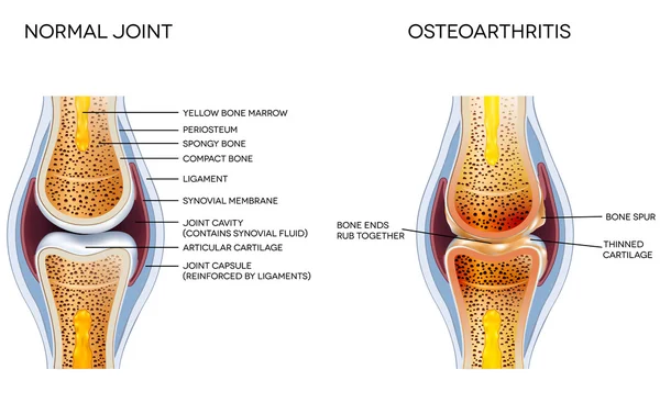
Osteoarthritis and normal joint anatomy
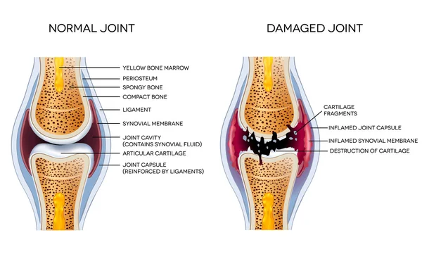
Damaged joint and healthy joint detailed diagram
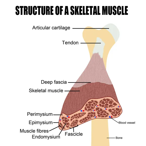
Structure of a skeletal muscle
Layer of a Long bone. Humerus. Tube.
Layer Long Bone Humerus Tube
Spongy vs compact bone comparison with anatomical structure outline diagram
Structure of the human knee joint with the name and description of all sites. Lateral view. Medical science anatomy poster. Vector illustration isolated on white background.
Vertebral Column of Human Body Anatomy infograpic diagram including all vertebra cervical thoracic lumbar sacral and coccygeal for medical science education and healthcare
Vertebral Column Human Body Anatomy Infograpic Diagram Including All Vertebra Cervical Thoracic Lumbar Sacral Coccygeal Medical Science Education Healthcare
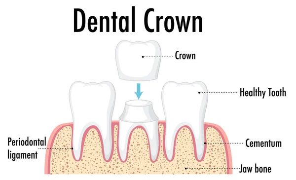
Infographic of human in dental crown illustration
Infographic Human Dental Crown Illustration
Fracture repair as educational bone remodeling formation vector illustration
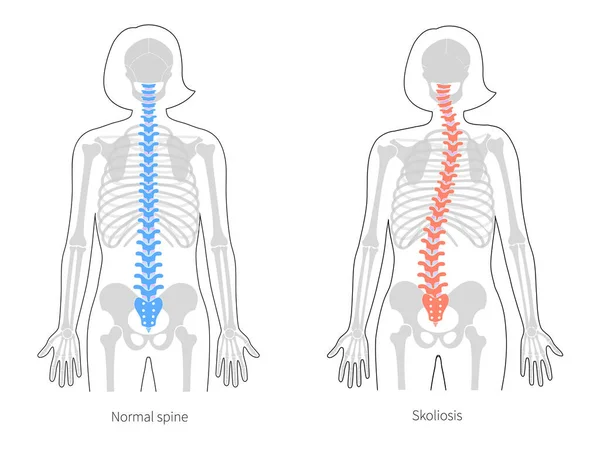
Scoliosis flat vector illustration
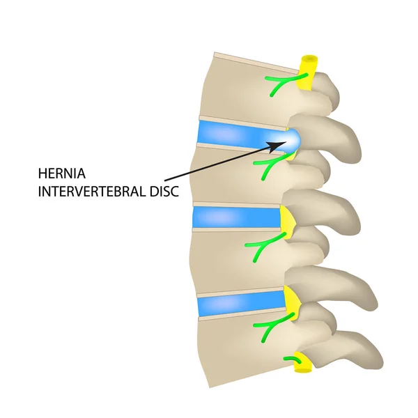
A hernia of the intervertebral disc. Vector illustration on isolated background.
Frontal view x ray of bones the of hand with anotations.
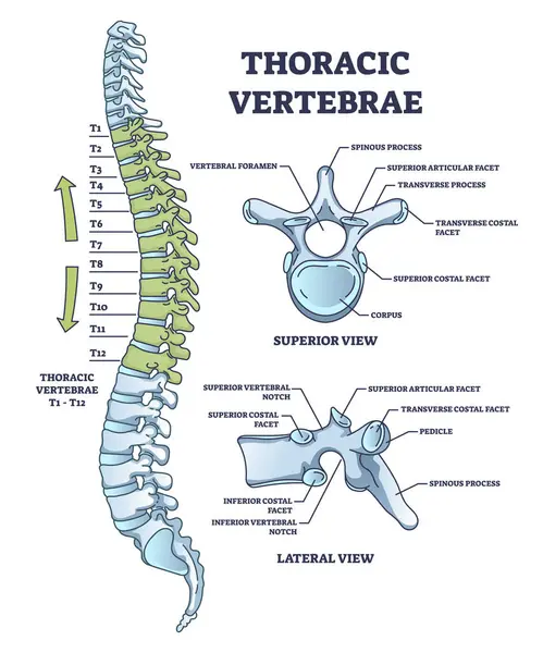
Thoracic vertebrae location and medical structure description outline diagram. Labeled educational scheme with anatomical backbone parts and detailed superior or lateral bone view vector illustration
Thoracic Vertebrae Location Medical Structure Description Outline Diagram Labeled Educational Scheme Anatomical Backbone Parts Detailed Superior Lateral Bone View Vector Illustration
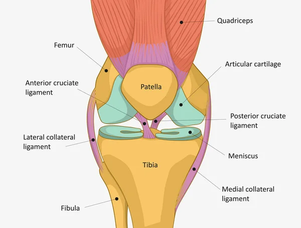
Collateral ligaments with knee anatomical skeletal structure outline diagram. Labeled educational joint part description with anterior and posterior cruciate ligament vector illustration. Leg anatomy
Collateral Ligaments Knee Anatomical Skeletal Structure Outline Diagram Labeled Educational Joint Part Description Anterior Posterior Cruciate Ligament Vector Illustration Leg Anatomy
Vector skeleton flat illustration
The skeleton kneels and prays. 3D. A religious man. Vector illustration.
Skeleton Kneels Prays Religious Man Vector Illustration
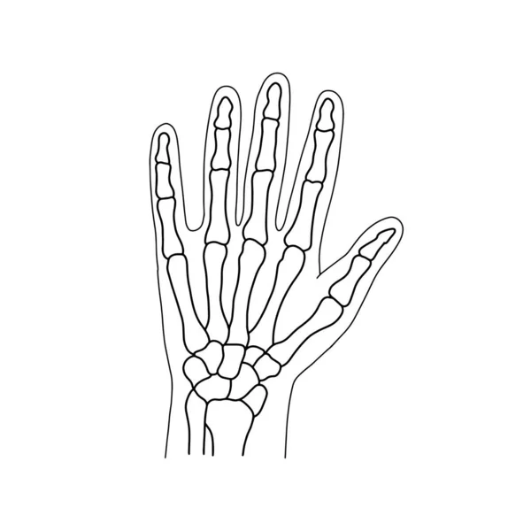
Skeletons of the human hand. Drawn by lines on white background. Vector Stock illustration.
Skeletons Human Hand Drawn Lines White Background Vector Stock Illustration
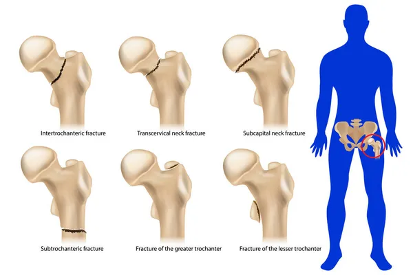
Femoral neck fracture. Types of hip fractures. Subtrochanteric, Intertrochanteric, Transcervical and Subcapital neck fracture, Fracture of the greater and lesser trochanter.
Foot Bone Anatomy Vector Illustration
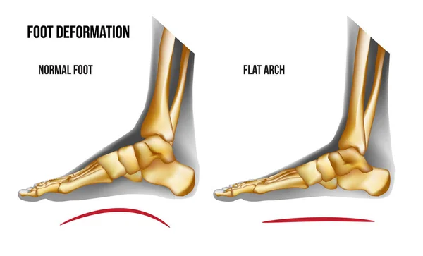
Anatomy flat foot arch medial view
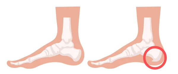
Heel spur illustration. Design elements on the theme of plantar fasciitis. Vector set.
Heel Spur Illustration Design Elements Theme Plantar Fasciitis Vector Set
Page