Osso frontale Stock Vector Images

305 Osso frontale vector art & graphics are available under a royalty-free license

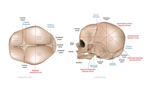
Anatomy of the Newborn Skull. Lateral view and view from the top. Cranial sutures
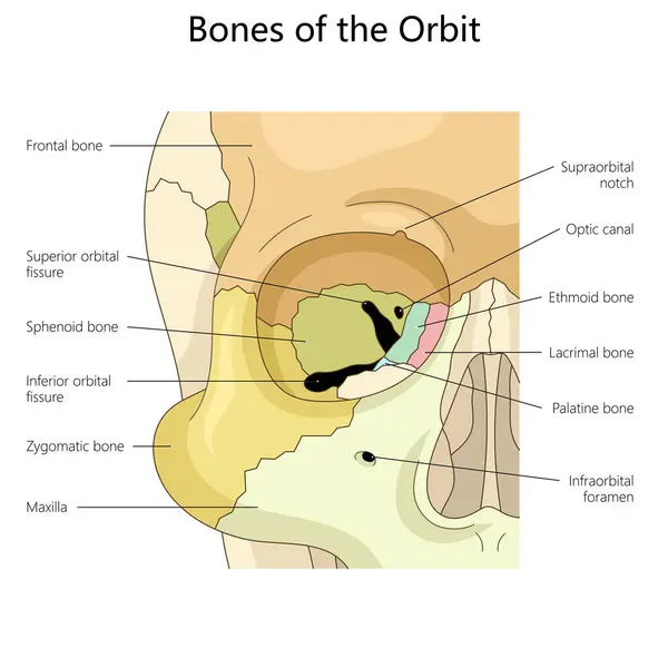
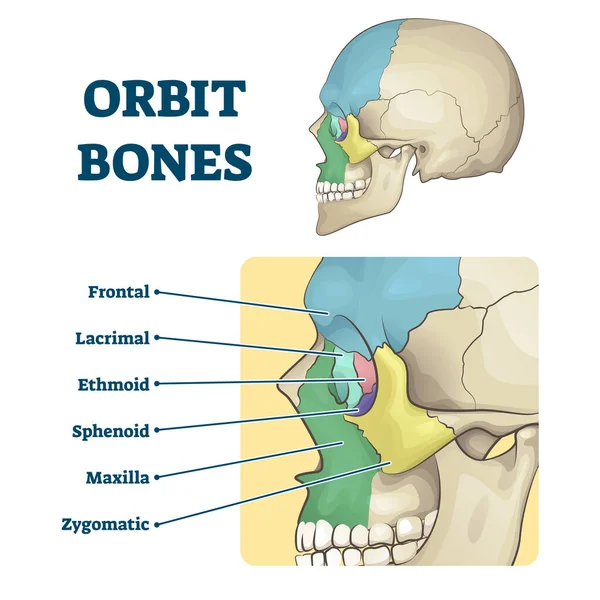
Bones of the orbit, highlighting key structures such as the frontal, zygomatic, and ethmoid bones structure diagram hand drawn schematic vector illustration. Medical science educational illustration
Bones Orbit Highlighting Key Structures Frontal Zygomatic Ethmoid Bones Structure Diagram Hand Drawn Schematic Vector Illustration Medical Science Educational Illustration
Human anatomy
Human anatomy
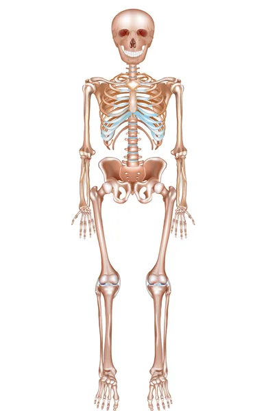
Vector glassy anatomy skeleton
Vector Glassy Anatomy Skeleton
Human Skull Diagram Illustration
Illustration of skull sutures diagram
Illustration Skull Sutures Diagram
Human anatomy
Human anatomy
Developmental stages of fontanelle closure in infants, detailing sutures and bone structure of the skull diagram hand drawn schematic vector illustration. Medical science educational illustration
Developmental Stages Fontanelle Closure Infants Detailing Sutures Bone Structure Skull Diagram Hand Drawn Schematic Vector Illustration Medical Science Educational Illustration
Human vs Chimpanzee Skull Anatomy Comparison Diagram
Human Chimpanzee Skull Anatomy Comparison Diagram
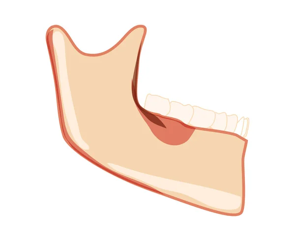
Mandible of Skeleton Human head side view with teeth. Skull lower jaw model with teeth row. Chump realistic flat natural
Anatomy of the knee joint front view. Knee Muscles and Ligaments Parts, knee tendons
Anatomy Knee Joint Front View Knee Muscles Ligaments Parts Knee Tendons
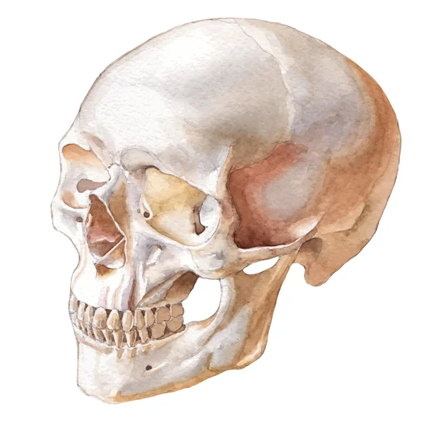
Side view of Human Skull
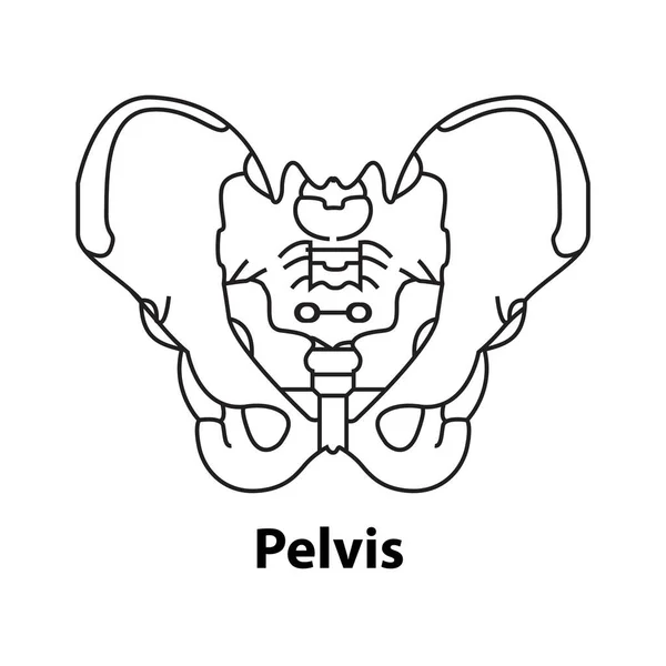
Human skeleton anatomy icon, pelvis bones vector. Body structure element isolated
Human Skeleton Anatomy Icon Pelvis Bones Vector Body Structure Element Isolated
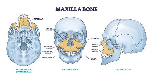
Maxilla bone detailed structure and facial skeleton anatomy outline diagram. Labeled educational scheme with medical inferior view without mandible, anterior and lateral skull view vector illustration
Maxilla Bone Detailed Structure Facial Skeleton Anatomy Outline Diagram Labeled Educational Scheme Medical Inferior View Mandible Anterior Lateral Skull View Vector Illustration
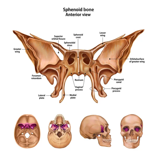
Sphenoid bone. With the name and description of all sites.
Sphenoid Bone Name Description All Sites
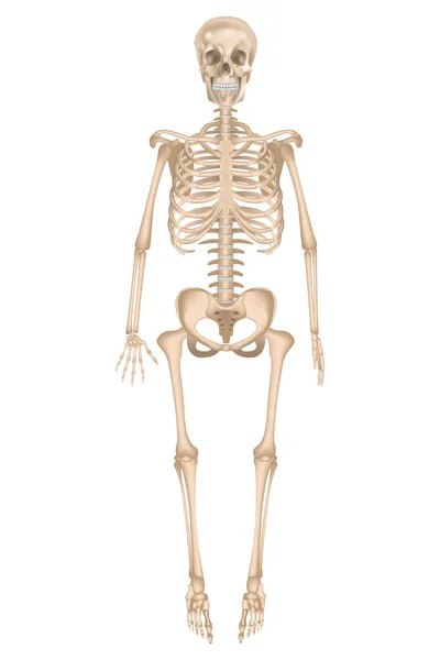
Human Skeleton Anatomy. Vector illustration.
Human Skeleton Anatomy Vector Illustration
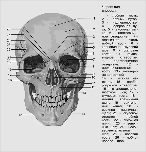
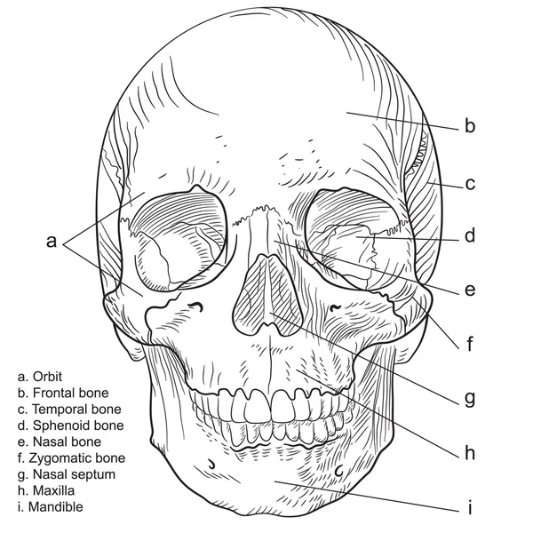
Medical Education Chart of Biology Human Skull Diagram. Vector. Front aspect white background basic medical education
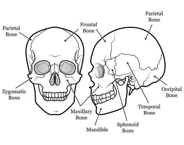
Human skull bones structure diagram schematic vector illustration. Medical science educational illustration
Human Skull Bones Structure Diagram Schematic Vector Illustration Medical Science Educational Illustration
Anatomical structure of human body, presented in of skeleton
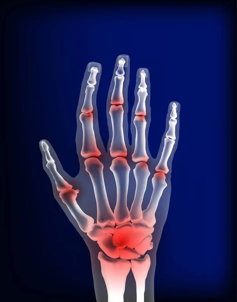
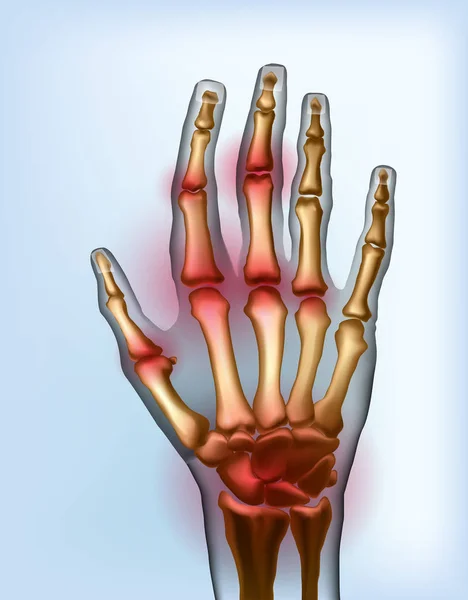
Osteoarthritis image sore inflammation joints of bones the of hand.
Medical illustration comparing a normal ankle, with a dislocated ankle, with annotations.
Medical Illustration Comparing Normal Ankle Dislocated Ankle Annotations
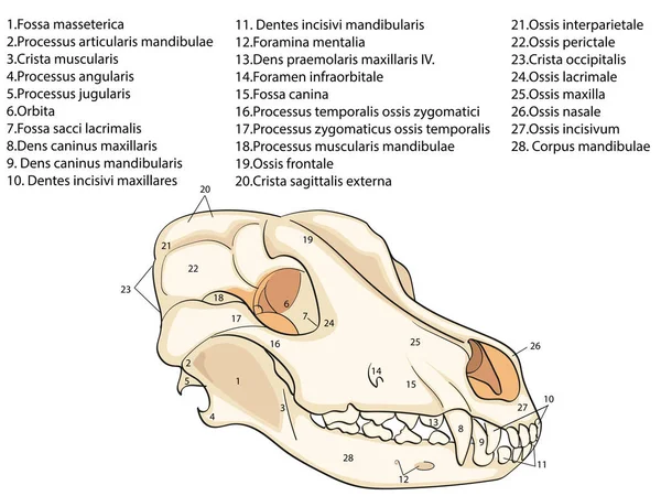
The skull of a dog. Structure of the bones of the head, anatomical design. In Latin
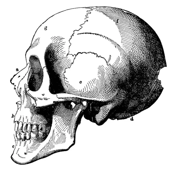
A typical representation of the human skull, with its parts labelled as a, b, c, d, e, f and g, representing, nasal bone, superior maxillary, inferior maxillary, occipital, temporal, parietal and frontal bone, respectively, vintage line drawing or en
Typical Representation Human Skull Its Parts Labelled Representing Nasal Bone Superior Maxillary Inferior Maxillary Occipital Temporal Parietal Frontal Bone Respectively Vintage Line Drawing
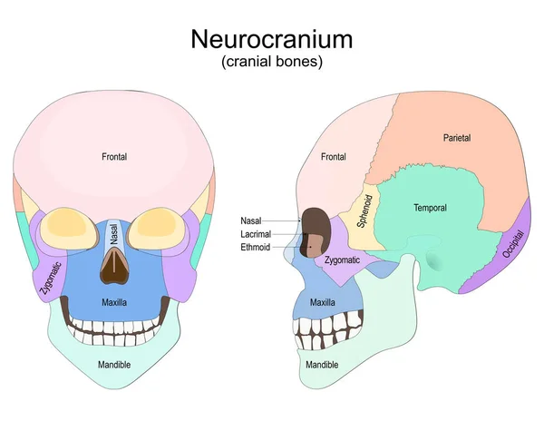
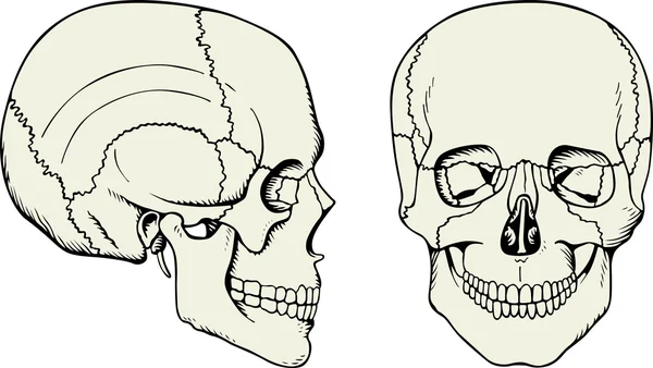
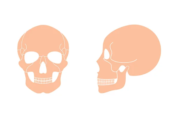
Human skull from the front and side view. Neurocranium. cranial bones. Vector poster for medical, and education use
Human Skull Front Side View Neurocranium Cranial Bones Vector Poster Medical Education Use

Radius and ulna bone anatomy with arm skeletal structure outline diagram. Labeled educational scheme with upper body parts and hand long bones vector illustration. Detailed physiological description.
Radius Ulna Bone Anatomy Arm Skeletal Structure Outline Diagram Labeled Educational Scheme Upper Body Parts Hand Long Bones Vector Illustration Detailed Physiological Description
Human rib cage. x-ray blue realistic torso. Human silhouette on dark background. Vector poster easy to edit
Human Rib Cage Ray Blue Realistic Torso Human Silhouette Dark Background Vector Poster Easy Edit
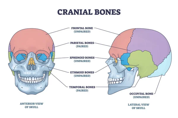
Cranial bones anatomy and skull skeleton medical division outline diagram. Labeled educational frontal, parietal, sphenoid, ethmoid and temporal bone vector illustration. Paired and unpaired examples.
Cranial Bones Anatomy Skull Skeleton Medical Division Outline Diagram Labeled Educational Frontal Parietal Sphenoid Ethmoid Temporal Bone Vector Illustration Paired Unpaired Examples
Medical Education Chart of Biology Human Skull Diagram. Vector. Front aspect white background basic medical education
3D Illustration Human Vertebral Column Spine Anatomy
Illustration Human Vertebral Column Spine Anatomy
Endoprosthetics, hip joint prosthesis. Vector 3D illustration. Medical poster
Endoprosthetics Hip Joint Prosthesis Vector Illustration Medical Poster
Scalene muscle structure with medical location on neck outline diagram. Labeled educational anatomical sprain scheme with middle, anterior and posterior parts of muscular system vector illustration.
Scalene Muscle Structure Medical Location Neck Outline Diagram Labeled Educational Anatomical Sprain Scheme Middle Anterior Posterior Parts Muscular System Vector Illustration
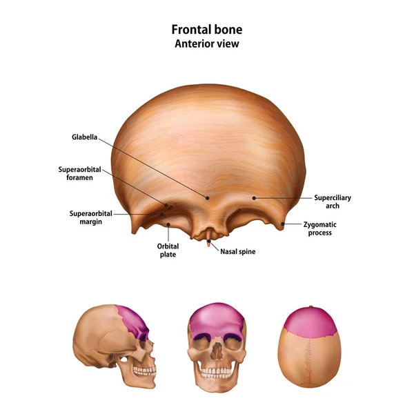
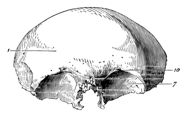
Frontal bone. With the name and description of all sites.
Frontal Bone Name Description All Sites
Medical Education Chart of Biology Human Skull Diagram. Vector. Front aspect white background basic medical education
Human skeleton
Human skeleton
This illustration represents Frontal Bone of the Human Skull, vintage line drawing or engraving illustration.
Illustration Represents Frontal Bone Human Skull Vintage Line Drawing Engraving Illustration
Illustration with skull.
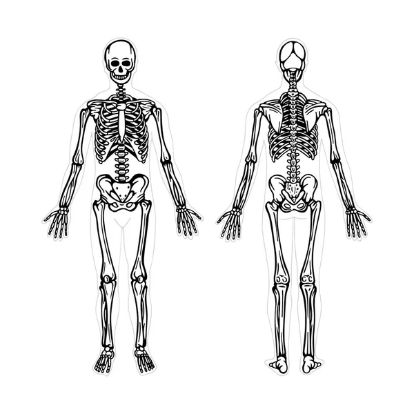
Skeleton. Human skeleton realistic vector illustration. Human skeleton front and back view. Bony system. Part of set.
Skeleton Human Skeleton Realistic Vector Illustration Human Skeleton Front Back View Bony System Part Set
Frontal view x ray of bones the of hand.
Frontal view image sore osteoarthritis joints of bones the of hand.
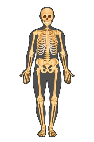
Human skeleton in full growth. Detailed anatomy. Vector illustration
Human Skeleton Full Growth Detailed Anatomy Vector Illustration
Human skull
Human skull
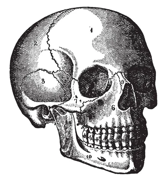
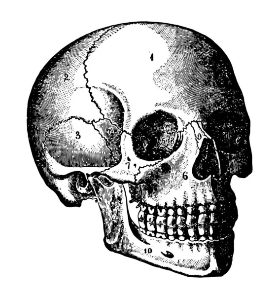
A diagram in which complete structure of human skull which is defined clearly, vintage line drawing or engraving illustration.
Diagram Which Complete Structure Human Skull Which Defined Clearly Vintage Line Drawing Engraving Illustration
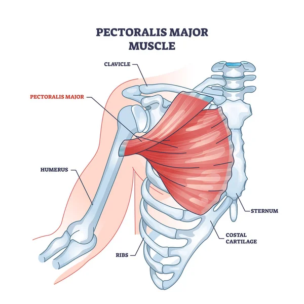
Pectoralis major muscle as human chest muscular anatomy outline diagram. Labeled educational medical scheme with skeletal system and musculature in human body breast and ribs area vector illustration.
Pectoralis Major Muscle Human Chest Muscular Anatomy Outline Diagram Labeled Educational Medical Scheme Skeletal System Musculature Human Body Breast Ribs Area Vector Illustration
Human skull
Human skull
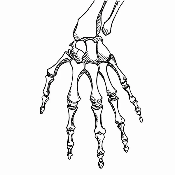
Skeleton hand vector. Sketch with hand bones isolated on white background.
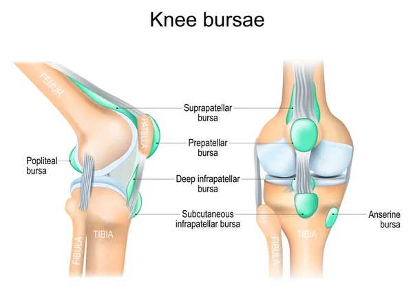
Knee bursae. synovial pockets or sacs that surround the knee joint cavity. Synovial joint anatomy. Frontal and side view of human knee joint. Vector illustration
Knee Bursae Synovial Pockets Sacs Surround Knee Joint Cavity Synovial Joint Anatomy Frontal Side View Human Knee Joint Vector Illustration
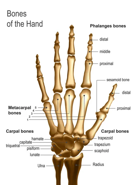
Frontal view image of bones the of hand with anotations.
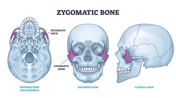
Zygomatic bone location with human skull skeleton anatomy outline diagram. Labeled educational scheme with cheekbone from inferior, anterior and lateral view vector illustration. Head parts structure.
Zygomatic Bone Location Human Skull Skeleton Anatomy Outline Diagram Labeled Educational Scheme Cheekbone Inferior Anterior Lateral View Vector Illustration Head Parts Structure
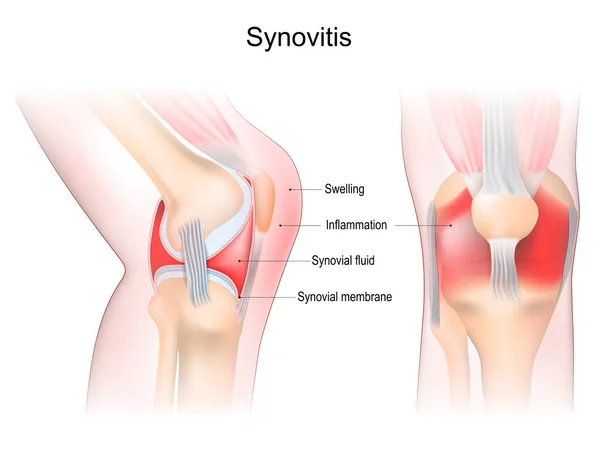
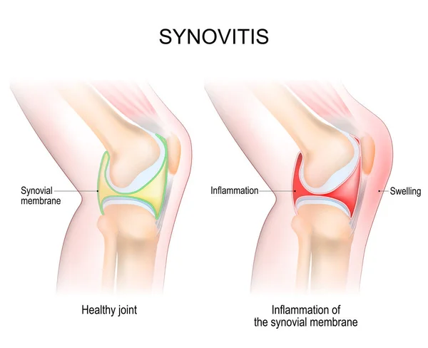
Synovitis of a Knee. Close-up of joint with inflammation of the synovial membrane. Signs and symptoms of the disease. Synovial joint anatomy. Frontal and side view of human knee joint. Vector illustration
Synovitis Knee Close Joint Inflammation Synovial Membrane Signs Symptoms Disease Synovial Joint Anatomy Frontal Side View Human Knee Joint Vector Illustration
Skull Chart
Skull Chart
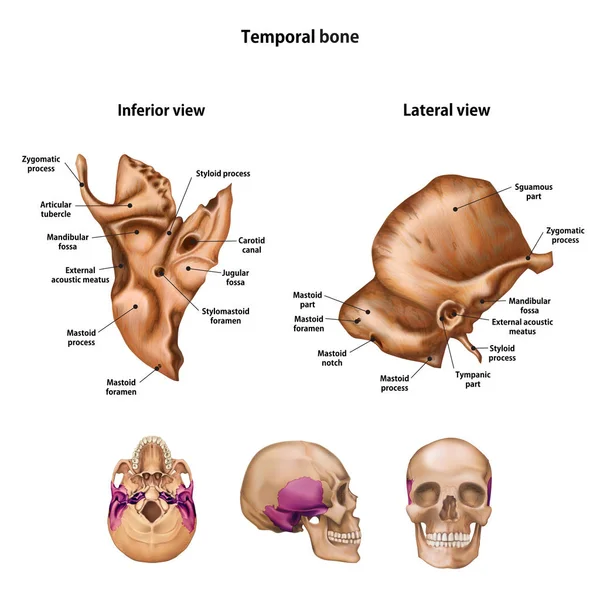
Temporal bone. With the name and description of all sites.
Temporal Bone Name Description All Sites
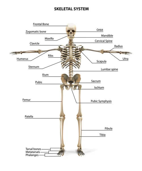
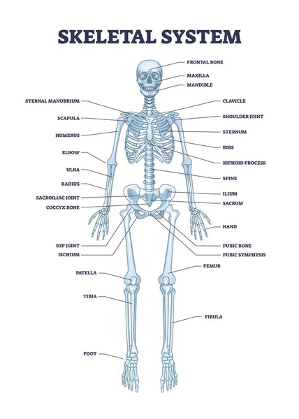
Skeletal system with body skeleton structure and anatomy outline diagram. Labeled educational medical physiology with skull, spine, ribs, hand and leg bones vector illustration. Biological human model
Skeletal System Body Skeleton Structure Anatomy Outline Diagram Labeled Educational Medical Physiology Skull Spine Ribs Hand Leg Bones Vector Illustration Biological Human Model
Skeleton. Human skeleton hand drawn vector illustration. Human skeleton front and back view. Bony system. Part of set.
Skeleton Human Skeleton Hand Drawn Vector Illustration Human Skeleton Front Back View Bony System Part Set
Orbit bones labeled educational skeletal division scheme vector illustration
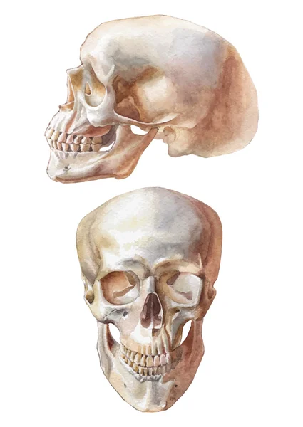
Illustration with two skulls
Human skull frontal
Human skull frontal
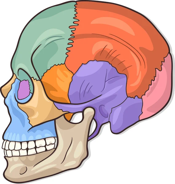
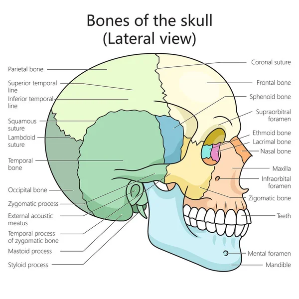
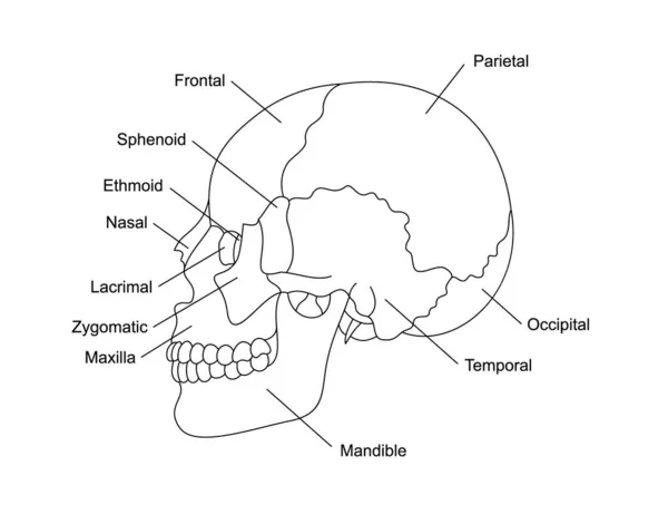
Human skull bones structure lateral view diagram schematic vector illustration. Medical science educational illustration
Human Skull Bones Structure Lateral View Diagram Schematic Vector Illustration Medical Science Educational Illustration
Medical illustration of the main parts of the bones of the foot in anterior view, with annotations.
Medical Illustration Main Parts Bones Foot Anterior View Annotations
The skull of a dog. Structure of the bones of the head, anatomical design. In Latin
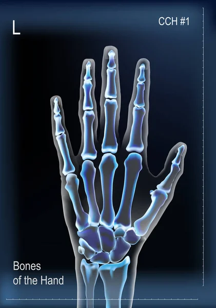
Frontal view x ray of bones the of hand with anotations.
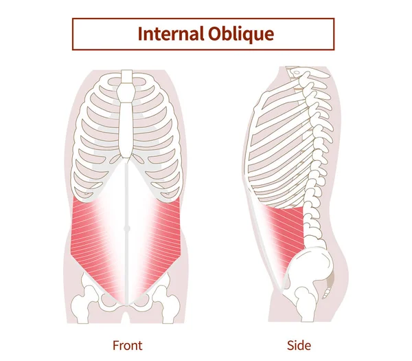
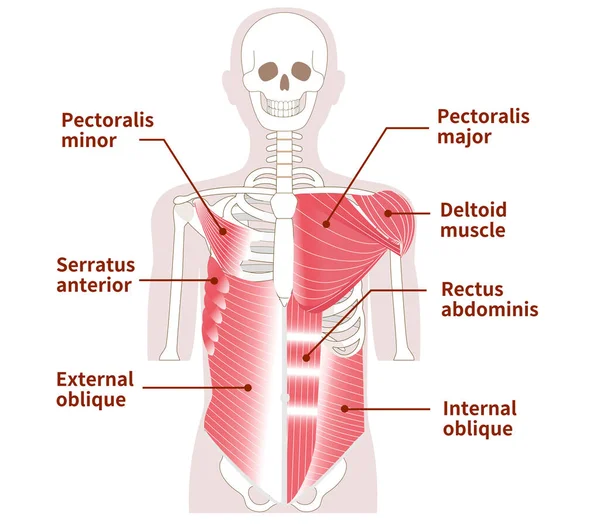
Abdominal muscle groups Illustrative illustrations of the internal abdominal oblique muscles Lateral and frontal views
Abdominal Muscle Groups Illustrative Illustrations Internal Abdominal Oblique Muscles Lateral Frontal Views
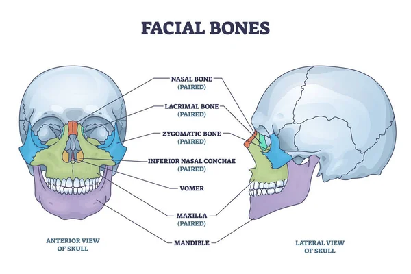
Facial bones with anterior and lateral view of human skull outline diagram. Skeletal anatomy with labeled educational medical scheme vector illustration. Nasal, lacrimal and zygomatic face parts.
Facial Bones Anterior Lateral View Human Skull Outline Diagram Skeletal Anatomy Labeled Educational Medical Scheme Vector Illustration Nasal Lacrimal Zygomatic Face Parts
Bones and ligaments of the pelvis
The temporal Bone of the human skull, situated at the sides and base of the skull, with its parts labelled, vintage line drawing or engraving illustration.
Temporal Bone Human Skull Situated Sides Base Skull Its Parts Labelled Vintage Line Drawing Engraving Illustration
The head is the part of the body that contains the brain and the organs of the special senses, vintage line drawing or engraving illustration.
Head Part Body Contains Brain Organs Special Senses Vintage Line Drawing Engraving Illustration
The head is the part of the body that contains the brain and the organs of the special senses, vintage line drawing or engraving illustration.
Head Part Body Contains Brain Organs Special Senses Vintage Line Drawing Engraving Illustration
Human skull bones anatomy. Cranial colored parts structure. Outline sutures. Zygomatic, nasal, maxilla, temporal lacrimal mandible frontal parietal occipital. Brain protection. Vector illustration.
Human Skull Bones Anatomy Cranial Colored Parts Structure Outline Sutures Zygomatic Nasal Maxilla Temporal Lacrimal Mandible Frontal Parietal Occipital Brain Protection Vector Illustration
Pig skeleton
Pig skeleton
Fetal Skull Dimensions. Superior view and Lateral view of the fetal skull showing the sutures, fontanelles, and transverse diameters. Anatomy of the Newborn Skull.
Fetal Skull Dimensions Superior View Lateral View Fetal Skull Showing Sutures Fontanelles Transverse Diameters Anatomy Newborn Skull
Large muscles in the abdomen, chest, and shoulders Outer and inner muscles
Large Muscles Abdomen Chest Shoulders Outer Inner Muscles
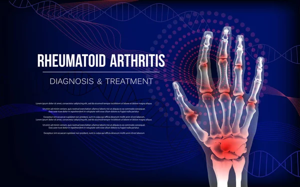
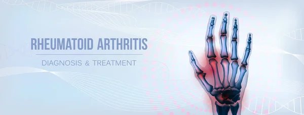
Rheumatoid arthritis osteoarthritis banner inflammation joints of bones of hand.
Set of human skull illustrations in outline and silhouette, front and side views, isolated on white background, graphic illustration. Concept of anatomy, tattoo, gothic, halloween design elements.
Set Human Skull Illustrations Outline Silhouette Front Side Views Isolated White Background Graphic Illustration Concept Anatomy Tattoo Gothic Halloween Design Elements
Pattern with human skull
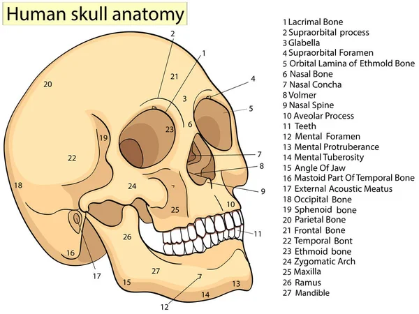
Human skull bones anatomy with descriptions. Cranial parts structure. Human internal organ illustration. Zygomatic, nasal, maxilla, temporal, frontal head parts.
Human Skull Bones Anatomy Descriptions Cranial Parts Structure Human Internal Organ Illustration Zygomatic Nasal Maxilla Temporal Frontal Head Parts
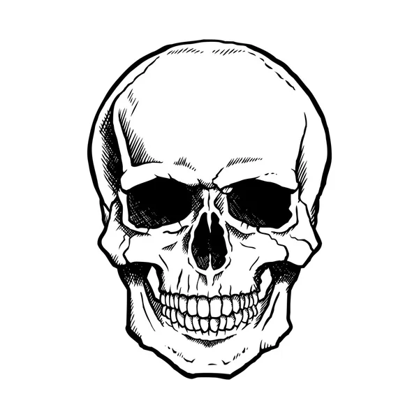
Black and white human skull with jaw
Human skull anatomy,
Synovitis of a Knee. Close-up of normal joint, and knee with inflammation of the synovial membrane. Signs and symptoms of the disease. side view of human knee joint. Vector illustration
Synovitis Knee Close Normal Joint Knee Inflammation Synovial Membrane Signs Symptoms Disease Side View Human Knee Joint Vector Illustration
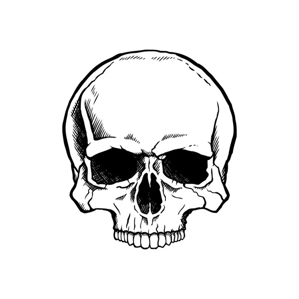
Black and white human skull
Different Types of Skull Sutures with Anterior, Posterior, Lateral, Superior View, Educational Medical Poster Design Healthcare Scheme, cavity, sagittal, lambdoid, parietomastoid, occipitomastoid.
Different Types Skull Sutures Anterior Posterior Lateral Superior View Educational Medical Poster Design Healthcare Scheme Cavity Sagittal Lambdoid Parietomastoid Occipitomastoid
Horizontal rheumatoid arthritis hand sore joints concept for social media
Anatomy of the Newborn Skull. Lateral and superior view. Cranial sutures and fontanels.
Anatomy Newborn Skull Lateral Superior View Cranial Sutures Fontanels
Colored human skull and crossbones
Skull human with a smile. Color vintage vector engraving illustration. For poster and tattoo biker club. Hand drawn design element isolated on white background
Skull Human Smile Color Vintage Vector Engraving Illustration Poster Tattoo Biker Club Hand Drawn Design Element Isolated White Background
Sutures of the skull as human head bone medical division outline diagram. Labeled educational anatomical scheme with squamous, coronal, lamboid, sagittal and occipitomastoid parts vector illustration.
Sutures Skull Human Head Bone Medical Division Outline Diagram Labeled Educational Anatomical Scheme Squamous Coronal Lamboid Sagittal Occipitomastoid Parts Vector Illustration
Colored human skull
Colored human skull
Colored human skull with ribbon banner
Human skull anatomy.
Illustration of anatomical skull
Vomer bone with facial skeleton and frontal nasal cavity outline diagram. Labeled educational nose skeletal structure vector illustration. Perpendicular or cribriform plate, sphenoid and maxilla.
Vomer Bone Facial Skeleton Frontal Nasal Cavity Outline Diagram Labeled Educational Nose Skeletal Structure Vector Illustration Perpendicular Cribriform Plate Sphenoid Maxilla
Vector black and white illustration skull
Vector Black and White Human Skull Drawing
Colored human skull with jaw
Colored human skull and crossbones
Human skull low poly art. Polygonal skull on a white background.
Human Skull Low Poly Art Polygonal Skull White Background
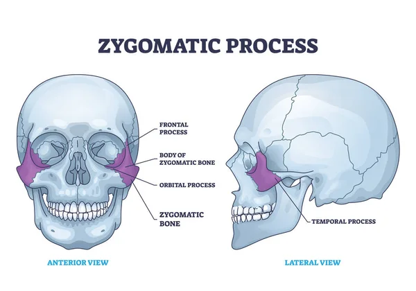
Zygomatic process as human cheek bone skeleton anatomy outline diagram. Labeled educational cheekbone location and skull parts structure vector illustration. Frontal, orbital and temporal process.
Zygomatic Process Human Cheek Bone Skeleton Anatomy Outline Diagram Labeled Educational Cheekbone Location Skull Parts Structure Vector Illustration Frontal Orbital Temporal Process
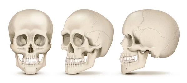
Realistic human skulls front and side views set isolated on white background vector illustration
Realistic Human Skulls Front Side Views Set Isolated White Background Vector Illustration
Line skull vector illustration. Spooky lighting from bellow. Frontal view of human skull made by white vertical lines on black background.
Line Skull Vector Illustration Spooky Lighting Bellow Frontal View Human Skull Made White Vertical Lines Black Background
White skull
White skull
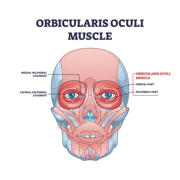
Orbicularis oculi muscle as face muscular system for eyelids outline diagram
Skull smiling with bandana and glasses for motorcycle. Black vintage vector illustration. For poster and tattoo biker club. Hand drawn design element isolated on white background.
Page