Sinoviale Stock Vector Images

244 Sinoviale vector art & graphics are available under a royalty-free license
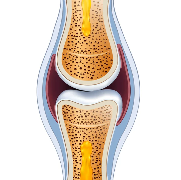
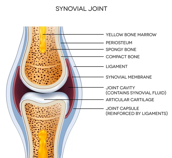
Normal synovial joint anatomy
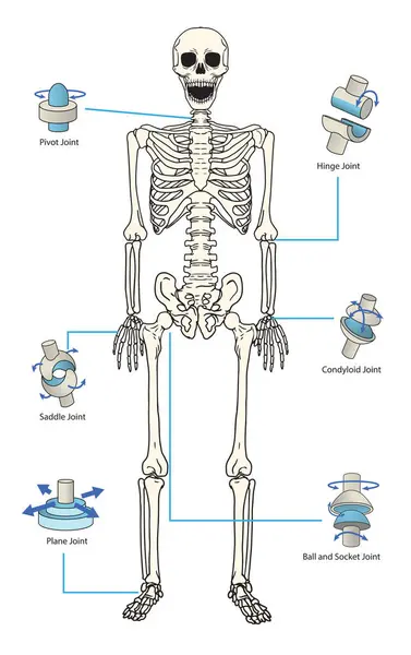
Types of synovial joints movement classification for body outline diagram. Labeled educational anatomical division with ellipsoid, hinge, saddle, pivot and ball socket bone joints vector illustration.
Types Synovial Joints Movement Classification Body Outline Diagram Labeled Educational Anatomical Division Ellipsoid Hinge Saddle Pivot Ball Socket Bone Joints Vector Illustration
Labeled joint anatomy
Modern graphic vector illustration
Modern Graphic Vector Illustration
Knee joint bone logo vector illustration
Bursitis inflammation concept
Types of location of the intervertebral hernia. Vector illustration on isolated background.
Articulation icon vector on white background, articulation trendy filled icons from People collection
Articulation Icon Vector White Background Articulation Trendy Filled Icons People Collection
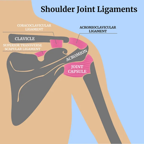
Cross-sectional anatomy of human muscles and bones. Ligaments of the shoulder joint, posterior. Minimal color drawing. Medical poster. Vector illustration.
Cross Sectional Anatomy Human Muscles Bones Ligaments Shoulder Joint Posterior Minimal Color Drawing Medical Poster Vector Illustration
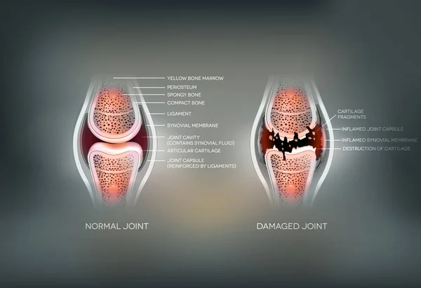
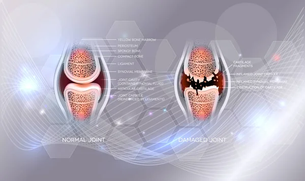
Damaged joint and normal joint colorful design
Damaged joint and normal joint
Hip dislocations, rheumatoid arthritis of the foot, arm concept vector set. Tiny doctors, rheumatologists treat rheumatism, osteoarthritis. World arthritis day in October.
Hip Dislocations Rheumatoid Arthritis Foot Arm Concept Vector Set Tiny Doctors Rheumatologists Treat Rheumatism Osteoarthritis World Arthritis Day October
Types of Joints diagram
Types Joints Diagram
Bakers cyst or popliteal cyst. Traumatology and orthopedics
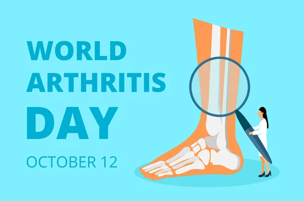
World arthritis day in October. Tiny doctors treat rheumatism, osteoarthritis. Healthcare flat concept vector
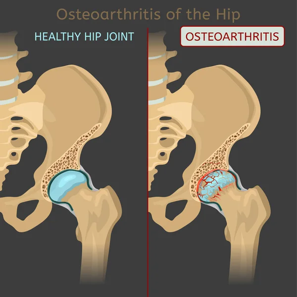
Hip Osteoarthritis Image
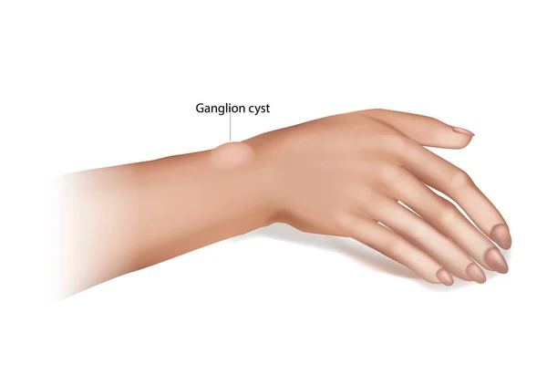
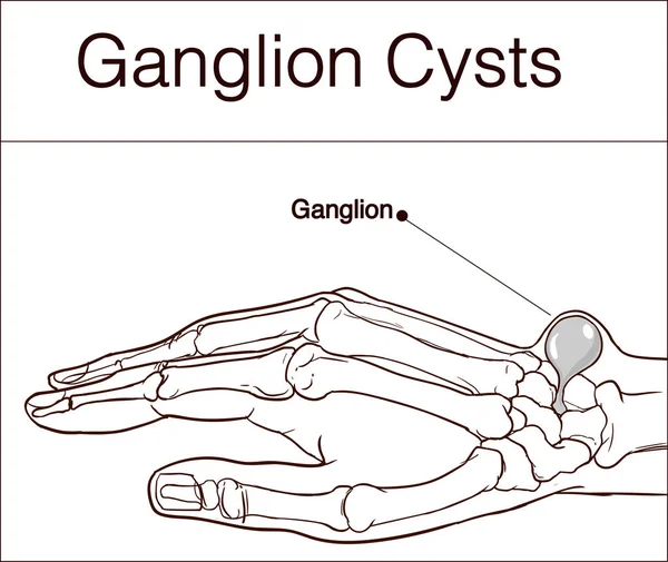
Ganglion Cyst of the Wrist and Hand. Synovial cyst or a Gideon s Disease, or a Bible Cyst, or a Bible Bump.
Set of icons of the joints and their treatment Cartilage damage, arthritis, osteoarthritis, restoration of cartilage pain relief icons. Flat icons in a round frames.
Set Icons Joints Treatment Cartilage Damage Arthritis Osteoarthritis Restoration Cartilage Pain Relief Icons Flat Icons Frames
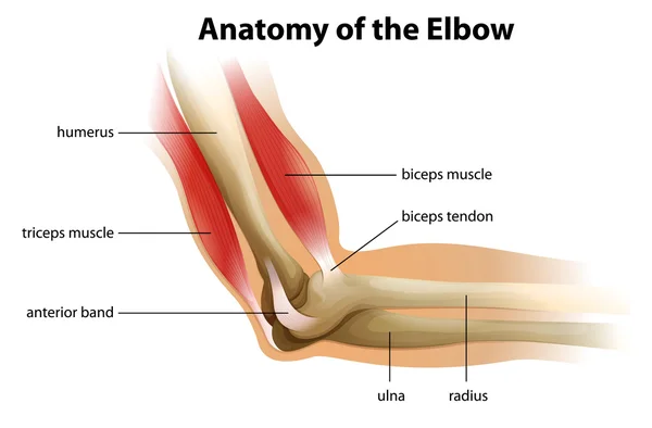
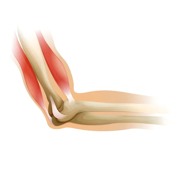
Anatomy of the Elbow
Meniscal tear. Torn meniscus. Knee injury. Cartilage damage. Meniscal repair. Sports injury. Head of tibia seen from above. Vector illustration
Meniscal Tear Torn Meniscus Knee Injury Cartilage Damage Meniscal Repair Sports Injury Head Tibia Seen Vector Illustration
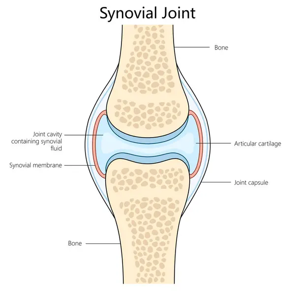
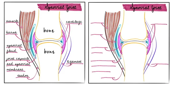
Human synovial joint structure diagram hand drawn schematic vector illustration. Medical science educational illustration
Human Synovial Joint Structure Diagram Hand Drawn Schematic Vector Illustration Medical Science Educational Illustration
The knee joint joins the thigh with the leg and consists of two articulations: one between the femur and tibia (tibiofemoral joint),and one between the femur and patella. It is the largest joint in the human body. Illustration anatomy body.
Knee Joint Joins Thigh Leg Consists Two Articulations One Femur Tibia Tibiofemoral Joint One Femur Patella Largest Joint Human Body Illustration Anatomy Body
Anatomy of the human elbow
Osteochondrosis of the cervical vertebrae
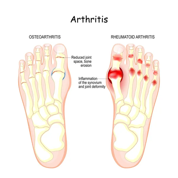
Osteoarthritis, rheumatoid arthritis, and posttraumatic arthritis. comparison of human foot with different forms of arthritis. inflamed synovium and joint deformity
Osteoarthritis Rheumatoid Arthritis Posttraumatic Arthritis Comparison Human Foot Different Forms Arthritis Inflamed Synovium Joint Deformity
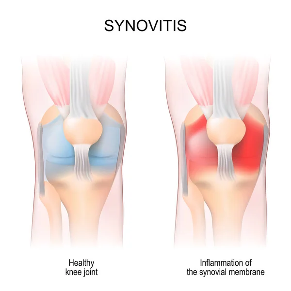
Synovitis. Human Knee joint anatomy. Healthy joint, and Inflammation of the synovial membrane. front view. anterior aspects. Vector illustration
Synovitis Human Knee Joint Anatomy Healthy Joint Inflammation Synovial Membrane Front View Anterior Aspects Vector Illustration
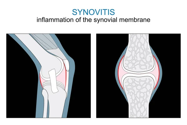
Types of Synovitis. inflammation of the synovial membrane of a Knee and Synovial joint. monochrome flat vector like x-ray illustration.
Types Synovitis Inflammation Synovial Membrane Knee Synovial Joint Monochrome Flat Vector Ray Illustration
Rheumatologist concept for website. Tiny doctors treat rheumatism, osteoarthritis. Arthritis flat concept vector on the blue background for national healthcare day
Bone Joints Vector Icon
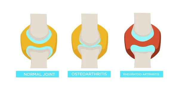
Osteoarthritis rheumatoid arthritis and normal joint vector rheumatology medicine and treatment bones disease or injury healthcare and illness human skeleton part medical problem skeletal system
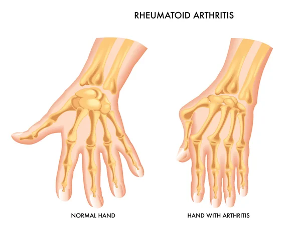
Osteoarthritis Rheumatoid Arthritis Normal Joint Vector Rheumatology Medicine Treatment Bones Disease Injury Healthcare Illness Human Skeleton Part Medical Problem Skeletal System
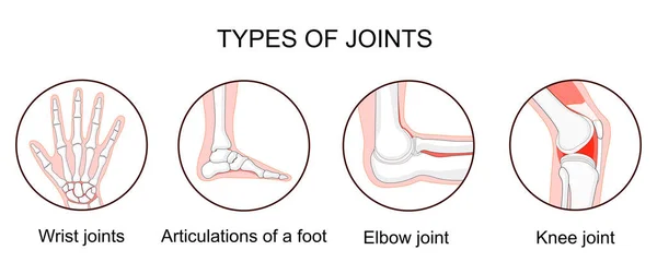
Illustration of leg knee or arm and hand joints
Bone Joints Vector Icon
HEALTHY JOINT
HEALTHY JOINT
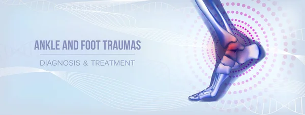
Horizontal ankle and foot traumas banner for social media
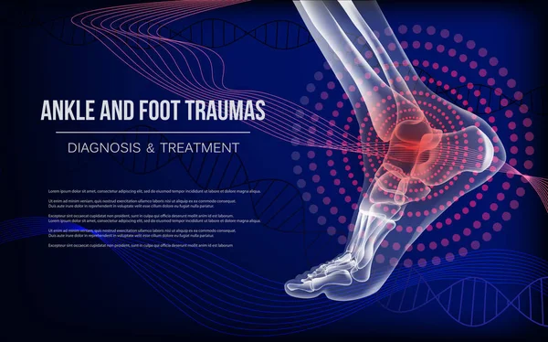
Horizontal dark blue banner for ankle and foot joints traumas
Comparison of normal and osteoarthritic elbow joints
Comparison Normal Osteoarthritic Elbow Joints
Fluid-filled cyst causing knee tightness
Fluid Filled Cyst Causing Knee Tightness
Normal bone and bone with osteoporosis
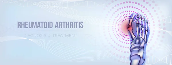
Rheumatoid Arthritis
Synovial Fluid Minimal Sophisticated Branding Design Asset
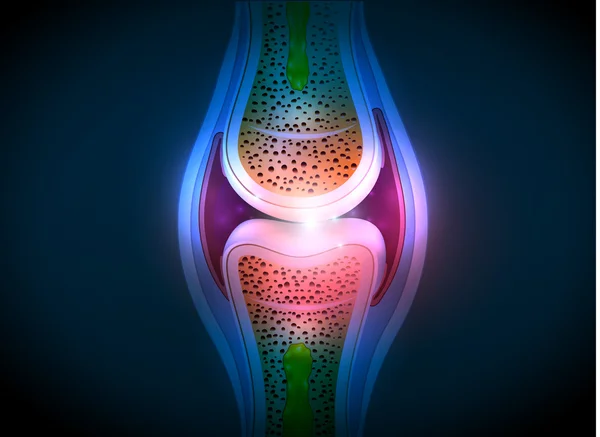
Synovial joint anatomy abstract bright design
Human elbow
Human elbow
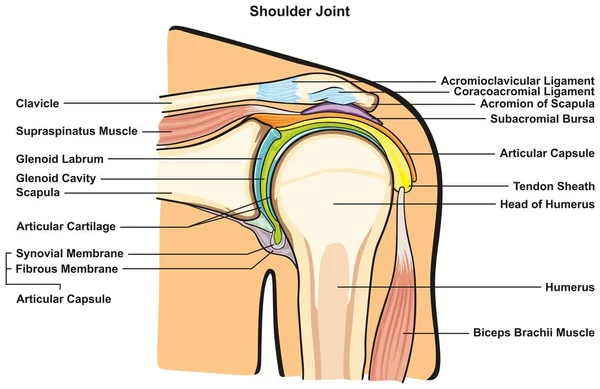
Shoulder Joint of Human Body Anatomy infographic diagram with all parts including bones ligaments muscles bursa cavity capsule cartilage membrane for medical science education and health care
Shoulder Joint Human Body Anatomy Infographic Diagram All Parts Including Bones Ligaments Muscles Bursa Cavity Capsule Cartilage Membrane Medical Science Education Health Care
Inflammation of knee bursae causing pain and swelling
Inflammation Knee Bursae Causing Pain Swelling
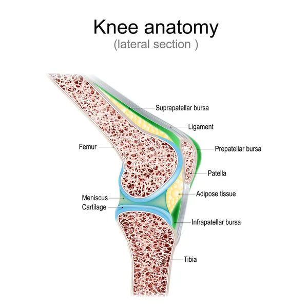
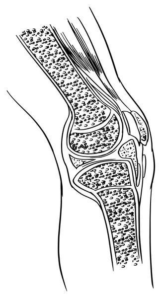
Knee anatomy. Side view. lateral section of joint with ligaments, meniscus, and bursae. knee joint cavity. Cross section of leg bones. detailed diagram. Vector poster
Knee Anatomy Side View Lateral Section Joint Ligaments Meniscus Bursae Knee Joint Cavity Cross Section Leg Bones Detailed Diagram Vector Poster
Illustration of Rheumatoid Arthi
Elbow joint minimally invasive surgery. Arthroscopy medical procedure. Anatomy of humerus, ulna and radius bones. Arm pain, fracture, osteoarthritis or rheumatoid arthritis x ray vector illustration
Elbow Joint Minimally Invasive Surgery Arthroscopy Medical Procedure Anatomy Humerus Ulna Radius Bones Arm Pain Fracture Osteoarthritis Rheumatoid Arthritis Ray Vector Illustration
3D Isometric Flat Vector Conceptual Illustration of Knee Problems
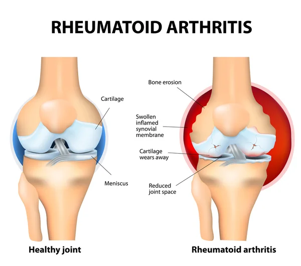
Rheumatoid arthritis infographic showing knee joint damage, inflamed synovial membrane, cartilage loss, bone erosion, exposed bone and reduced joint space, vector illustration
Rheumatoid Arthritis Infographic Showing Knee Joint Damage Inflamed Synovial Membrane Cartilage Loss Bone Erosion Exposed Bone Reduced Joint Space Vector Illustration
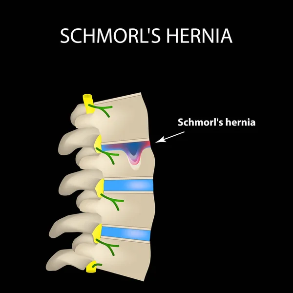
Hernia Schmorl. Intervertebral disc. Side view. Spine. Infographics. Vector illustration on a black background
Vector illustration of a Ganglion cyst
Bone logo vector template
Juvenile arthritis awareness month concept vector. Rheumatoid arthritis of foot, arm. Treat rheumatism, osteoarthritis, make ultrasound, x ray. World arthritis day, polyarthritis sign.
Juvenile Arthritis Awareness Month Concept Vector Rheumatoid Arthritis Foot Arm Treat Rheumatism Osteoarthritis Make Ultrasound Ray World Arthritis Day Polyarthritis Sign
Hip joint injection
Hip joint injection
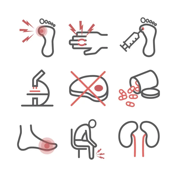
Gout. Symptoms, Treatment. Line icons set. Vector signs for web graphics
Gout Symptoms Treatment Line Icons Set Vector Signs Web Graphics
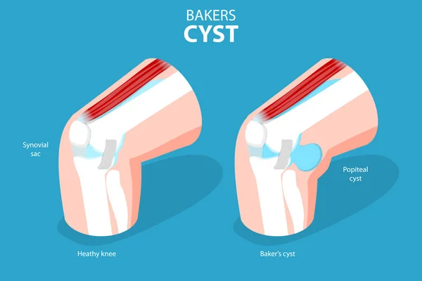
3D Isometric Flat Vector Conceptual Illustration of Bakers CYST, Traumatology and Prthopedics
Isometric Flat Vector Conceptual Illustration Bakers Cyst Traumatology Prthopedics
Meniscus structure. Knee joint anatomy. Cartilage and Ligaments. Vector illustration
Meniscus Structure Knee Joint Anatomy Cartilage Ligaments Vector Illustration
Hernia Schmorl. Intervertebral disc. Side view. Spine. Infographics Vector illustration on isolated background
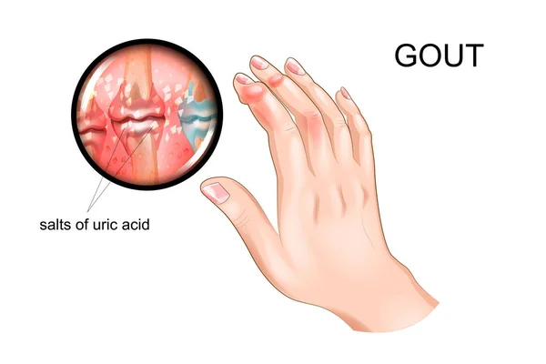
Gout, arthritis, tofus.
Gout, arthritis of fingers
Rheumatoid arthritis bones concept
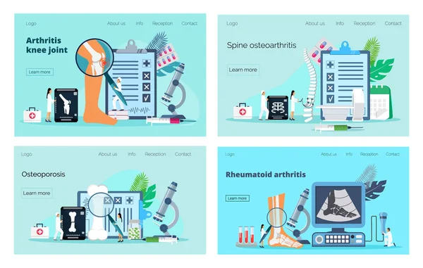
Arthritis knee joint, osteoporosis concept. Tiny doctors treat rheumatism, osteoarthritis, spin, bones, make x ray scan.
Health Care and Medical Poster Brochure Flyer design Layout template
Elbow joint minimally invasive surgery. Arthroscopy medical procedure. Anatomy of humerus, ulna and radius bones. Arm pain, fracture, osteoarthritis or rheumatoid arthritis x ray vector illustration
Elbow Joint Minimally Invasive Surgery Arthroscopy Medical Procedure Anatomy Humerus Ulna Radius Bones Arm Pain Fracture Osteoarthritis Rheumatoid Arthritis Ray Vector Illustration
Osteoarthritis. Arthritis of a knee joint. Pain within a knee. Degenerative joint disease. Isometric Flat vector illustration
Osteoarthritis Arthritis Knee Joint Pain Knee Degenerative Joint Disease Isometric Flat Vector Illustration
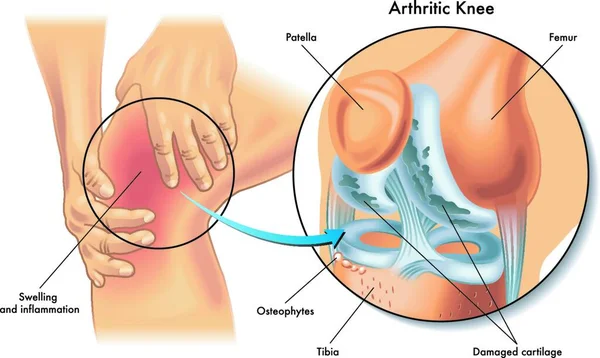
Rheumatoid arthritis. Pain in the joint. degenerative joint disease. The cartilage becomes worn out. This leads to inflammation, swelling, and joint pain.
Rheumatoid Arthritis Pain Joint Degenerative Joint Disease Cartilage Becomes Worn Out Leads Inflammation Swelling Joint Pain
Damaged and healthy joint design
Gout. Symptoms, Treatment. Line icons set. Vector signs for web graphics
Gout Symptoms Treatment Line Icons Set Vector Signs Web Graphics
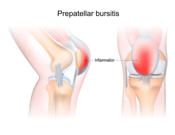
Bursitis. Prepatellar bursitis. inflammation of sacs with synovial fluid. Front and side view of human knee joint. Lateral and anterior section of the knee. vector illustration.
Bursitis Prepatellar Bursitis Inflammation Sacs Synovial Fluid Front Side View Human Knee Joint Lateral Anterior Section Knee Vector Illustration
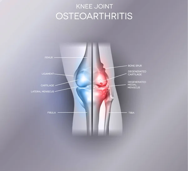
Knee osteoarthritis. degenerative joint disease. Cartilage becomes worn. Vector poster
Knee Osteoarthritis Degenerative Joint Disease Cartilage Becomes Worn Vector Poster
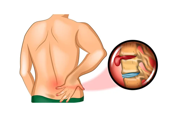
Back pain. damage to the spine
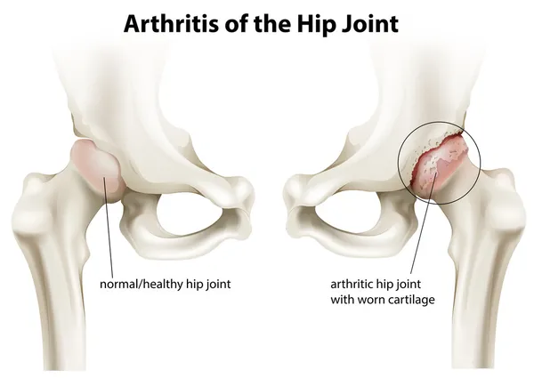
Arthritis of the hip joint
Hip Joint Anatomy with Acetabular Labrum and Ligamentum Teres, Anterior and Lateral Pelvis Medical Illustration. Human Pelvis and Hip Joint Anatomical Structure
Hip Joint Anatomy Acetabular Labrum Ligamentum Teres Anterior Lateral Pelvis Medical Illustration Human Pelvis Hip Joint Anatomical Structure
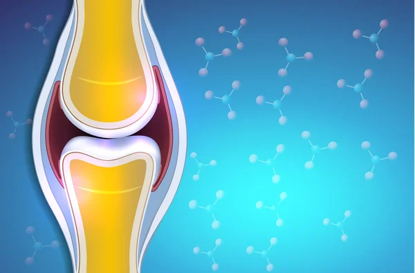
Illustration of a synovial joint
Human elbow joint polygonal vector illustration. Arm bones anatomy concept. Medical abstract low poly design on a white background
Human Elbow Joint Polygonal Vector Illustration Arm Bones Anatomy Concept Medical Abstract Low Poly Design White Background
Vector drawing of a synovial joint
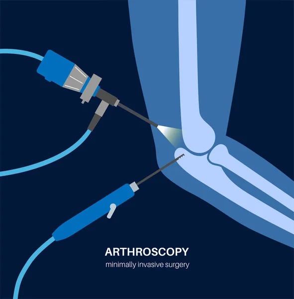
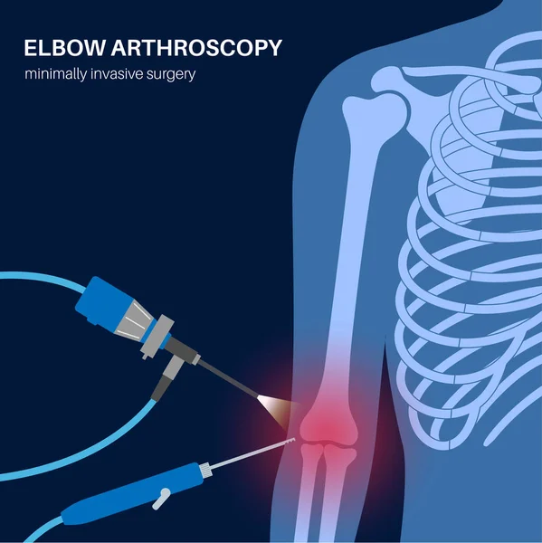
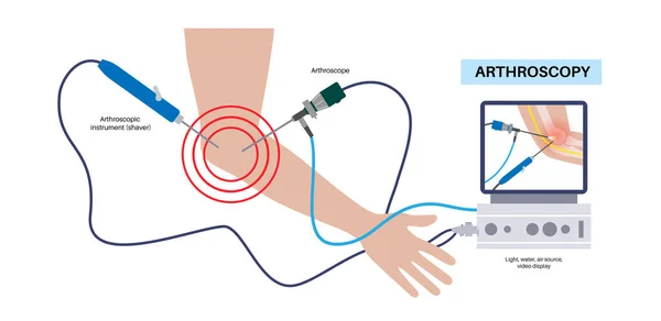
Elbow joint minimally invasive surgery. Arthroscopy medical procedure. Arthroscope and arthroscopic instrument. Arm pain, fracture, osteoarthritis or rheumatoid arthritis treatment, x ray flat vector
Elbow Joint Minimally Invasive Surgery Arthroscopy Medical Procedure Arthroscope Arthroscopic Instrument Arm Pain Fracture Osteoarthritis Rheumatoid Arthritis Treatment Ray Flat Vector
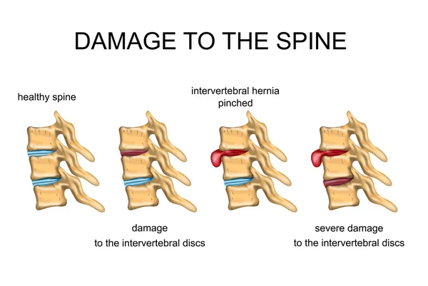
DAMAGE TO THE SPINE
DAMAGE TO THE SPINE
Broken or Sprained Wrist Diagram vector illustration isolated white background
Broken Sprained Wrist Diagram Vector Illustration Isolated White Background
Normal synovial joint anatomy
Rheumatoid Arthritis Vector Medical Scheme
Rheumatoid Arthritis RA. Inflammatory type of arthritis that affects of knee. Auto immune disease. The immune system mistakenly attacks healthy joint tissue. Joint deformity. Isometric Flat vector illustration
Rheumatoid Arthritis Inflammatory Type Arthritis Affects Knee Auto Immune Disease Immune System Mistakenly Attacks Healthy Joint Tissue Joint Deformity Isometric Flat Vector Illustration
Structure ofthe joint
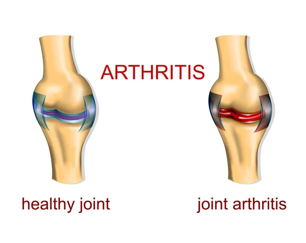
JOINT ARTHRITIS
JOINT ARTHRITIS
Horizontal turf toe foot traumas banner
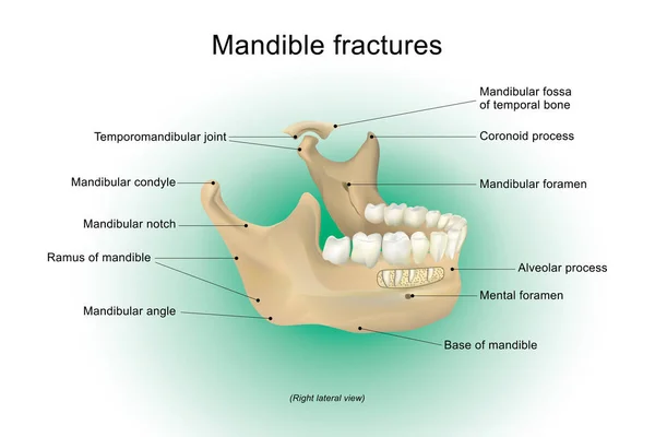
Mandible fractures. Anatomy Vector design, Illustration.
Meniscal tear. Knee anatomy. Close-up of Torn meniscus and cartilage damage. Sports knee injury. Vector illustration
Meniscal Tear Knee Anatomy Close Torn Meniscus Cartilage Damage Sports Knee Injury Vector Illustration
Stages of Osteoarthritis. knee joint anatomy Vector illustration
Stages Osteoarthritis Knee Joint Anatomy Vector Illustration
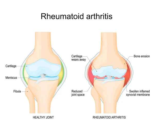
Rheumatoid arthritis. A comparison between a healthy knee and joint with Bone erosion, Cartilage wears, Reduced joint space and Swollen inflamed synovial membrane. Vector illustration with labels. diagnostic images to aid patient and doctor
Rheumatoid Arthritis Comparison Healthy Knee Joint Bone Erosion Cartilage Wears Reduced Joint Space Swollen Inflamed Synovial Membrane Vector Illustration Labels Diagnostic Images Aid Patient Doctor
Prepatellar bursitis. Knee joint inflammation. Vector illustration. Medical poster. Schematic diagram
Prepatellar Bursitis Knee Joint Inflammation Vector Illustration Medical Poster Schematic Diagram
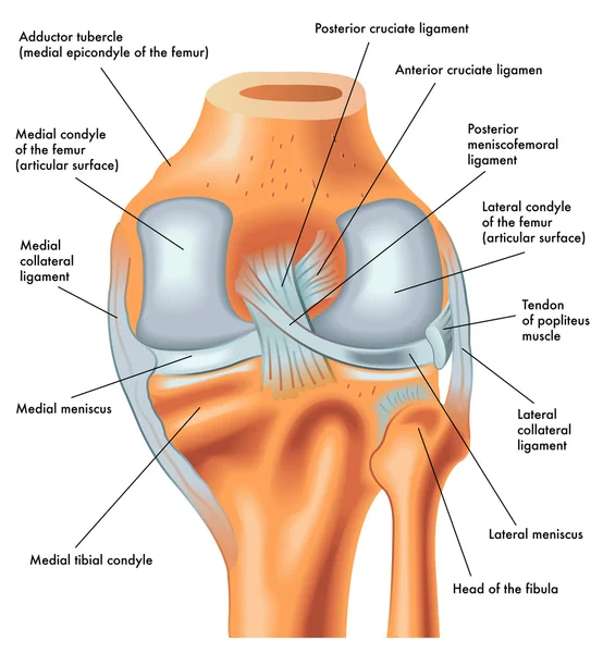
Posterior view of the right knee in extension
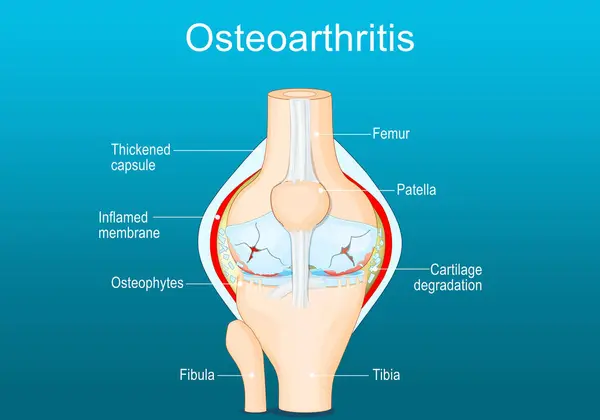
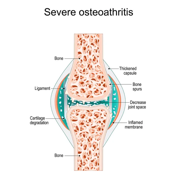
Severe osteoarthritis. Synovial joint with bone Spurs, Inflamed membrane, Decreased joint space, and Cartilage degradation. vector illustration
Severe Osteoarthritis Synovial Joint Bone Spurs Inflamed Membrane Decreased Joint Space Cartilage Degradation Vector Illustration
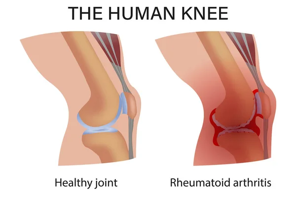
Normal Joint and Rheumatoid Arthritis
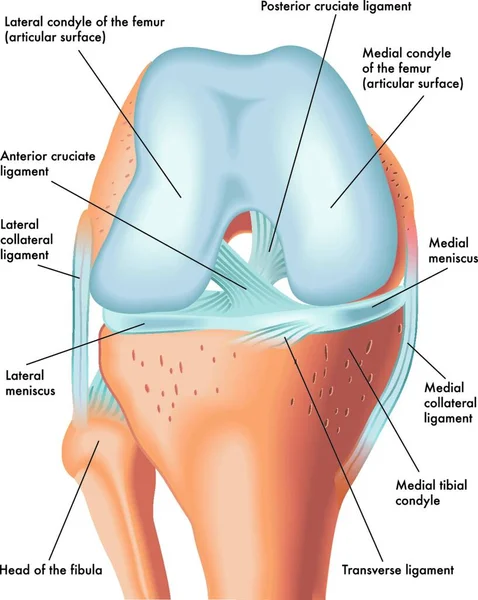
Anatomy of the front of the right knee in flexion
Anatomy Front Right Knee Flexion
Anatomy of the knee joint
Bone joints, Isolated Vector icon that can be easily modified or edit
Bone Joints Isolated Vector Icon Can Easily Modified Edit
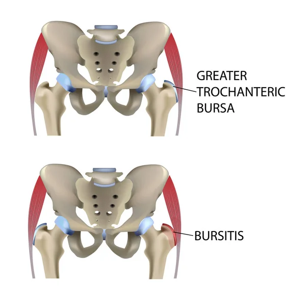
Hip bursitis, vector medical illustration vector
Hip Bursitis Vector Medical Illustration Vector
Knee joint bone logo vector illustration
Types of joints. Biomechanical and anatomical classification. Set icons with human elbow, knee, wrist joints, and Articulations of a foot. bones and joints. medical poster for clinic. vector illustration.
Types Joints Biomechanical Anatomical Classification Set Icons Human Elbow Knee Wrist Joints Articulations Foot Bones Joints Medical Poster Clinic Vector Illustration
Bone Joints Colored Vector Icon
Page