Pelvic Stock Video Footage

1,430 Pelvic videos are available under a royalty-free license
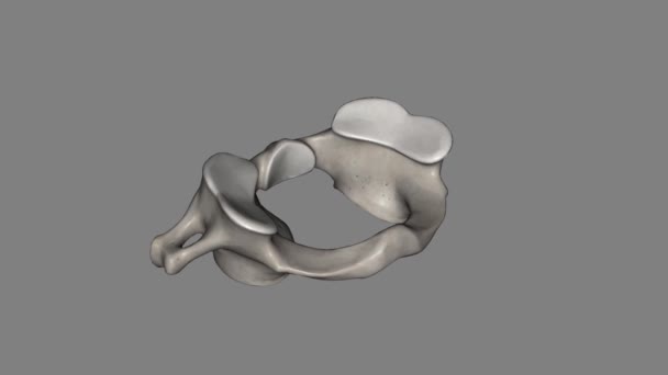
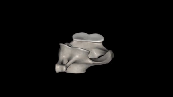
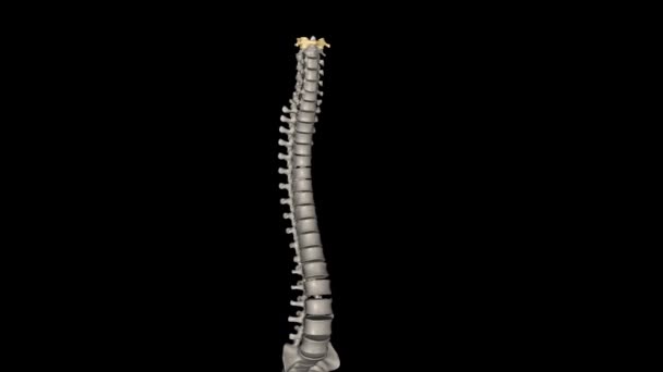
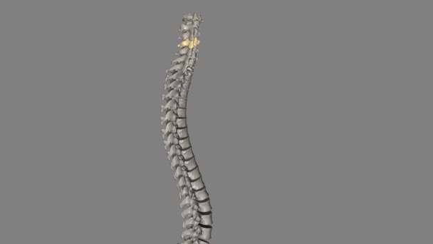
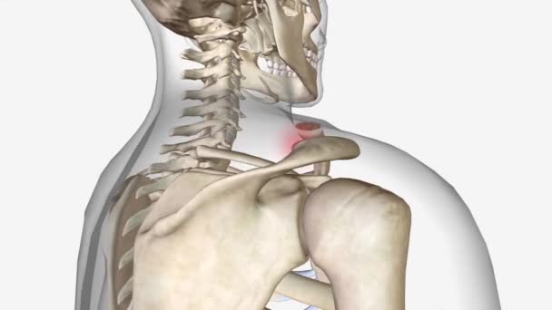
The atlas (plural: atlases) is the first cervical vertebra, commonly called C1 .
Atlas Plural Atlases First Cervical Vertebra Commonly Called
00:21
Degenerative spondylolisthesis (DS) is a disorder that causes the slip of one vertebral body over the one below due to degenerative changes in the spine .
Degenerative Spondylolisthesis Disorder Causes Slip One Vertebral Body One Due Degenerative Changes Spine
00:12
Woman pilates. Core strengthening. Young athlete doing pelvic floor exercise in shoulder bridge yoga pose controlling breath in gym.
Woman Pilates Core Strengthening Young Athlete Doing Pelvic Floor Exercise Shoulder Bridge Yoga Pose Controlling Breath Gym
00:16
Woman, pain and hands on stomach on sofa for menstruation, ovarian cysts and pelvic congestion syndrome or miscarriage. Girl, home and abdominal ache for endometriosis and gastrointestinal disorder
Woman Pain Hands Stomach Sofa Menstruation Ovarian Cysts Pelvic Congestion Syndrome Miscarriage Girl Home Abdominal Ache Endometriosis Gastrointestinal Disorder
00:12
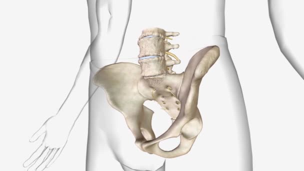
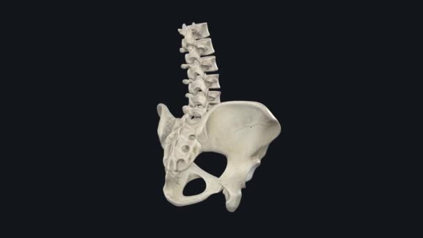
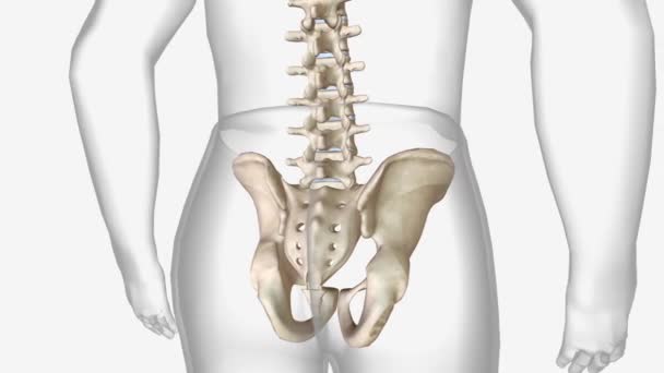
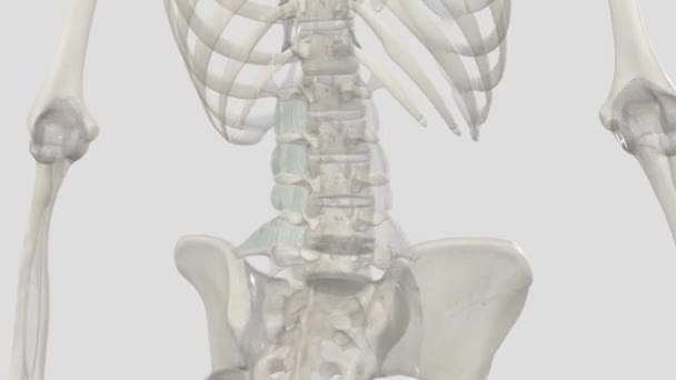
Human skeleton: pelvis and sacrum. Medically accurate 3D animation
00:18
Wooden cubes spell word Endometriosis highlighting chronic problem. Gynecology model with uterus stands near cup of capsules on table drawing focus
Wooden Cubes Spell Word Endometriosis Highlighting Chronic Problem Gynecology Model Uterus Stands Cup Capsules Table Drawing Focus
00:13
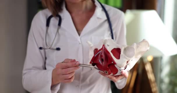
Close up of female doctor pointing at plastic model of female reproductive system in clinic. Gynecological education with visible uterus and vaginal structure. Medical training scene. High quality 4k footage
Close Female Doctor Pointing Plastic Model Female Reproductive System Clinic Gynecological Education Visible Uterus Vaginal Structure Medical Training Scene High Quality Footage
00:16
Hypopressive pilates exercises for pelvic floor. Fitness breathing physiotherapy core training. Young hispanic woman trainer doing fitness at home during the lockdown
00:15
The atlas (plural: atlases) is the first cervical vertebra, commonly called C1 .
Atlas Plural Atlases First Cervical Vertebra Commonly Called
00:21
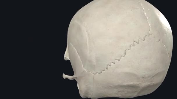
Neurocranium as a whole bone complex. See multiple interactive high quality pictures demonstrating different views of the skull bones surrounding the brain.
Neurocranium Whole Bone Complex See Multiple Interactive High Quality Pictures Demonstrating Different Views Skull Bones Surrounding Brain
00:16
Sketch style minimal pelvis icon animation - smooth progressive reveal'
Sketch Style Minimal Pelvis Icon Animation Smooth Progressive Reveal
00:08
Upper limb bones in human skeletal system.3d animation
Upper Limb Bones Human Skeletal System Animation
00:59
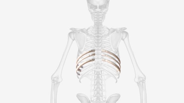
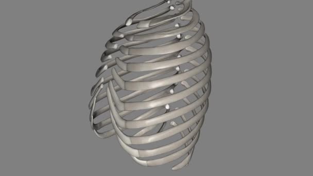
The ribs partially enclose and protect the chest cavity, where many vital organs .
Ribs Partially Enclose Protect Chest Cavity Many Vital Organs
00:23
Joint capsule is a sac-like fibrous capsule continuous with the periosteum of the articulating bones .
Joint Capsule Sac Fibrous Capsule Continuous Periosteum Articulating Bones
00:16
Endometriosis awareness month for better public health
Endometriosis Awareness Month Better Public Health
00:11
A medical professional uses a spine model to explain the anatomy and function of the sacroiliac joint
Medical Professional Uses Spine Model Explain Anatomy Function Sacroiliac Joint
00:13
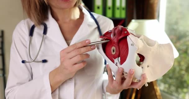
Gynecologist shows model of female pelvic organs with muscles. Pelvic bone structure and typical diseases and injuries
Gynecologist Shows Model Female Pelvic Organs Muscles Pelvic Bone Structure Typical Diseases Injuries
00:05
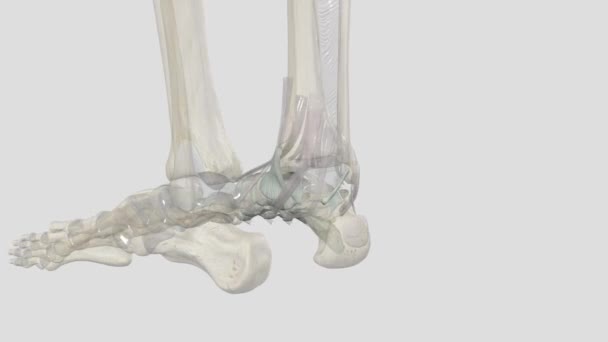
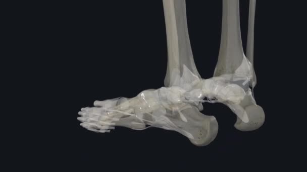
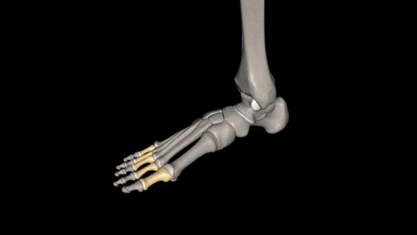
The intermetatarsal joints are articulations formed between the metatarsal bones and are stabilized by intermetatarsal interosseus ligaments
Intermetatarsal Joints Articulations Formed Metatarsal Bones Stabilized Intermetatarsal Interosseus Ligaments
00:14
The buccal fat pad, also known as Bichat's fat pad,
Buccal Fat Pad Also Known Bichat Fat Pad
00:16
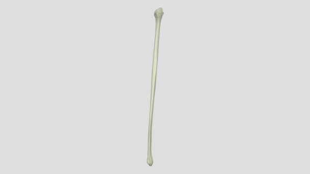
The fibula is a slender, cylindrical leg bone that is located on the posterior portion of the limb .
Fibula Slender Cylindrical Leg Bone Located Posterior Portion Limb
00:10
Woman in sportswear doing hip bridge exercise. Joyful attractive young woman lying on floor in living room and doing hip bridge exercise.
00:20
Chlamydia word made of wooden blocks with stethoscope. Medical and healthcare concepts
Chlamydia Word Made Wooden Blocks Stethoscope Medical Healthcare Concepts
00:08
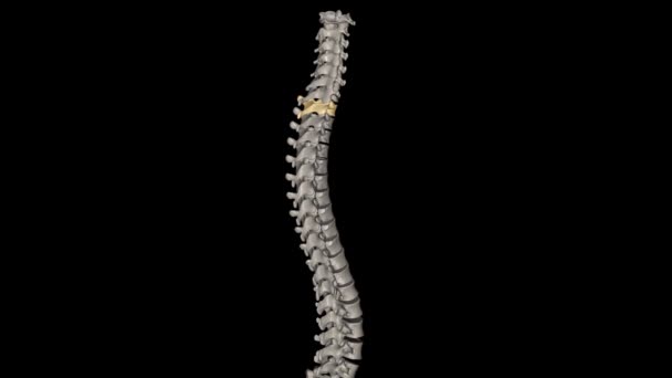
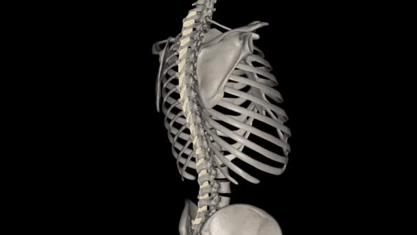
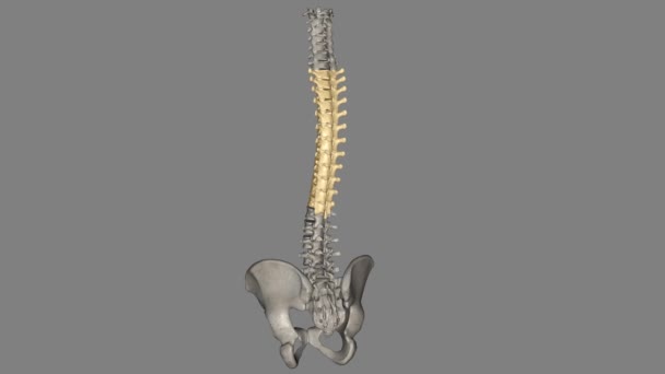
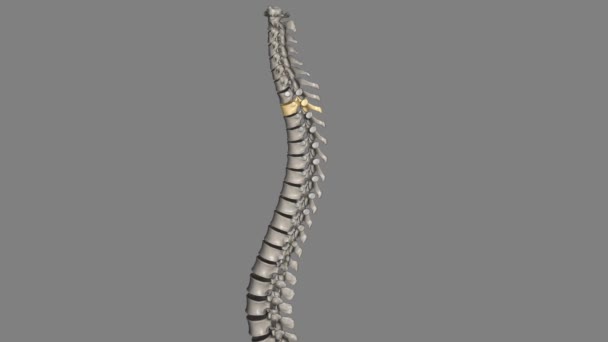
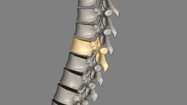
Twelve vertebrae are located in the thoracic spine and are numbered T-1 to T-12 T6 .
Twelve Vertebrae Located Thoracic Spine Numbered
00:12
206 Bones in the adult human body.3d illustration
206 Bones Adult Human Body Illustration
00:35
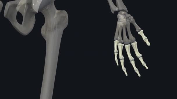
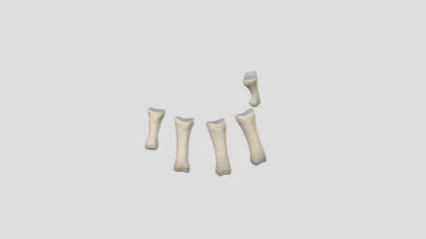
Phalanges. The 14 bones that are found in the fingers of each hand and also in the toes of each foot .
Phalanges Bones Found Fingers Each Hand Also Toes Each Foot
00:13
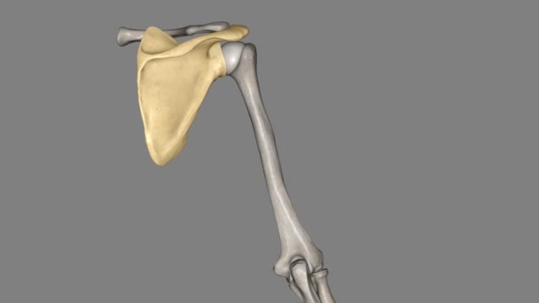
The scapula is a flat, triangular-shaped bone .
Scapula Flat Triangular Shaped Bone
00:11
A young pretty sports Female kneads her ankles, knees and pelvic joints in the fresh air in the middle of a dirt road outside the city. Slow-motion.
00:30
Word Endometriosis on wooden blocks against blurry doctor showing uterus image. Female reproductive system disease diagnostic and treatment
Word Endometriosis Wooden Blocks Blurry Doctor Showing Uterus Image Female Reproductive System Disease Diagnostic Treatment
00:14
The atlas (plural: atlases) is the first cervical vertebra, commonly called C1.
Atlas Plural Atlases First Cervical Vertebra Commonly Called
00:07
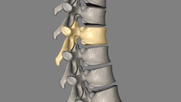
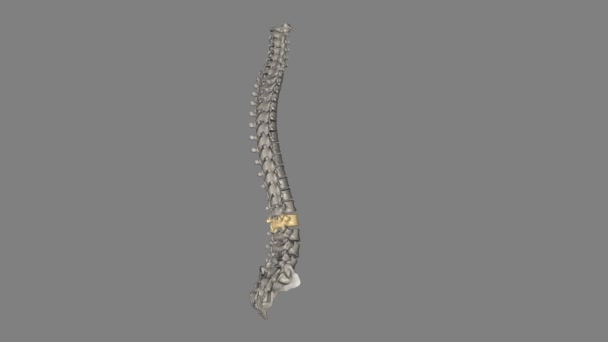
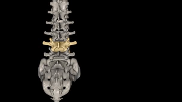
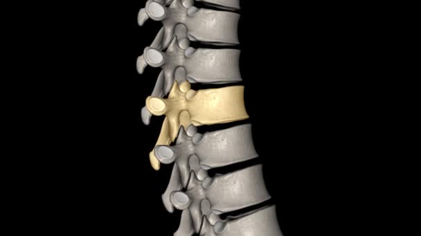
The L4 and L5 are the lowest vertebrae, or spinal bones, in the lumbar spine .
Lowest Vertebrae Spinal Bones Lumbar Spine
00:11
The role of the interspinous ligament is to limit flexion (bending forwards) through restricting separation of the spinous processes of the vertebral column
Role Interspinous Ligament Limit Flexion Bending Forwards Restricting Separation Spinous Processes Vertebral Column
00:09
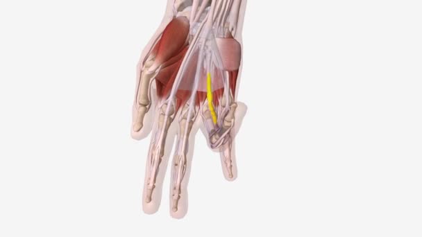
Dupuytren is Contracture Central Cord .
Dupuytren Contracture Central Cord
00:08
Sporty Millennial Woman Exercising With Fitball Over White Background In Studio
00:15
"PCOS is an ovarian cancer subtype that has metastasized to the uterus, fallopian tubes, or womb.
Pcos Ovarian Cancer Subtype Has Metastasized Uterus Fallopian Tubes Womb
00:12
In vertebrates, thoracic vertebrae compose the middle segment of the vertebral column, between the cervical vertebrae and the lumbar vertebrae
Vertebrates Thoracic Vertebrae Compose Middle Segment Vertebral Column Cervical Vertebrae Lumbar Vertebrae
00:10
Pregnant woman adjusting maternity support belt around baby bump at home, prenatal support accessory for reducing lower back and pelvic discomfort during pregnancy
Pregnant Woman Adjusting Maternity Support Belt Baby Bump Home Prenatal Support Accessory Reducing Lower Back Pelvic Discomfort Pregnancy
00:28
Text Colposcopy on wooden blocks and uterus model for education on desk. Woman doctor documents gynecological examination on clinical workspace
Text Colposcopy Wooden Blocks Uterus Model Education Desk Woman Doctor Documents Gynecological Examination Clinical Workspace
00:12
The muscles in your upper arm and forearm allow you to move your arms, hands, fingers and thumbs.
Muscles Your Upper Arm Forearm Allow You Move Your Arms Hands Fingers Thumbs
00:15
Pelvic Nerves system
00:15
The fourth cervical vertebra (C4) is centrally located in the cervical (or neck) region of the spinal column .
Fourth Cervical Vertebra Centrally Located Cervical Neck Region Spinal Column
00:10
The proximal phalanx of the fingers is the proximal, or first bone, in the fingers when counting from the hand to the tip of the finger
Proximal Phalanx Fingers Proximal First Bone Fingers Counting Hand Tip Finger
00:19
Muscles of the Pelvis and Perineum anal sphincter, internal, encircles the anal canal, encircles the anal canal .
Muscles Pelvis Perineum Anal Sphincter Internal Encircles Anal Canal Encircles Anal Canal
00:14
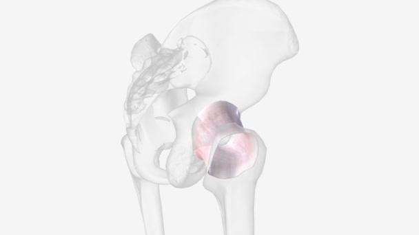
The hip joint is a ball and socket joint that is the point of articulation between the head of the femur and the acetabulum
Hip Joint Ball Socket Joint Point Articulation Head Femur Acetabulum
00:10
Tendon sheaths consist of two layers: a fibrous layer, made of tight collagenous tissue, and a synovial layer.
Tendon Sheaths Consist Two Layers Fibrous Layer Made Tight Collagenous Tissue Synovial Layer
00:14
The ligamenta flava are a series of ligaments that connect the ventral parts of the laminae of adjacent vertebrae .
Ligamenta Flava Series Ligaments Connect Ventral Parts Laminae Adjacent Vertebrae
00:08
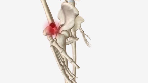
Human Skeleton and painful Hip Joint Pain
Human Skeleton Painful Hip Joint Pain
00:12
The hip joint is a ball and socket joint that is the point of articulation between the head of the femur and the acetabulum
Hip Joint Ball Socket Joint Point Articulation Head Femur Acetabulum
00:10
Woman stomach very sick and ache in front of green screen, girl is a disease and so much pain, gestures sadness
Woman Stomach Very Sick Ache Front Green Screen Girl Disease Much Pain Gestures Sadness
00:11
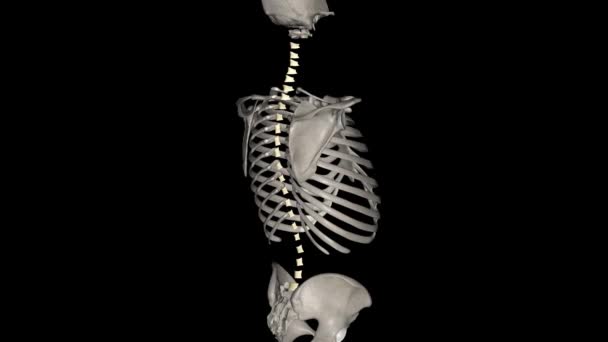
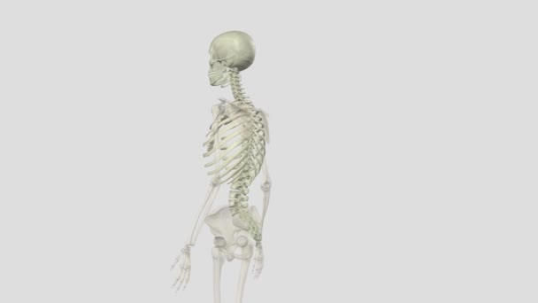
Human skeleton. Thorax and skull.
00:09
An experienced Doctor in the hospital carefully looks at an x-ray of a kidney with a stone. oxalate stone on x-ray
Experienced Doctor Hospital Carefully Looks Ray Kidney Stone Oxalate Stone Ray
00:09
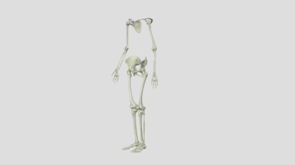
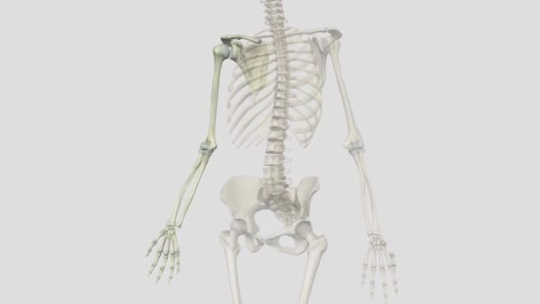
The appendicular skeleton is comprised of the upper and lower extremities, which include the shoulder girdle and pelvis
Appendicular Skeleton Comprised Upper Lower Extremities Which Include Shoulder Girdle Pelvis
00:12
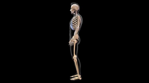
Axial skeleton is made up of the 80 bones within the central core of your body .
Axial Skeleton Made Bones Central Core Your Body
00:08
The hip is the area on each side of the pelvis .
Hip Area Each Side Pelvis
00:12
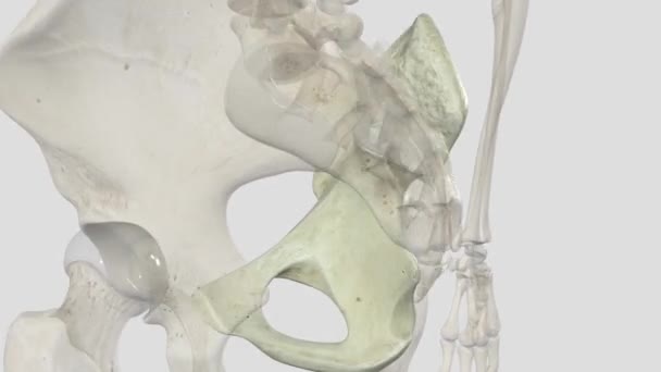
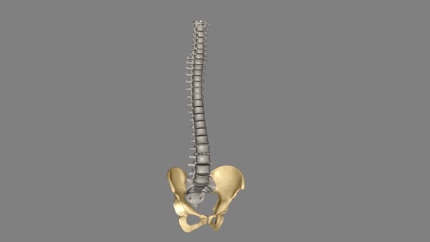
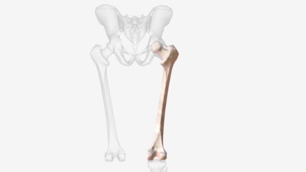
Pelvic bone structure
00:15
The femur is the only bone in the thigh and the longest bone in the body .
Femur Only Bone Thigh Longest Bone Body
00:20
The doctor is typing on the computer keyboard making a diagnosis of the disease, An experienced doctor in the hospital carefully looks at the x-ray of the kidney with a stone. oxalate stone on x-ray
Doctor Typing Computer Keyboard Making Diagnosis Disease Experienced Doctor Hospital Carefully Looks Ray Kidney Stone Oxalate Stone Ray
00:16
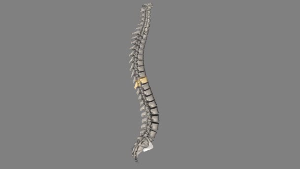
The L2 vertebra is located in the lower curvature of the spine, and the portion of the spinal cord that it protects is known as the lumbar cord.
Vertebra Located Lower Curvature Spine Portion Spinal Cord Protects Known Lumbar Cord
00:10
Female gynecologist shows location of pelvis with muscles. Pain pelvic bones causes diagnosis and treatment
Female Gynecologist Shows Location Pelvis Muscles Pain Pelvic Bones Causes Diagnosis Treatment
00:10
Twelve vertebrae are located in the thoracic spine and are numbered T-1 to T-12 .
Twelve Vertebrae Located Thoracic Spine Numbered
00:11
Pain in lower abdomen caused woman sit on bench, acute menstrual ache, closeup
Pain Lower Abdomen Caused Woman Sit Bench Acute Menstrual Ache Closeup
00:11
Vertical video. Over-the-shoulder shot. A gynecologist applies a clear gel from a blue plastic bottle to an ultrasound probe and then places a condom on it. Ultrasound of the pelvic organs
Vertical Video Shoulder Shot Gynecologist Applies Clear Gel Blue Plastic Bottle Ultrasound Probe Places Condom Ultrasound Pelvic Organs
00:10
Thoracic Vertebral, T8 Twelve vertebrae are located in the thoracic spine and are numbered T-1 to T-12
Thoracic Vertebral Twelve Vertebrae Located Thoracic Spine Numbered
00:13
A woman suffers from pain in the pelvic organs. The gynecologist examines the patient. Painful location, female disease, cystitis, or urinary incontinence. The doctor will palpate to find out the source of the pain
Woman Suffers Pain Pelvic Organs Gynecologist Examines Patient Painful Location Female Disease Cystitis Urinary Incontinence Doctor Palpate Find Out Source Pain
00:11
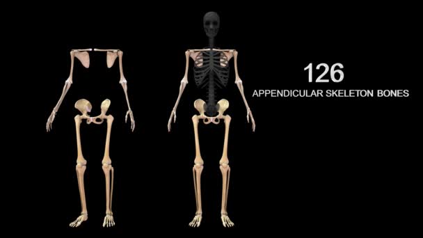
Appendicular skeleton in human skeletal system.3d illustration
Appendicular Skeleton Human Skeletal System Illustration
00:27
The L4 and L5 are the lowest vertebrae, or spinal bones, in the lumbar spine .
Lowest Vertebrae Spinal Bones Lumbar Spine
00:11
Ankylosing spondylitis is a type of arthritis that causes inflammation in the joints and ligaments of the spine .
Ankylosing Spondylitis Type Arthritis Causes Inflammation Joints Ligaments Spine
00:10
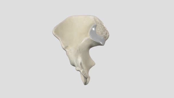
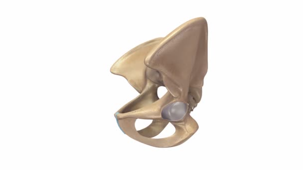
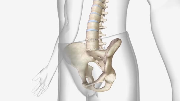
Detailed view of human pelvic bone
Detailed View Human Pelvic Bone
00:11
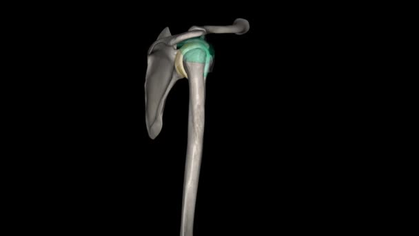
Joint capsule of the glenohumeral joint .
Joint Capsule Glenohumeral Joint
00:08
The proximal, or first bone, in the fingers when counting from the hand to the tip of the finger .
Proximal First Bone Fingers Counting Hand Tip Finger
00:09
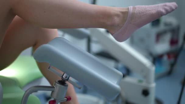
Close-up side view slim female Caucasian legs spread on gynecological chair. Unrecognizable young woman consulting gynecologist waiting for doctor in hospital indoors
Close Side View Slim Female Caucasian Legs Spread Gynecological Chair Unrecognizable Young Woman Consulting Gynecologist Waiting Doctor Hospital Indoors
00:06
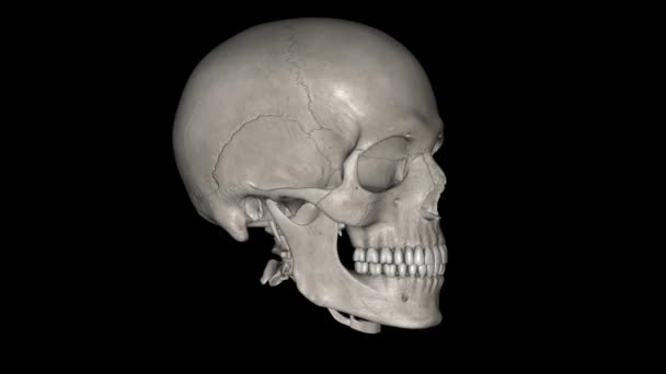
The skull is anterior to the spinal column and is the bony structure that encases the brain.
Skull Anterior Spinal Column Bony Structure Encases Brain
00:24
Ankylosing spondylitis is a type of arthritis that causes inflammation in the joints and ligaments of the spine .
Ankylosing Spondylitis Type Arthritis Causes Inflammation Joints Ligaments Spine
00:08
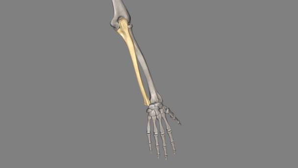
The ulna is one of two bones that make up the forearm, the other being the radius .
Ulna One Two Bones Make Forearm Other Being Radius
00:11
Plastic anatomical model showing the stages of a fetus moving through the birth canal
Plastic Anatomical Model Showing Stages Fetus Moving Birth Canal
00:14
Young woman doctor in hijab holds anatomical model of pelvic bone. Physician squeezes fist to show correct execution of Kegel exercises at lesson
Young Woman Doctor Hijab Holds Anatomical Model Pelvic Bone Physician Squeezes Fist Show Correct Execution Kegel Exercises Lesson
00:19
Female doctor writes prescription for Candidiasis treatment in medical office. Reproductive system anatomical model put on table in clinical setting
Female Doctor Writes Prescription Candidiasis Treatment Medical Office Reproductive System Anatomical Model Put Table Clinical Setting
00:17
Thoracic Vertebral, T9 Twelve vertebrae are located in the thoracic spine and are numbered T-1 to T-12
Thoracic Vertebral Twelve Vertebrae Located Thoracic Spine Numbered
00:10
Distal radius fractures are the most common orthopaedic injury and generally result from fall on an outstretched hand .
Distal Radius Fractures Most Common Orthopaedic Injury Generally Result Fall Outstretched Hand
00:13
Clavicle fracture is a break in the collarbone, one of the main bones in the shoulder .
Clavicle Fracture Break Collarbone One Main Bones Shoulder
00:09
The intertransverse ligaments are weak, sheet-like ligaments interconnecting adjacent transverse processes in the thoracic spine, and adjacent accessory processes in the lumbar spine.
Intertransverse Ligaments Weak Sheet Ligaments Interconnecting Adjacent Transverse Processes Thoracic Spine Adjacent Accessory Processes Lumbar Spine
00:10
Model of female pelvis with clock in clinic. Diagnosis of pelvic organs in women
Model Female Pelvis Clock Clinic Diagnosis Pelvic Organs Women
00:10
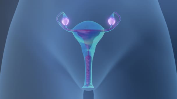
Mechanism of reproduction in women
Mechanism Reproduction Women
00:12
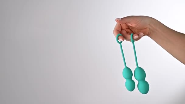
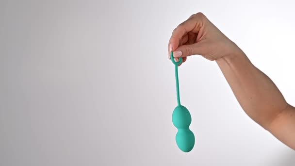
A faceless woman demonstrates a set of turquoise vaginal balls. Girl holding a kegel trainer for training pelvic floor muscles on a white background.
00:10
Faceless woman demonstrates a turquoise vaginal ball. Girl holding a kegel trainer for training pelvic floor muscles on a white background.
00:14
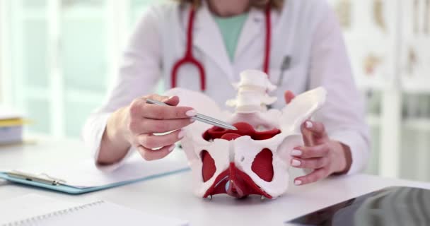
Female gynecologist shows location of pelvis with muscles. Doctor demonstrates the pelvic bones and pelvic muscles
Female Gynecologist Shows Location Pelvis Muscles Doctor Demonstrates Pelvic Bones Pelvic Muscles
00:15
Female gynecologist shows location of pelvis with muscles. Pain pelvic bones causes diagnosis and treatment
Female Gynecologist Shows Location Pelvis Muscles Pain Pelvic Bones Causes Diagnosis Treatment
00:08
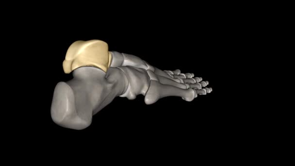
The talus talus bone, astragalus or ankle bone is one of the group of foot bones known as the tarsus .
Talus Talus Bone Astragalus Ankle Bone One Group Foot Bones Known Tarsus
00:12
Close up of female doctor pointing at plastic model of female reproductive system in clinic. Gynecological education with visible uterus and vaginal structure. Medical training scene. High quality 4k footage
Close Female Doctor Pointing Plastic Model Female Reproductive System Clinic Gynecological Education Visible Uterus Vaginal Structure Medical Training Scene High Quality Footage
00:19
The upper extremity or arm is a functional unit of the upper body .
Upper Extremity Arm Functional Unit Upper Body
00:10
Human ribs are flat bones that form part of the rib cage to help protect internal organs. Humans usually have 24 ribs, in 12 pairs .
Human Ribs Flat Bones Form Part Rib Cage Help Protect Internal Organs Humans Usually Have Ribs Pairs
00:09
Twelve vertebrae are located in the thoracic spine and are numbered T-1 to T-12 Thoracic Vertebral, T7
Twelve Vertebrae Located Thoracic Spine Numbered Thoracic Vertebral
00:11
Axial MRI image of the pelvis showing the lower abdomen, vertebral body, and surrounding muscles. Useful for radiology references, anatomy study, medical education, and healthcare visuals.
Axial Mri Image Pelvis Showing Lower Abdomen Vertebral Body Surrounding Muscles Useful Radiology References Anatomy Study Medical Education Healthcare Visuals
00:41
Doctor gynecologist performing an examination
00:44
Gynecologist shows bones of female pelvis closeup. Strengthening muscles of pelvic floor and gynecological diseases
Gynecologist Shows Bones Female Pelvis Closeup Strengthening Muscles Pelvic Floor Gynecological Diseases
00:08
Speckled sandperch (Parapercis hexophtalma) stands on its pelvic fins and turning its eyes to examine the surroundings, close up.
Speckled Sandperch Parapercis Hexophtalma Stands Its Pelvic Fins Turning Its Eyes Examine Surroundings Close
00:14
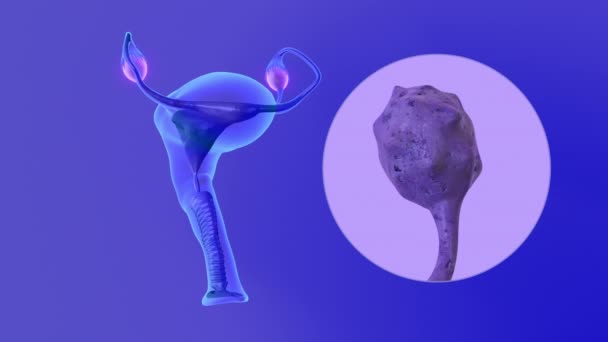
Infected ovaries in the female reproductive system
Infected Ovaries Female Reproductive System
00:12
The true ribs are the ribs that directly articulate with the sternum with their costal cartilages they are the first seven ribs .
True Ribs Ribs Directly Articulate Sternum Costal Cartilages First Seven Ribs
00:08
3D animation of human pelvic bones anatomy in colorful X-ray style, showing coxals, sacrum, coccyx, lumbar vertebrae, and femurs with vibrant detail.
Animation Human Pelvic Bones Anatomy Colorful Ray Style Showing Coxals Sacrum Coccyx Lumbar Vertebrae Femurs Vibrant Detail
00:16
Doctor measures model of pelvic bones with female reproductive organs using tape-line. Woman in medical uniform sits at table in clinic slow motion
Doctor Measures Model Pelvic Bones Female Reproductive Organs Using Tape Line Woman Medical Uniform Sits Table Clinic Slow Motion
00:10
Gynecologist shows location of the female pelvis with muscles. Pelvic floor muscle dysfunction and chronic pelvic pain syndrome in woman
Gynecologist Shows Location Female Pelvis Muscles Pelvic Floor Muscle Dysfunction Chronic Pelvic Pain Syndrome Woman
00:12
Page