Pelvis Stock Video Footage

2,073 Pelvis videos are available under a royalty-free license

Anatomy of the Gluteus Maximus Muscles
Anatomy Gluteus Maximus Muscles
00:10
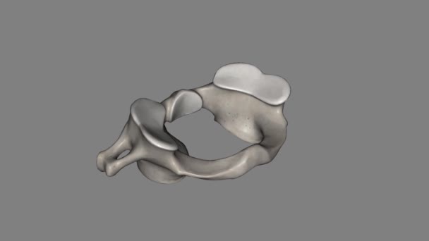
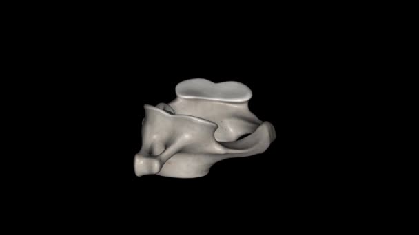
The atlas (plural: atlases) is the first cervical vertebra, commonly called C1 .
Atlas Plural Atlases First Cervical Vertebra Commonly Called
00:21
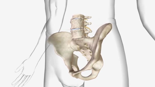
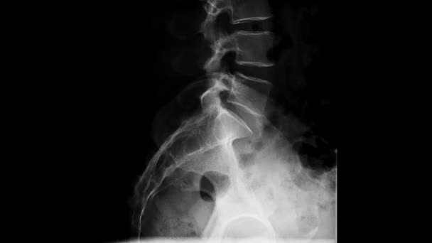
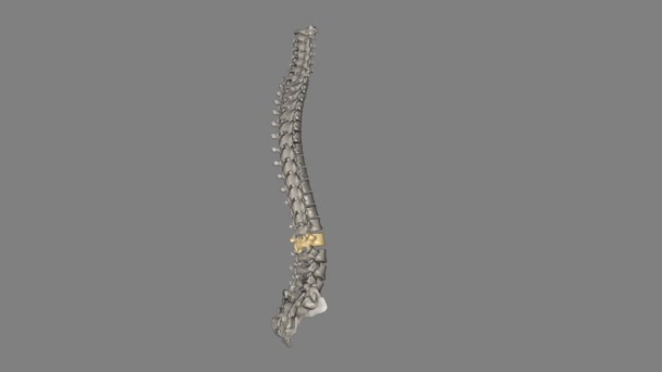
Degenerative spondylolisthesis (DS) is a disorder that causes the slip of one vertebral body over the one below due to degenerative changes in the spine .
Degenerative Spondylolisthesis Disorder Causes Slip One Vertebral Body One Due Degenerative Changes Spine
00:12
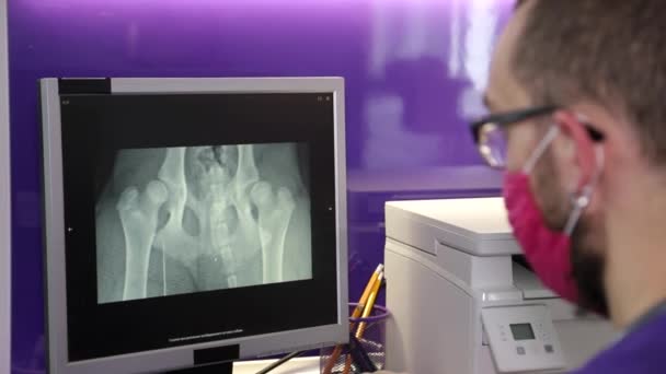
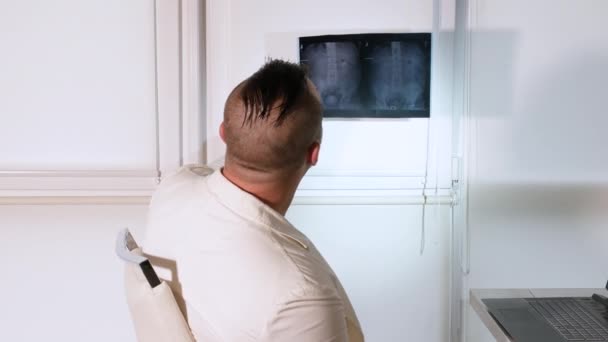
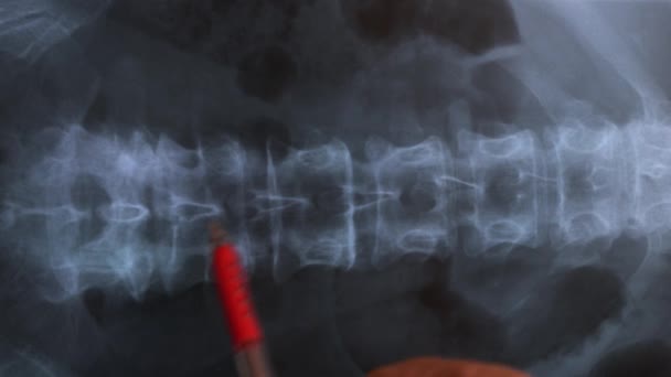
Doctor radiologically analyzing the pelvic bone with x-ray.Hand of a doctor analyzing and pointing with a pen to the affected area.
Doctor Radiologically Analyzing Pelvic Bone Ray Hand Doctor Analyzing Pointing Pen Affected Area
00:21
Knowledgeable university professor teaching human anatomy to a female student, pointing at a pelvis x ray on a large digital screen in a classroom
Knowledgeable University Professor Teaching Human Anatomy Female Student Pointing Pelvis Ray Large Digital Screen Classroom
00:18
Minimal studio scene pregnant woman exercising on Pilates reformer machine. High quality 4k footage
Minimal Studio Scene Pregnant Woman Exercising Pilates Reformer Machine High Quality Footage
00:26
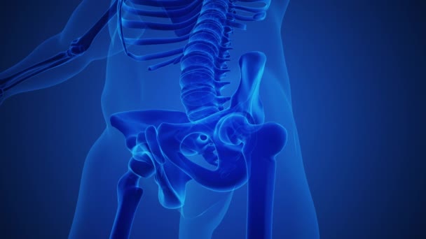
Skeletal system in human body. X Ray. Loop
Skeletal System Human Body Ray Loop
00:10
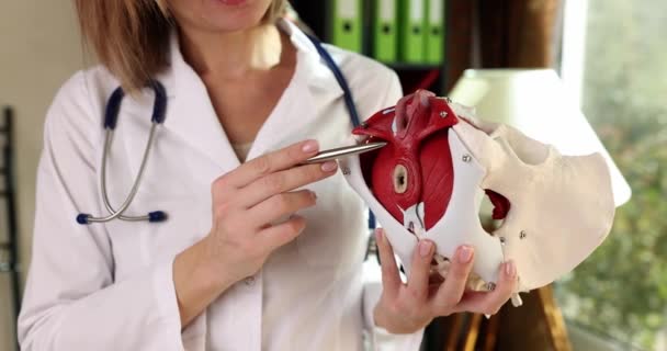
Gynecologist shows model of female pelvic organs with muscles. Pelvic bone structure and typical diseases and injuries
Gynecologist Shows Model Female Pelvic Organs Muscles Pelvic Bone Structure Typical Diseases Injuries
00:05
Female client on blue mat, trainer lifting foot, guiding knee, overlay tracing hip rotation for PT. Therapist, participant, mobility, stretching, rehabilitation, studio, stabilityball
Female Client Blue Mat Trainer Lifting Foot Guiding Knee Overlay Tracing Hip Rotation Therapist Participant Mobility Stretching Rehabilitation Studio Stabilityball
00:10
Skeleton with osteoarthritis hip joint injury
Skeleton Osteoarthritis Hip Joint Injury
00:12
Human Skeleton Sacral Bone Anatomy Loopable 3D Illustration For Medical Concept
Human Skeleton Sacral Bone Anatomy Loopable Illustration Medical Concept
00:12
Montage of doctors at work
00:19
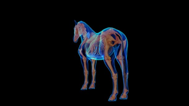
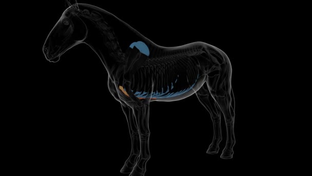
Horse skeleton anatomy for medical concept 3D animation
Horse Skeleton Anatomy Medical Concept Animation
00:12
The atlas (plural: atlases) is the first cervical vertebra, commonly called C1 .
Atlas Plural Atlases First Cervical Vertebra Commonly Called
00:21
A senior woman does an exercise of the internal muscles of the body. She raises her pelvis while lying on the ground. Hands bend at the shoulders. Restoration of muscles and ligaments of the body.
Senior Woman Does Exercise Internal Muscles Body She Raises Her Pelvis While Lying Ground Hands Bend Shoulders Restoration Muscles Ligaments Body
00:17
Base partner lifting hips and holding partner mounting on teal mats, group building core fitness. Workout, coordination, synchronization, teamwork, movement, strength, balance
Base Partner Lifting Hips Holding Partner Mounting Teal Mats Group Building Core Fitness Workout Coordination Synchronization Teamwork Movement Strength Balance
00:10
Sketch style minimal pelvis icon animation - smooth progressive reveal'
Sketch Style Minimal Pelvis Icon Animation Smooth Progressive Reveal
00:08
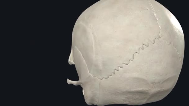
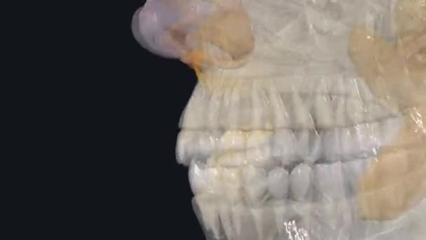
Neurocranium as a whole bone complex. See multiple interactive high quality pictures demonstrating different views of the skull bones surrounding the brain.
Neurocranium Whole Bone Complex See Multiple Interactive High Quality Pictures Demonstrating Different Views Skull Bones Surrounding Brain
00:16
Amazing aerial view of Krasny Yar village, Russia. Krasny Yar is the capital of the Primorsky Udege - indigenous small peoples of the far north. The village stands on the river Bikin, the tributary of the great Amur. In the village works the Bikin
Amazing Aerial View Krasny Yar Village Russia Krasny Yar Capital Primorsky Udege Indigenous Small Peoples Far North Village Stands River Bikin Tributary Great Amur Village Works Bikin
00:26
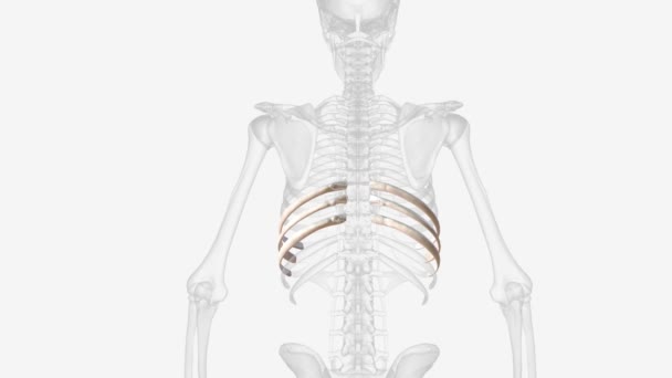
The ribs partially enclose and protect the chest cavity, where many vital organs .
Ribs Partially Enclose Protect Chest Cavity Many Vital Organs
00:23
Dog skeleton Anatomy For Medical Concept 3D Illustration
Dog Skeleton Anatomy Medical Concept Illustration
00:12
Shop of goods for home. A young woman chooses a large plastic pelvis
00:25
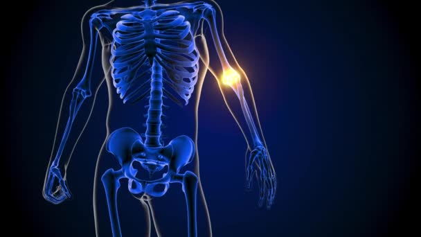
Elbow joint pain anatomy for medical concept 3D animation
Elbow Joint Pain Anatomy Medical Concept Animation
00:12
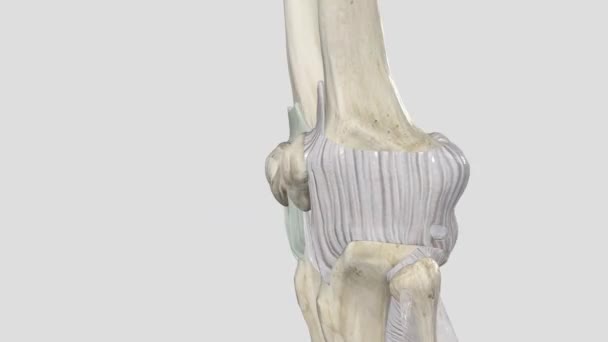
Joint capsule is a sac-like fibrous capsule continuous with the periosteum of the articulating bones .
Joint Capsule Sac Fibrous Capsule Continuous Periosteum Articulating Bones
00:16
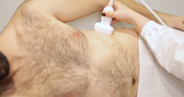
Doctor examining males kedneys with ultrasound scan in clinic
00:16
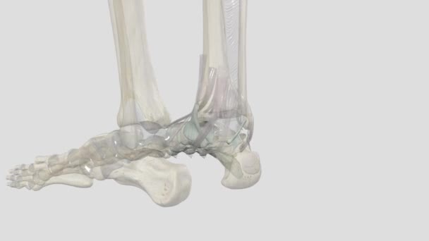
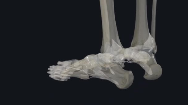
The intermetatarsal joints are articulations formed between the metatarsal bones and are stabilized by intermetatarsal interosseus ligaments
Intermetatarsal Joints Articulations Formed Metatarsal Bones Stabilized Intermetatarsal Interosseus Ligaments
00:14
Hand washing laundry in a bowl
Hand Washing Laundry Bowl
00:18
The buccal fat pad, also known as Bichat's fat pad,
Buccal Fat Pad Also Known Bichat Fat Pad
00:16
Bottom portrait of sporty woman with hands resting on knees and gracefully swaying pelvis while positioned in center of pedestrian street. Female focusing on enhancing buttocks and leg muscles.
Bottom Portrait Sporty Woman Hands Resting Knees Gracefully Swaying Pelvis While Positioned Center Pedestrian Street Female Focusing Enhancing Buttocks Leg Muscles
00:12
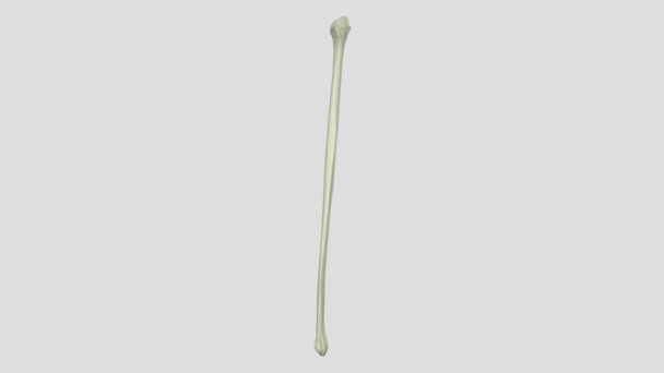
The fibula is a slender, cylindrical leg bone that is located on the posterior portion of the limb .
Fibula Slender Cylindrical Leg Bone Located Posterior Portion Limb
00:10
Woman in sportswear doing hip bridge exercise. Joyful attractive young woman lying on floor in living room and doing hip bridge exercise.
00:20
PELVIS bone skeleton x-ray scan in blue
00:14
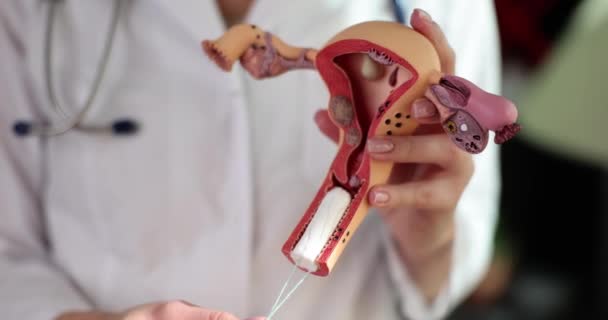
Doctor in medical uniform shows tampon in female pelvis model with reproductive organs. Woman pulls out tampon from model of body part slow motion
Doctor Medical Uniform Shows Tampon Female Pelvis Model Reproductive Organs Woman Pulls Out Tampon Model Body Part Slow Motion
00:05
Model of female pelvis with clock in clinic. Diagnosis of pelvic organs in women
Model Female Pelvis Clock Clinic Diagnosis Pelvic Organs Women
00:10
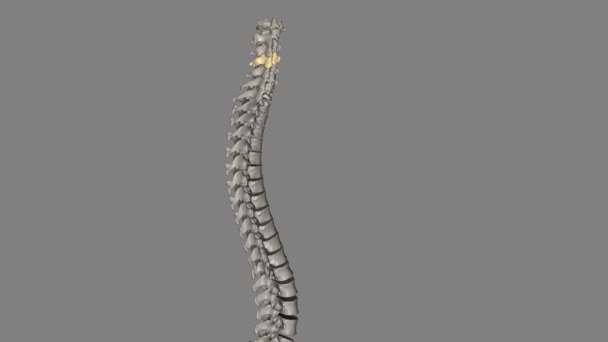
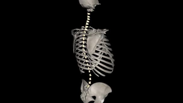
Twelve vertebrae are located in the thoracic spine and are numbered T-1 to T-12 T6 .
Twelve Vertebrae Located Thoracic Spine Numbered
00:12
Figure of man running with videos of circulation
00:21
Doctor examining liver of male patient with ultrasound scan in clinic
00:18
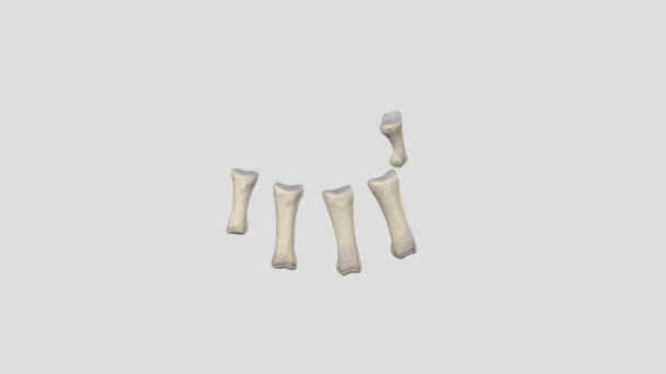
Phalanges. The 14 bones that are found in the fingers of each hand and also in the toes of each foot .
Phalanges Bones Found Fingers Each Hand Also Toes Each Foot
00:13
The scapula is a flat, triangular-shaped bone .
Scapula Flat Triangular Shaped Bone
00:11
Human skeleton rib cage anatomy 3D animation for medical concept
Human Skeleton Rib Cage Anatomy Animation Medical Concept
00:20
The atlas (plural: atlases) is the first cervical vertebra, commonly called C1.
Atlas Plural Atlases First Cervical Vertebra Commonly Called
00:07
Pretty woman lies on mat, raises her pelvis, body, hands. Concept of sport, wellness and health. Young smiling lady doing active fitness workout, satisfied fit woman.
Pretty Woman Lies Mat Raises Her Pelvis Body Hands Concept Sport Wellness Health Young Smiling Lady Doing Active Fitness Workout Satisfied Fit Woman
00:21
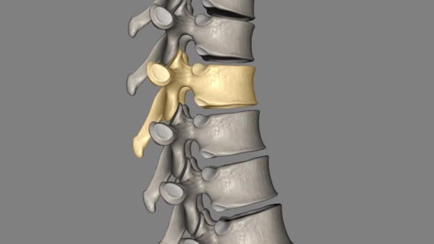
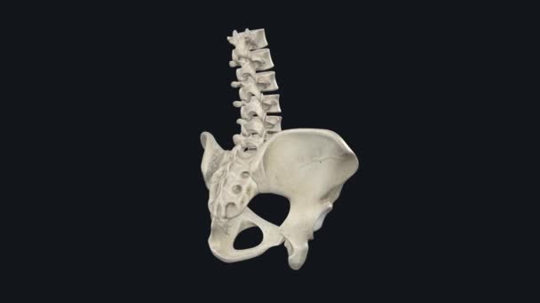
The L4 and L5 are the lowest vertebrae, or spinal bones, in the lumbar spine .
Lowest Vertebrae Spinal Bones Lumbar Spine
00:11
The role of the interspinous ligament is to limit flexion (bending forwards) through restricting separation of the spinous processes of the vertebral column
Role Interspinous Ligament Limit Flexion Bending Forwards Restricting Separation Spinous Processes Vertebral Column
00:09
Dupuytren is Contracture Central Cord .
Dupuytren Contracture Central Cord
00:08
Digitally generated human anatomy walking with binary codes and screens with medical images
Digitally Generated Human Anatomy Walking Binary Codes Screens Medical Images
00:09
Physiotherapist looking at spine model
00:08
Elderly woman doing stretching after a workout. A grayhaired woman sitting on her knees leans forward, pulls her pelvis back and stretches her arms forward, then stretches forward bending in her back
Elderly Woman Doing Stretching Workout Grayhaired Woman Sitting Her Knees Leans Forward Pulls Her Pelvis Back Stretches Her Arms Forward Stretches Forward Bending Her Back
00:33
Pretty woman kneels on mat, lifts her pelvis and body. Concept of sport, wellness and health. Young smiling lady doing active fitness workout, satisfied fit woman.
Pretty Woman Kneels Mat Lifts Her Pelvis Body Concept Sport Wellness Health Young Smiling Lady Doing Active Fitness Workout Satisfied Fit Woman
00:21
Illustrating human digestive system anatomy
Illustrating Human Digestive System Anatomy
00:11
3D Medical Animation of internal structure of kidney
00:06
Detailed transparent human skeleton with spinal surgery stabilization
Detailed Transparent Human Skeleton Spinal Surgery Stabilization
00:09
The medium shot of a male chiropractor after a massage makes a girl patient healthy exercises of the legs and pelvis in a dark room
00:18
Shoulder joint pain Anatomy For Medical Concept 3D Animation
Shoulder Joint Pain Anatomy Medical Concept Animation
00:15
In vertebrates, thoracic vertebrae compose the middle segment of the vertebral column, between the cervical vertebrae and the lumbar vertebrae
Vertebrates Thoracic Vertebrae Compose Middle Segment Vertebral Column Cervical Vertebrae Lumbar Vertebrae
00:10
Clavicle bones anatomy for medical concept 3D animation
Clavicle Bones Anatomy Medical Concept Animation
00:20
Physicians are performing a surgical procedure in pelvis area
00:14
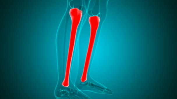
Human Skeleton Tibia Bone Anatomy Loopable 3D Illustration For Medical Concept
Human Skeleton Tibia Bone Anatomy Loopable Illustration Medical Concept
00:12
The muscles in your upper arm and forearm allow you to move your arms, hands, fingers and thumbs.
Muscles Your Upper Arm Forearm Allow You Move Your Arms Hands Fingers Thumbs
00:15
This 3d animation shows the obturator internus muscle movement
Animation Shows Obturator Internus Muscle Movement
00:06
The fourth cervical vertebra (C4) is centrally located in the cervical (or neck) region of the spinal column .
Fourth Cervical Vertebra Centrally Located Cervical Neck Region Spinal Column
00:10
Toned women doing sports exercise lying on exercise mats
00:11
Sternum bone horse skeleton anatomy for medical concept 3D animation
Sternum Bone Horse Skeleton Anatomy Medical Concept Animation
00:13
The proximal phalanx of the fingers is the proximal, or first bone, in the fingers when counting from the hand to the tip of the finger
Proximal Phalanx Fingers Proximal First Bone Fingers Counting Hand Tip Finger
00:19
MRI scan timelapse of various sides human body, spine, coccyx bones, pelvis, lumbar. Magnetic resonance imaging of skeleton parts, CT, x-ray, side view, stop motion animation. Seamless loop.
Mri Scan Timelapse Various Sides Human Body Spine Coccyx Bones Pelvis Lumbar Magnetic Resonance Imaging Skeleton Parts Ray Side View Stop Motion Animation Seamless Loop
00:06
Muscles of the Pelvis and Perineum anal sphincter, internal, encircles the anal canal, encircles the anal canal .
Muscles Pelvis Perineum Anal Sphincter Internal Encircles Anal Canal Encircles Anal Canal
00:14
Doctor uses anatomical model to explain male urinary system. Model labeled with parts, doctor points and explains how they work together for urinary function, ensuring patient comprehension.
Doctor Uses Anatomical Model Explain Male Urinary System Model Labeled Parts Doctor Points Explains How Work Together Urinary Function Ensuring Patient Comprehension
00:17
CT Abdomen - movie in sagittal plane w internal organs
Abdomen Movie Sagittal Plane Internal Organs
00:16
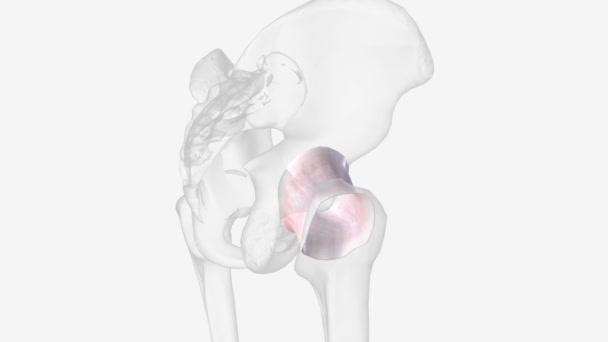
The hip joint is a ball and socket joint that is the point of articulation between the head of the femur and the acetabulum
Hip Joint Ball Socket Joint Point Articulation Head Femur Acetabulum
00:10
Tendon sheaths consist of two layers: a fibrous layer, made of tight collagenous tissue, and a synovial layer.
Tendon Sheaths Consist Two Layers Fibrous Layer Made Tight Collagenous Tissue Synovial Layer
00:14
This 3d animation shows the obturator internus muscle movement
Animation Shows Obturator Internus Muscle Movement
00:06
The ligamenta flava are a series of ligaments that connect the ventral parts of the laminae of adjacent vertebrae .
Ligamenta Flava Series Ligaments Connect Ventral Parts Laminae Adjacent Vertebrae
00:08
Human Skeleton and painful Hip Joint Pain
Human Skeleton Painful Hip Joint Pain
00:12
The hip joint is a ball and socket joint that is the point of articulation between the head of the femur and the acetabulum
Hip Joint Ball Socket Joint Point Articulation Head Femur Acetabulum
00:10
Veterinarian examines x-ray picture of dog in veterinary clinic, pet care
00:10
A rooster is in the courtyard of a rural house.
00:23
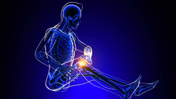
Knee joint pain Anatomy For Medical Concept 3D Animation sitting position
Knee Joint Pain Anatomy Medical Concept Animation Sitting Position
00:13
Bodybuilder trains lower back muscles in a fitness club
00:12
Human skeleton. Thorax and skull.
00:09
Sciatica nerve pain lower back through hips to leg
Sciatica Nerve Pain Lower Back Hips Leg
00:12
Running woman focused on leg bones
00:08
The close up shot of a male chiropractor after a massage makes a girl patient healthy exercises of the legs and pelvis in a dark room
00:18
Sciatica disease put a plaster on butt warming patch inflammation of the sciatic nerve. heating patch. sciatic nerve pain. High quality 4k footage
Sciatica Disease Put Plaster Butt Warming Patch Inflammation Sciatic Nerve Heating Patch Sciatic Nerve Pain High Quality Footage
00:22
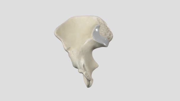
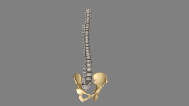
Human skeleton hip or pelvic bone anatomy 3D animation for medical concept
Human Skeleton Hip Pelvic Bone Anatomy Animation Medical Concept
00:20
Girl art makeup watercolor
00:22
An experienced Doctor in the hospital carefully looks at an x-ray of a kidney with a stone. oxalate stone on x-ray
Experienced Doctor Hospital Carefully Looks Ray Kidney Stone Oxalate Stone Ray
00:09
Amazing aerial view of Krasny Yar village, Russia. Krasny Yar is the capital of the Primorsky Udege - indigenous small peoples of the far north. The village stands on the river Bikin, the tributary of the great Amur. In the village works the Bikin
Amazing Aerial View Krasny Yar Village Russia Krasny Yar Capital Primorsky Udege Indigenous Small Peoples Far North Village Stands River Bikin Tributary Great Amur Village Works Bikin
00:27
The appendicular skeleton is comprised of the upper and lower extremities, which include the shoulder girdle and pelvis
Appendicular Skeleton Comprised Upper Lower Extremities Which Include Shoulder Girdle Pelvis
00:12
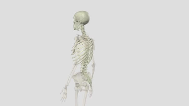
Axial skeleton is made up of the 80 bones within the central core of your body .
Axial Skeleton Made Bones Central Core Your Body
00:08
This 3d animation shows a side view of the adductor longus muscle movement
Animation Shows Side View Adductor Longus Muscle Movement
00:06
A man suffering from a urinary tract infection visits a doctor at the examination table in a hospital. Symptoms. Talk about enlarged prostate. Male reproductive system. Asian doctor.
Man Suffering Urinary Tract Infection Visits Doctor Examination Table Hospital Symptoms Talk Enlarged Prostate Male Reproductive System Asian Doctor
00:19
The hip is the area on each side of the pelvis .
Hip Area Each Side Pelvis
00:12
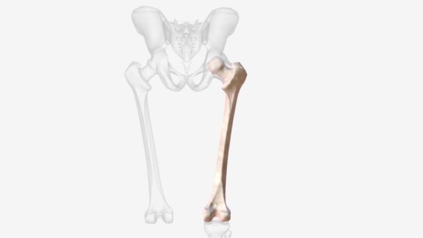
The femur is the only bone in the thigh and the longest bone in the body .
Femur Only Bone Thigh Longest Bone Body
00:20
The doctor is typing on the computer keyboard making a diagnosis of the disease, An experienced doctor in the hospital carefully looks at the x-ray of the kidney with a stone. oxalate stone on x-ray
Doctor Typing Computer Keyboard Making Diagnosis Disease Experienced Doctor Hospital Carefully Looks Ray Kidney Stone Oxalate Stone Ray
00:16
Doctor studying X-ray of the bones of the human vertebral column , 4K
Doctor Studying Ray Bones Human Vertebral Column
00:11
MRI scans set stop motion loop, human bones and joints, body, spine, ribs chest, coccyx, pelvis and organs. Animated stop motion mosaic of various body parts CT scan or x ray. Seamless loop.
Mri Scans Set Stop Motion Loop Human Bones Joints Body Spine Ribs Chest Coccyx Pelvis Organs Animated Stop Motion Mosaic Various Body Parts Scan Ray Seamless Loop
00:06
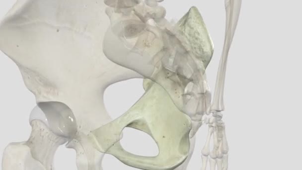
Human Skeleton Hip or Pelvic bone Anatomy 3D Illustration For Medical Concept
Human Skeleton Hip Pelvic Bone Anatomy Illustration Medical Concept
00:12
The L2 vertebra is located in the lower curvature of the spine, and the portion of the spinal cord that it protects is known as the lumbar cord.
Vertebra Located Lower Curvature Spine Portion Spinal Cord Protects Known Lumbar Cord
00:10
Female gynecologist shows location of pelvis with muscles. Pain pelvic bones causes diagnosis and treatment
Female Gynecologist Shows Location Pelvis Muscles Pain Pelvic Bones Causes Diagnosis Treatment
00:10
Doctor examining liver of male patient with ultrasound scan in clinic
00:15
Page