Pelvis Stock Video Footage

2,073 Pelvis videos are available under a royalty-free license
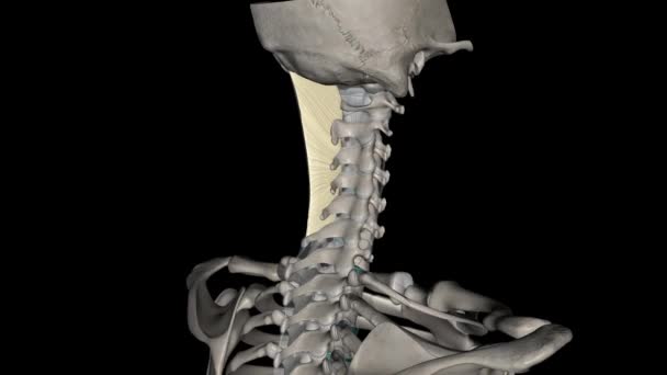
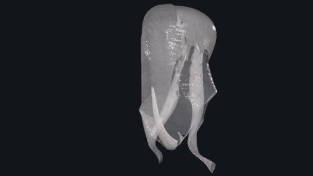
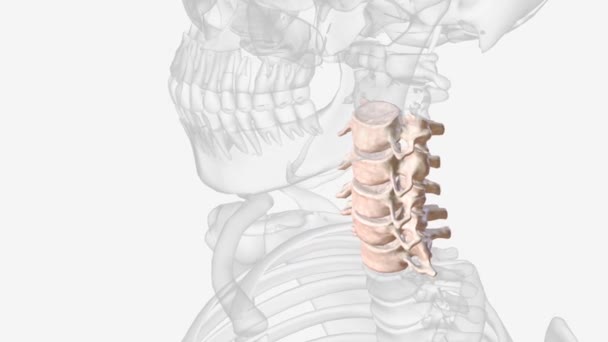
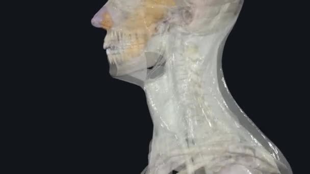
The nuchal ligament is a ligament at the back of the neck that is continuous with the supraspinous ligament.
Nuchal Ligament Ligament Back Neck Continuous Supraspinous Ligament
00:11
Orthopedic, Doctor Writing on Transparent Screen
00:08
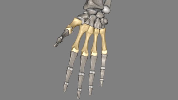
Metacarpal, any of several tubular bones between the wrist (carpal) bones and each of the forelimb digits in land vertebrates, corresponding to the metatarsal bones of the foot.
Metacarpal Any Several Tubular Bones Wrist Carpal Bones Each Forelimb Digits Land Vertebrates Corresponding Metatarsal Bones Foot
00:10
Pregnant woman training on Pilates reformer machine in studio fitness session. High quality 4k footage
Pregnant Woman Training Pilates Reformer Machine Studio Fitness Session High Quality Footage
00:28
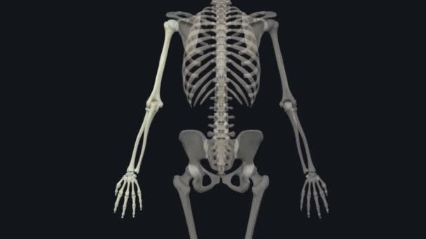
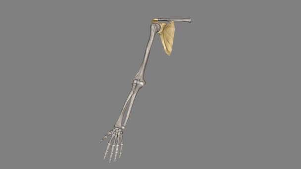
The free part of upper limb denotes the distal segment of the upper limb, which excludes the shoulder girdle (shoulder and axilla)
Free Part Upper Limb Denotes Distal Segment Upper Limb Which Excludes Shoulder Girdle Shoulder Axilla
00:14
Human skeleton Fibula Bone Anatomy Loopable 3D Illustration For Medical Concept
Human Skeleton Fibula Bone Anatomy Loopable Illustration Medical Concept
00:12
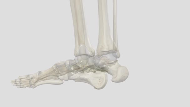
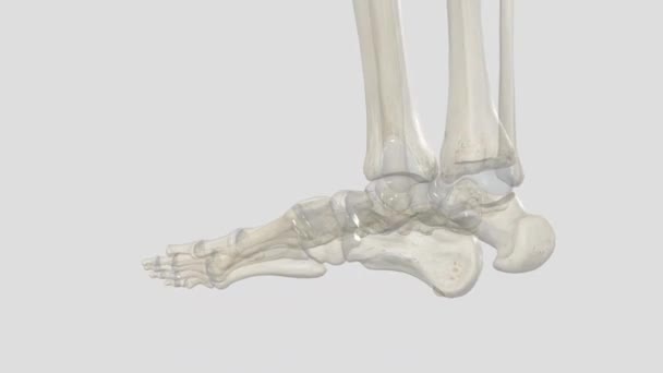
The human foot is a strong and complex mechanical structure containing 26 bones, 33 joints .
Human Foot Strong Complex Mechanical Structure Containing Bones Joints
00:09
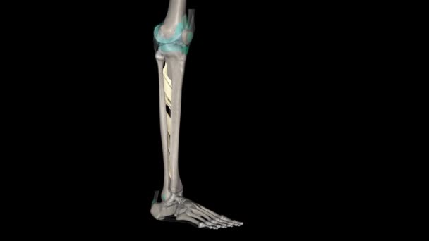
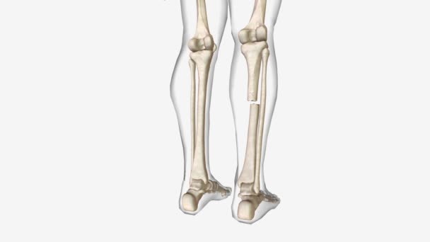
The interosseous membrane of the leg is also referred to as the middle tibiofibular ligament .
Interosseous Membrane Leg Also Referred Middle Tibiofibular Ligament
00:14
The sphenomandibular ligament (internal lateral ligament) is one of the three ligaments of the temporomandibular joint .
Sphenomandibular Ligament Internal Lateral Ligament One Three Ligaments Temporomandibular Joint
00:12
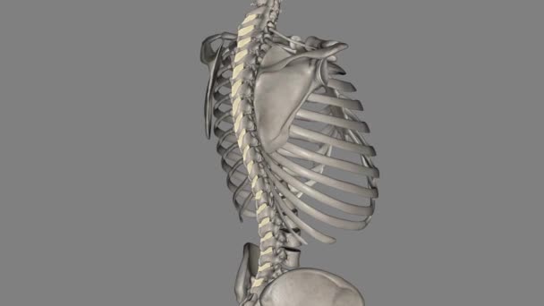
Thoracic Vertebral, T4 Twelve vertebrae are located in the thoracic spine and are numbered T-1 to T-12
Thoracic Vertebral Twelve Vertebrae Located Thoracic Spine Numbered
00:12
A rooster is in the courtyard of a rural house.
00:16
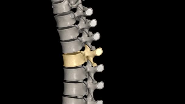
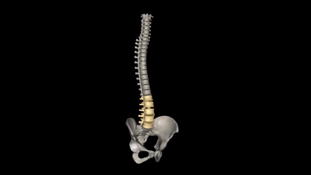
The lumbar vertebrae are, in human anatomy, the five vertebrae between the rib cage and the pelvis.
Lumbar Vertebrae Human Anatomy Five Vertebrae Rib Cage Pelvis
00:10
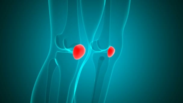
Human skeleton patella bone anatomy for medical concept 3d animation
Human Skeleton Patella Bone Anatomy Medical Concept Animation
00:20
The little girl washes the dishes with a rag in a dirty basin of water
00:32
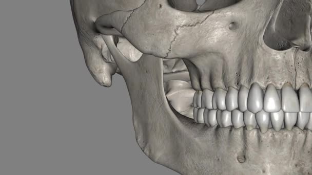
The maxilla, also known as the upper jaw, is a vital viscerocranium structure of the skull .
Maxilla Also Known Upper Jaw Vital Viscerocranium Structure Skull
00:09
4k animated x-ray simulated body parts
00:20
Human skeleton patella bone anatomy for medical concept 3d animation
Human Skeleton Patella Bone Anatomy Medical Concept Animation
00:20
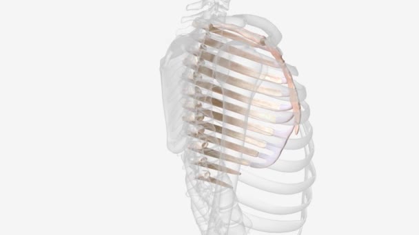
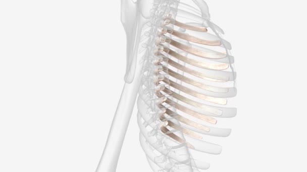
The first rib is the most superior of the twelve ribs .
First Rib Most Superior Twelve Ribs
00:31
Dog bath time. Animal care. Adorable cute young puppy white Pekingese take a bath in soapy suds full of bubbles in yellow plastic pelvis.
Dog Bath Time Animal Care Adorable Cute Young Puppy White Pekingese Take Bath Soapy Suds Full Bubbles Yellow Plastic Pelvis
00:13
Woman doing mermaid exercise on back spine corrector. Reformer pilates studio machine for fitness workouts in gym. Fit, healthy and strong authentical body. Fitness concept
Woman Doing Mermaid Exercise Back Spine Corrector Reformer Pilates Studio Machine Fitness Workouts Gym Fit Healthy Strong Authentical Body Fitness Concept
00:15
Man is cleaning terrarium with big cockroaches and moving insects into pelvis.
00:28
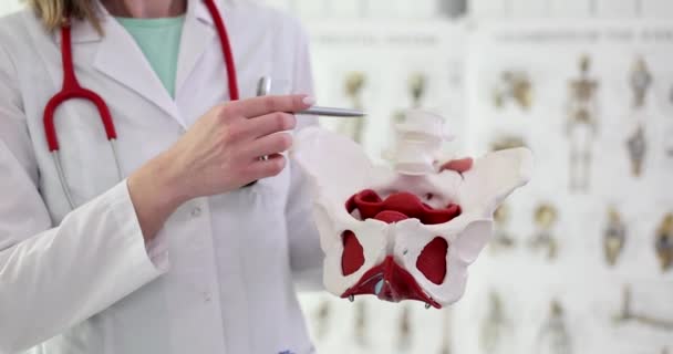
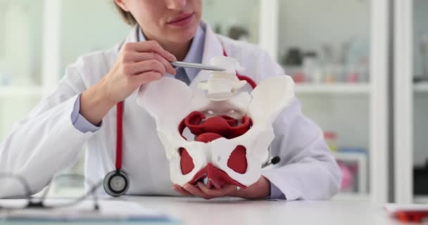
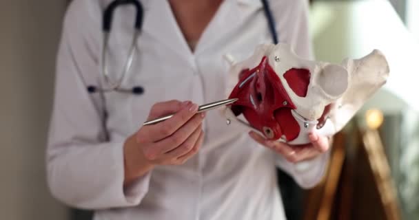
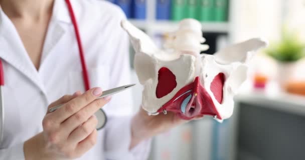
Doctor female gynecologist holds female pelvis with muscle model. Gynecology and woman health
Doctor Female Gynecologist Holds Female Pelvis Muscle Model Gynecology Woman Health
00:13
Many argentine cockroaches small and large crawl in the pelvis, closeup view.
00:16
Parietal abdominal fascia (endoabdominal fascia) may be the fascia that covers the abdominal cavity,
Parietal Abdominal Fascia Endoabdominal Fascia May Fascia Covers Abdominal Cavity
00:12
Revolving human skeleton figures
00:19
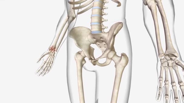
Comminuted fractures are a type of broken bone .
Comminuted Fractures Type Broken Bone
00:10
Metatarsal Bones Dog skeleton Anatomy For Medical Concept 3D Illustration
Metatarsal Bones Dog Skeleton Anatomy Medical Concept Illustration
00:12
Amazing aerial view of Krasny Yar village, Russia. Krasny Yar is the capital of the Primorsky Udege - indigenous small peoples of the far north. The village stands on the river Bikin, the tributary of the great Amur. In the village works the Bikin
Amazing Aerial View Krasny Yar Village Russia Krasny Yar Capital Primorsky Udege Indigenous Small Peoples Far North Village Stands River Bikin Tributary Great Amur Village Works Bikin
00:16
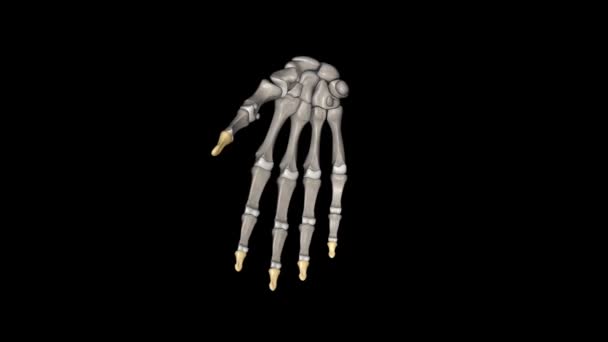
The thumb distal phalanx is a short bone with a rounded tuft at the end that makes a joint with the proximal phalanx .
Thumb Distal Phalanx Short Bone Rounded Tuft End Makes Joint Proximal Phalanx
00:12
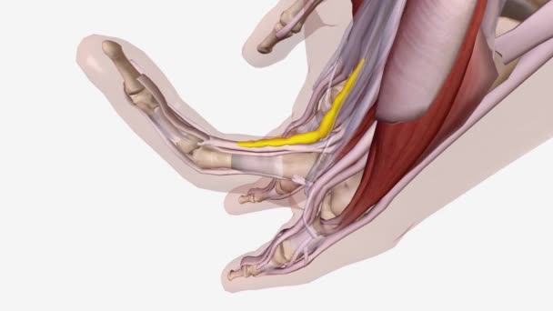
Dupuytren is Contracture Central Cord .
Dupuytren Contracture Central Cord
00:14
Warming patch on sciatic nerve. heating patch. sciatic nerve pain. hip joint treatment
Warming Patch Sciatic Nerve Heating Patch Sciatic Nerve Pain Hip Joint Treatment
00:20
Woman doctor in work uniform shows structure of human pelvis on artificial model. Part of body and explanation from female specialist about functionality in hospital
Woman Doctor Work Uniform Shows Structure Human Pelvis Artificial Model Part Body Explanation Female Specialist Functionality Hospital
00:15
Montage of medical clips
00:14
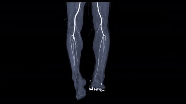
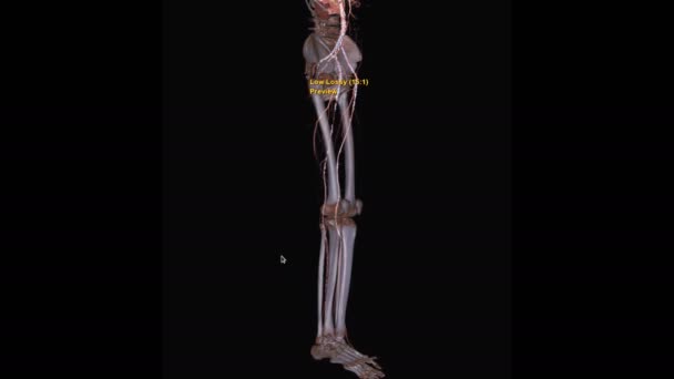
CTA femoral artery runn off angiography view turn around on the screen for detect peripheral arterial disease.
Cta Femoral Artery Runn Angiography View Turn Screen Detect Peripheral Arterial Disease
00:15
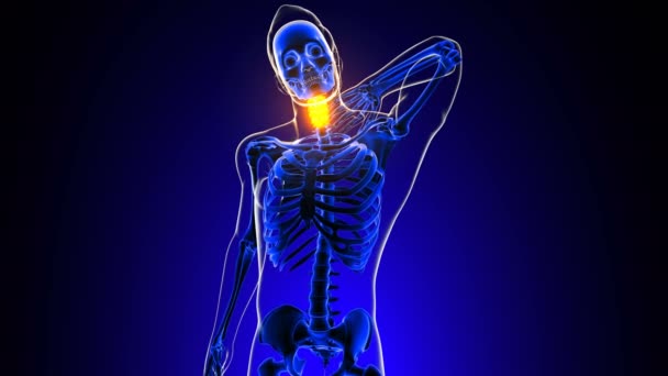
Cervical spine neck joint pain Anatomy For Medical Concept 3D Animation
Cervical Spine Neck Joint Pain Anatomy Medical Concept Animation
00:13
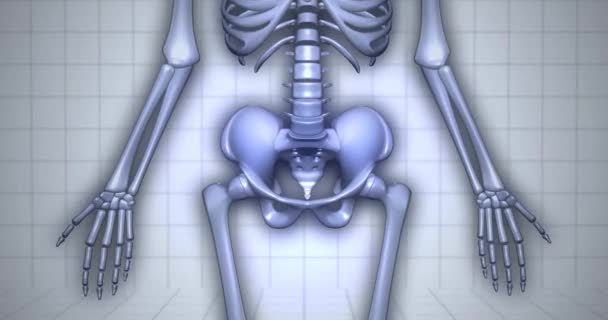
3D Human Anatomy Skeleton Visualization - Hip Bones
Human Anatomy Skeleton Visualization Hip Bones
00:18
Doctor points to pelvic bones of anatomical model of small pelvis. Inflammatory diseases of pelvic organs
Doctor Points Pelvic Bones Anatomical Model Small Pelvis Inflammatory Diseases Pelvic Organs
00:15
Human Skeleton System Pelvis Bone Joints Anatomy. 3D
Human Skeleton System Pelvis Bone Joints Anatomy
00:12
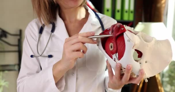
The doctor shows the anatomical model of the human pelvis, a close-up, slowmotion. Muscles, female health
Doctor Shows Anatomical Model Human Pelvis Close Slowmotion Muscles Female Health
00:06
Many goose in yard near the drinker
00:23
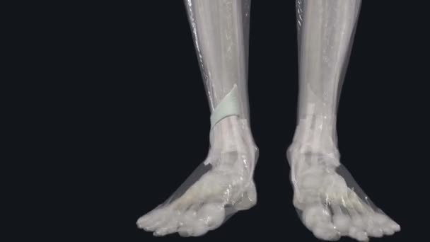
The extensor retinaculum refers to the set of ligaments inside the ankle that connect the tibia and fibula .
Extensor Retinaculum Refers Set Ligaments Ankle Connect Tibia Fibula
00:15
Base partner lifting hips and holding partner mounting on teal mats, group building core fitness. Workout, coordination, synchronization, teamwork, movement, strength, balance
Base Partner Lifting Hips Holding Partner Mounting Teal Mats Group Building Core Fitness Workout Coordination Synchronization Teamwork Movement Strength Balance
00:10
CTA femoral artery runn off MIP method turn around on the screen for detect peripheral arterial disease.
Cta Femoral Artery Runn Mip Method Turn Screen Detect Peripheral Arterial Disease
00:20
Interosseous membrane of the leg .
Interosseous Membrane Leg
00:10
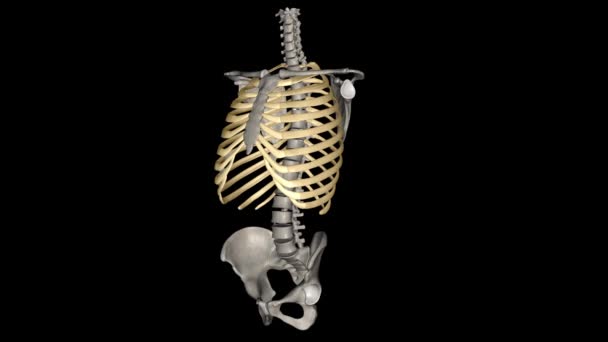
Human ribs are flat bones that form part of the rib cage to help protect internal organs. Humans usually have 24 ribs, in 12 pairs .
Human Ribs Flat Bones Form Part Rib Cage Help Protect Internal Organs Humans Usually Have Ribs Pairs
00:10
Detailed anatomy of the vertebral column in the human skeletal system with labeled spine regions and bone structure animation.
Detailed Anatomy Vertebral Column Human Skeletal System Labeled Spine Regions Bone Structure Animation
00:05
The scapula is a flat, triangular-shaped bone .
Scapula Flat Triangular Shaped Bone
00:11
Young girl washing her long hair in the pelvis in the courtyard
00:06
Many goose in yard near the drinker
00:20
Woman in sportswear doing hip bridge exercise. Joyful attractive young woman lying on floor in living room and doing hip bridge exercise.
00:42
Doctor uses anatomical model to explain male urinary system. Model labeled with parts, doctor points and explains how they work together for urinary function, ensuring patient comprehension.
Doctor Uses Anatomical Model Explain Male Urinary System Model Labeled Parts Doctor Points Explains How Work Together Urinary Function Ensuring Patient Comprehension
00:16
Pelvis in human body. X Ray. Loop
Pelvis Human Body Ray Loop
00:12
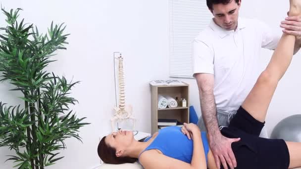
Physiotherapist stretching a leg of a woman
00:15
Cooking cherry jam. Cooking jam. Cooking cherry jam in the pelvis with a kitchen spatula. Man mixes cherry jam. Slow motion. 4k footage.
Cooking Cherry Jam Cooking Jam Cooking Cherry Jam Pelvis Kitchen Spatula Man Mixes Cherry Jam Slow Motion Footage
00:15
The close up shot of a male chiropractor after a massage makes a girl patient healthy exercises of the legs and pelvis in a dark room
00:18
Hands of a specialist doctor holding a kidney The concept of taking care of the organs in the body
Hands Specialist Doctor Holding Kidney Concept Taking Care Organs Body
00:08
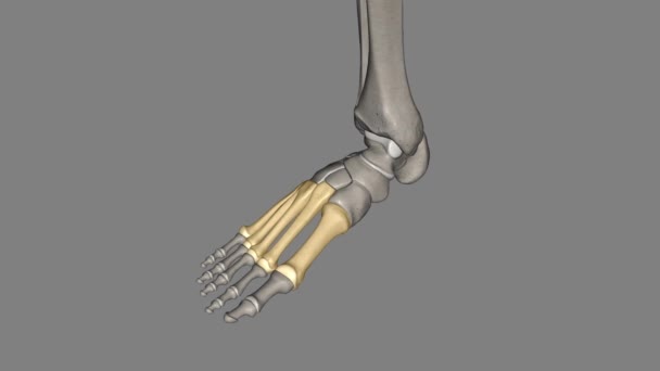
The metatarsal bones are the bones of the forefoot that connect the distal aspects of the cuneiform .
Metatarsal Bones Bones Forefoot Connect Distal Aspects Cuneiform
00:10
The role of the interspinous ligament is to limit flexion (bending forwards) through restricting separation of the spinous processes of the vertebral column
Role Interspinous Ligament Limit Flexion Bending Forwards Restricting Separation Spinous Processes Vertebral Column
00:09
Digital animation of the human brain and spinal cord beside DNA helix. Interface codes can be seen in the foreground
Digital Animation Human Brain Spinal Cord Dna Helix Interface Codes Can Seen Foreground
00:10
Vertebral column anatomy for medical concept 3D animation
Vertebral Column Anatomy Medical Concept Animation
00:20
Hip joint. Exercise for rehabilitation
00:11
Instructor cueing warrior II in fitness class, students adjusting alignment with floating glitter. Meditation, wellness, fitness, harmony, mindfulness, serenity, stretching
Instructor Cueing Warrior Fitness Class Students Adjusting Alignment Floating Glitter Meditation Wellness Fitness Harmony Mindfulness Serenity Stretching
00:12
Degenerative spondylolisthesis (DS) is a disorder that causes the slip of one vertebral body over the one below due to degenerative changes in the spine .
Degenerative Spondylolisthesis Disorder Causes Slip One Vertebral Body One Due Degenerative Changes Spine
00:09
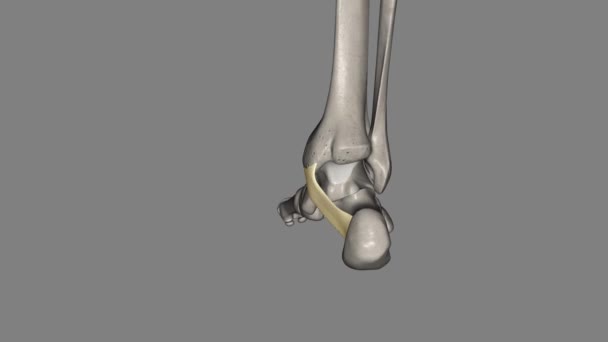
The flexor retinaculum of the foot extends from the medial malleolus above, to the calcaneus below .
Flexor Retinaculum Foot Extends Medial Malleolus Calcaneus
00:09
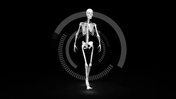
Digital animation of a skeleton walking on a dark background with digital circles rotating around it
Digital Animation Skeleton Walking Dark Background Digital Circles Rotating
00:20
Human Skeleton Sacral Bone Anatomy 3D Illustration For Medical Concept
Human Skeleton Sacral Bone Anatomy Illustration Medical Concept
00:12
Nurse using digital interface to look at x-rays
00:14
Medical education demonstrating the birthing process using an anatomical model of a baby and pelvis
Medical Education Demonstrating Birthing Process Using Anatomical Model Baby Pelvis
00:14
Open fracture, also called a compound fracture, is a fracture in which there is an open wound or break in the skin near the site of the broken bone .
Open Fracture Also Called Compound Fracture Fracture Which Open Wound Break Skin Site Broken Bone
00:13
Vertebrae C3 - C7 .3d medical
Vertebrae Medical
00:28
The athletic woman is doing pilates. She is warming up by raising hands and moving pelvis in the studio.
00:14
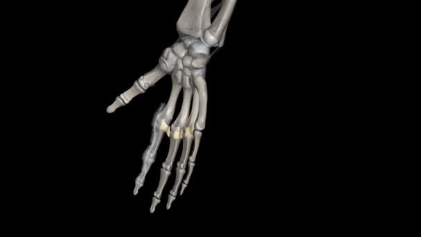
The deep transverse metacarpal ligament connects the palmar surfaces of metacarpophalangeal joints of all the fingers of the hand except the thumb
Deep Transverse Metacarpal Ligament Connects Palmar Surfaces Metacarpophalangeal Joints All Fingers Hand Thumb
00:07
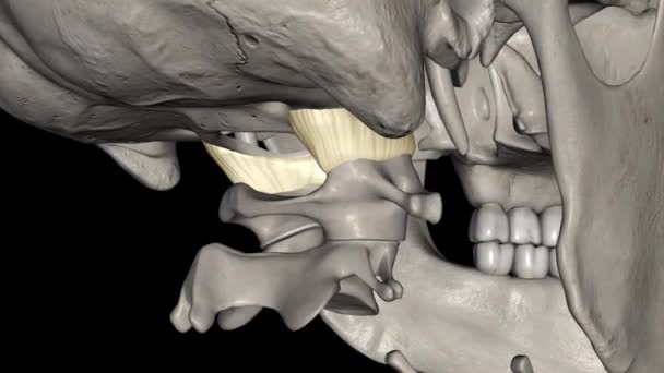
Capsule of Atlanto occipital joint .
Capsule Atlanto Occipital Joint
00:12
Human Skeleton Patella Bone Anatomy Loopable 3D Illustration For Medical Concept
Human Skeleton Patella Bone Anatomy Loopable Illustration Medical Concept
00:12
Doctor holds model of pelvic bone. Effective treatment of chronic pelvic pain concept
Doctor Holds Model Pelvic Bone Effective Treatment Chronic Pelvic Pain Concept
00:11
Human skeleton Skull Temporal Bone Anatomy 3D Illustration For Medical Concept
Human Skeleton Skull Temporal Bone Anatomy Illustration Medical Concept
00:12
Male Stomach During Workout
00:28
Asian woman starting side plank on rug, sweeping arm overhead, bending knee to stretch at home. Wellness, flexibility, indoor exercise, modern lifestyle, serenity, health, home fitness
Asian Woman Starting Side Plank Rug Sweeping Arm Overhead Bending Knee Stretch Home Wellness Flexibility Indoor Exercise Modern Lifestyle Serenity Health Home Fitness
00:17
Man in funny bright retro vintage tracksuit lies on floor raises pelvis and legs doing exercises eating fast food and soda while working out at home with help of female instructor controlling workout
00:58
Argentine cockroaches small and large crawl in the pelvis, closeup view.
00:16
Painful pelvis and cervical medical background
Painful Pelvis Cervical Medical Background
00:10
Ompartment syndrome is an increase in pressure inside a muscle, which restricts blood flow and causes pain .
Ompartment Syndrome Increase Pressure Muscle Which Restricts Blood Flow Causes Pain
00:13
Joint capsule of the elbow joint .
Joint Capsule Elbow Joint
00:10
Woman exercises Glute bridge on a reformer bed machine with trainer in natural sunlight, focusing on pelvis and hips.
Woman Exercises Glute Bridge Reformer Bed Machine Trainer Natural Sunlight Focusing Pelvis Hips
00:10
Pelvic MRI cross-section highlighting muscles, bones and surrounding soft tissues in a single axial slice. Useful for medical education, anatomy study
Pelvic Mri Cross Section Highlighting Muscles Bones Surrounding Soft Tissues Single Axial Slice Useful Medical Education Anatomy Study
00:17
The knee is the largest and most complex joint in the body, holding together the thigh bone, shin bone, fibula (on the outer side of the shin), and kneecap .
Knee Largest Most Complex Joint Body Holding Together Thigh Bone Shin Bone Fibula Outer Side Shin Kneecap
00:14
This layer of deep cervical fascia is a thin fascia limited to the anterior part of the neck .
Layer Deep Cervical Fascia Thin Fascia Limited Anterior Part Neck
00:15
The ribs partially enclose and protect the chest cavity, where many vital organs .
Ribs Partially Enclose Protect Chest Cavity Many Vital Organs
00:30
The second metatarsal bone is a long bone in the foot .
Second Metatarsal Bone Long Bone Foot
00:10
Skeleton walking across grid after animation start, showing rhythmic gait linking to DNA helix. Anatomy, biomechanical, genetics, visualization, glowing, neon, stride
Skeleton Walking Grid Animation Start Showing Rhythmic Gait Linking Dna Helix Anatomy Biomechanical Genetics Visualization Glowing Neon Stride
00:10
A young afro american doctor with x-ray of female lungs and pelvis is meeting with colleagues and discussing history of the disease patient. Hospital, profession, people and medicine concept
00:06
The metacarpals are long, thin bones that are located between the carpal bones in the wrist and the phalanges in the digits .
Metacarpals Long Thin Bones Located Carpal Bones Wrist Phalanges Digits
00:12
CTA femoral artery run off 3D MIP view with transparent bone turn around on black background.
Cta Femoral Artery Run Mip View Transparent Bone Turn Black Background
00:16
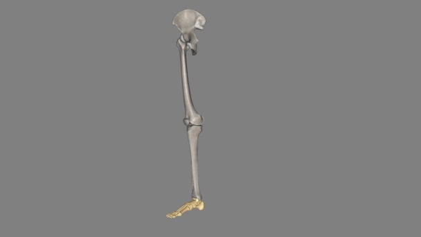
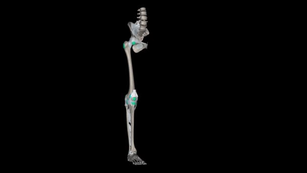
Lower Limb, Ligaments and Bones .
Lower Limb Ligaments Bones
00:12
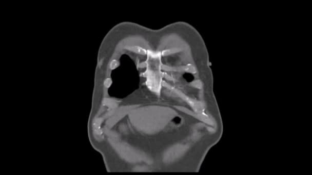
Tac in Portal phase in coronal of an Angiotac thorax
Tac Portal Phase Coronal Angiotac Thorax
00:08
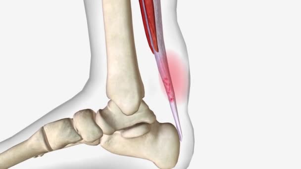
Sprained Ankle Symptoms Swelling and bruising .
Sprained Ankle Symptoms Swelling Bruising
00:06
Black silicone vibrator in water. Circles on water around sex toy. Vertical video
Black Silicone Vibrator Water Circles Water Sex Toy Vertical Video
00:09
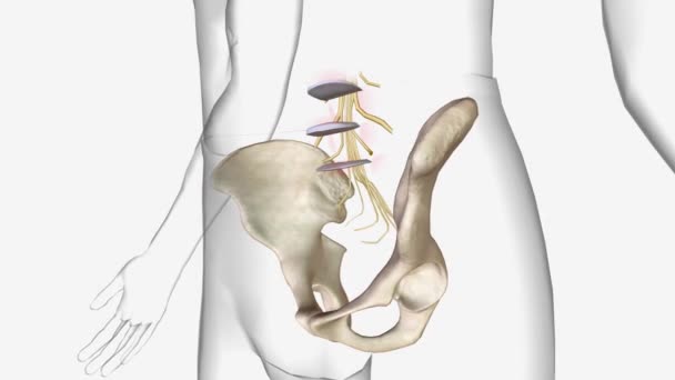
Pelvic Hip
Pelvic Hip
00:06
Human Skeleton Vertebral Column Lumbar Vertebrae Anatomy 3D Illustration For Medical Concept
Human Skeleton Vertebral Column Lumbar Vertebrae Anatomy Illustration Medical Concept
00:12
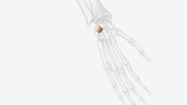
The pisiform is one of eight and smallest carpal bones that forms part of the wrist joint.
Pisiform One Eight Smallest Carpal Bones Forms Part Wrist Joint
00:10
Page