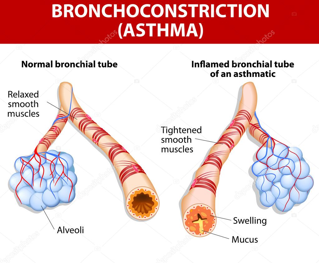
Inflamation of the bronchus causing asthma — Stock Vector

Inflamation of the bronchus causing asthma — Vector

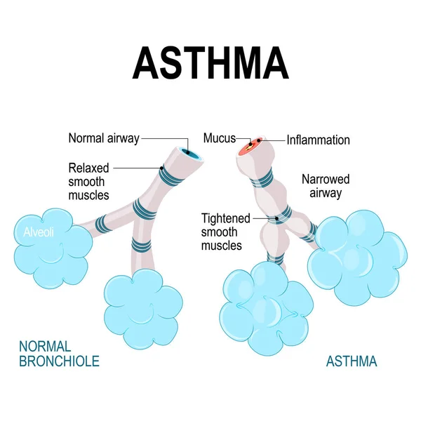
Asthma is a chronic inflammatory disease of the airways that is characterized by narrowing of the airways and dyspnea, wheezing, and coughing.

 — Vector by edesignua

Same Series:

Asthma. alveoli and bronchiole. normal and asthmatic. Part of Human lungs
Asthma. alveoli and bronchiole. — Stock Vector
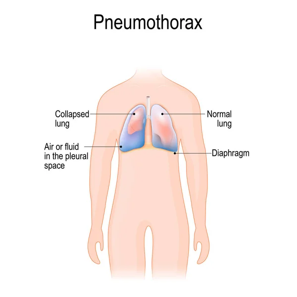
Collapsed lung. abnormal collection of air (pneumothorax) or fluid (pleural effusion) or pus (empyema) in the pleural space between the lung and the chest wall.
Pneumothorax — Stock Vector
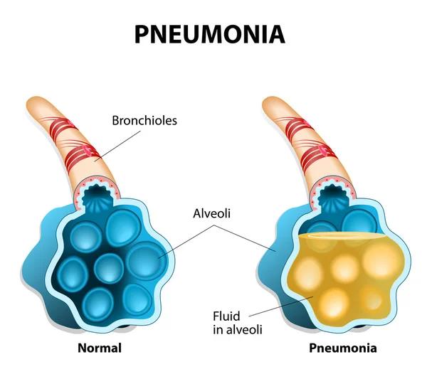
Pneumonia is a inflammatory condition of the lung. It is caused by infection with viruses, bacteria, parasites or fungi. The disease is characterized by the inflammation of the alveoli. Alveoli are filled with fluids.
Pneumonia. Illustration shows normal and infected alveoli. — Stock Vector
Pneumonia. Lung infection. Inflammatory of the respiratory system. Close-up of healthy alveolus and Alveoli with fluid, where oxygen unable to reach bloodstream. comparison and difference. Vector poster for medical use
Pneumonia Lung Infection Inflammatory Respiratory System Close Healthy Alveolus Alveoli — Stock Vector
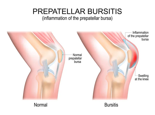
Prepatellar bursitis. inflammation of the prepatellar bursa. comparison of a human knee with Normal prepatellar bursa, and joint with Bursitis. vector poster
Prepatellar Bursitis Inflammation Prepatellar Bursa Comparison Human Knee Normal Prepatellar — Stock Vector
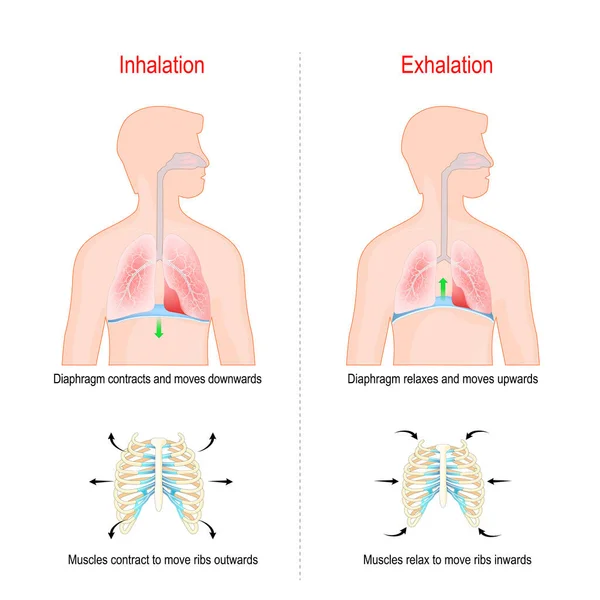
Cycle of breathing, inspiration and expiration. Role of Diaphragm and intercostal muscles (ribs and chest) in Gas exchange in lungs. respiratory system anatomy. vector illustration
Cycle Breathing Inspiration Expiration Role Diaphragm Intercostal Muscles Ribs Chest — Stock Vector
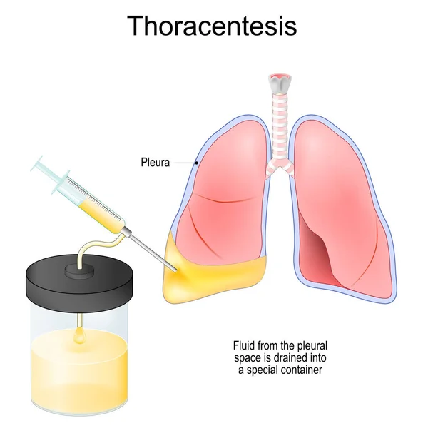
Thoracentesis. Thoracocentesis is an invasive medical procedure to remove fluid or air from the pleural space for diagnostic or therapy. Syringe, and Human lungs with Fluid that drained into a special glass or plastic container. Medical vector poster
Thoracentesis Thoracocentesis Invasive Medical Procedure Remove Fluid Air Pleural Space — Stock Vector
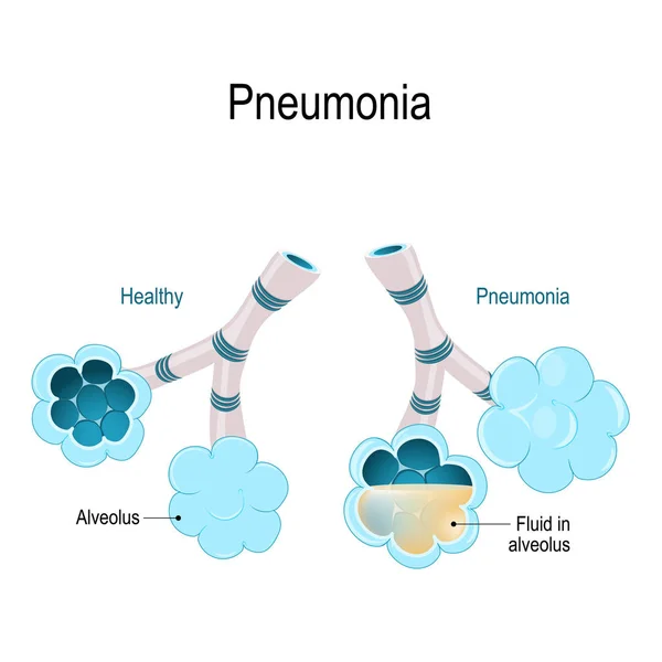
Pneumonia. Healthy alveoli and alveolus with pneumonia. Illustration shows normal and infected alveoli.
Pneumonia. Illustration shows normal and infected alveoli. — Stock Vector
Molecule haemoglobin. Each haemoglobin molecule can bind with 4 oxygen molecules. Vector diagram.
Molecule haemoglobin — Stock Vector
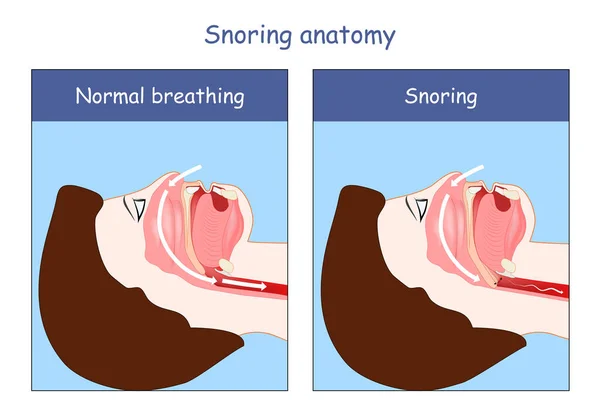
Snoring anatomy. Normal breathing and Snoring. air open and obstructed airways. Cross section of human's head, nose, mouth, trachea, and tongue.
Snoring Anatomy Normal Breathing Snoring Air Open Obstructed Airways Cross — Stock Vector
Ruminant stomach. The ruminant species have one stomach that is divided into four compartments: rumen, reticulum, omasum and abomasum. Ruminating mammals include cattle, goats, sheep, giraffes, yaks, deer, camels, llamas, antelope.
Ruminant stomach — Stock Vector
Coronary heart disease is a condition in which the heart's arteries become narrower. coronary artery disease. human vascular system on silhouettes of men. Cholesterol plaque in artery. Illustration with annotation
Coronary heart disease — Stock Vector
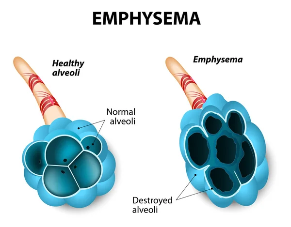
Emphysema. Damage to the air sacs in lungs. Chronic Obstructive Pulmonary Disease.
Emphysema — Stock Vector
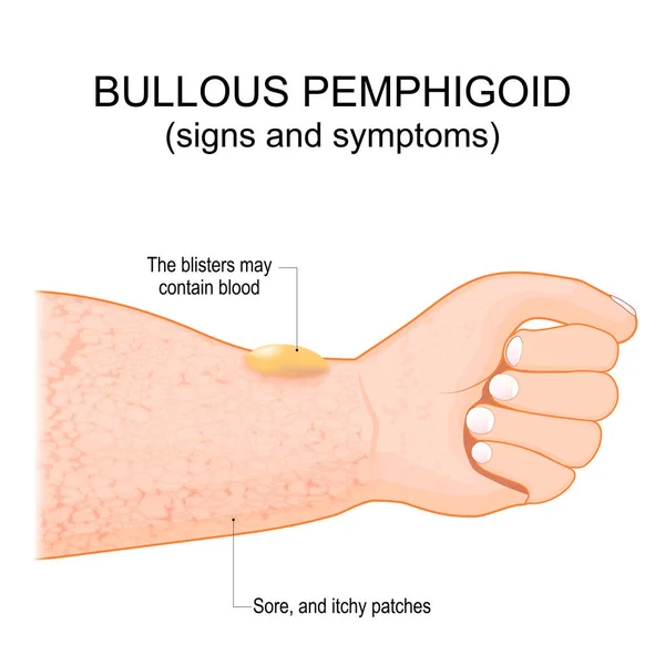
Bullous pemphigoid. autoimmune pruritic skin disease. skin condition of older people. It starts with an itchy rash than, blisters can form on the skin. vector poster
Bullous Pemphigoid Autoimmune Pruritic Skin Disease Skin Condition Older People — Stock Vector
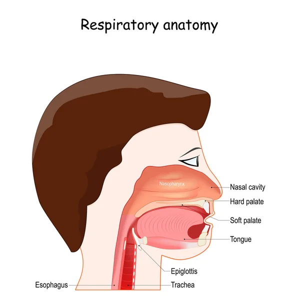
Upper respiratory system, with nasopharynx, oropharynx and laryngopharynx labeled, Epiglottis, Pharynx, and Larynx. Location, and Structure. Regions of Nose.
Upper Respiratory System Nasopharynx Oropharynx Laryngopharynx Labeled Epiglottis Pharynx Larynx — Stock Vector
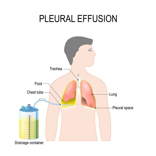
Pleural effusion. Diagram showing human silhouette with highlighted lungs, fluid buildup in the pleura, Chest Tube, and Drainage container. Treatment of tension hydrothorax (or hemothorax) insertion of chest tubes for invasive procedure to remove flu
Pleural effusion. Treatment of tension hydrothorax — Stock Vector

Similar Stock Videos:

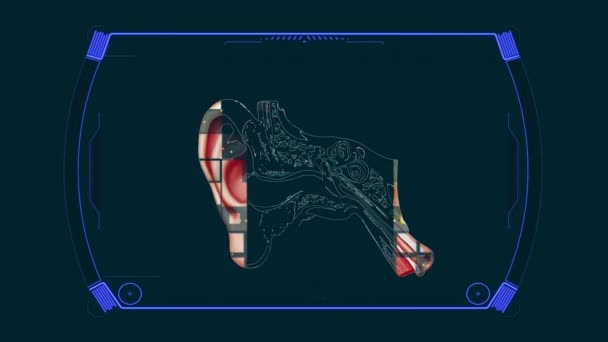
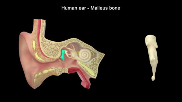
Anatomy Diagram Ear Structure
Anatomy Diagram Ear Structure — Stock Video
Polycystic ovarian syndrome 2D animation. Uterus, infertility, gynecology. For topics like pcos, disease, pelvic floor
Polycystic Ovarian Syndrome Animation Uterus Infertility Gynecology Topics Pcos Disease — Stock Video
Graphic animation showing the structure and composition of a coronavirus.
Graphic Animation Showing Structure Composition Coronavirus — Stock Video
Animated endocannabinoid system on red background. High quality 4k footage
Animated endocannabinoid system on red background — Stock Video
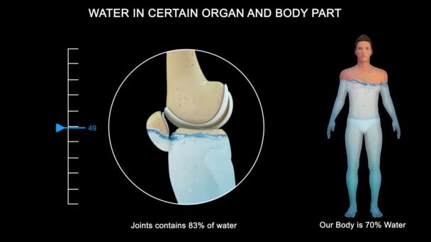
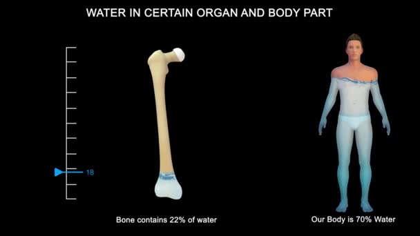
Some organs contain much more water than others. The brain and kidneys possess the highest percentage of water; the bones and teeth contain the lowest proportion.
Some Organs Contain Much More Water Others Brain Kidneys Possess — Stock Video
The human digestive system consists of the gastrointestinal tract plus the accessory organs of digestion. Digestion involves the breakdown of food into smaller and smaller components until they can be absorbed and assimilated into the body.
Human Digestive System Consists Gastrointestinal Tract Accessory Organs Digestion Digestion — Stock Video
3d medicine footage on white background with Stomach
3d anatomy concept — Stock Video
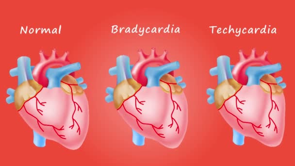
The heart is a muscular organ in humans and many other animals, which pumps blood through the blood vessels of the circulatory system. The heart is located in the middle compartment (mediastinum) of the chest.
Human Heart — Stock Video
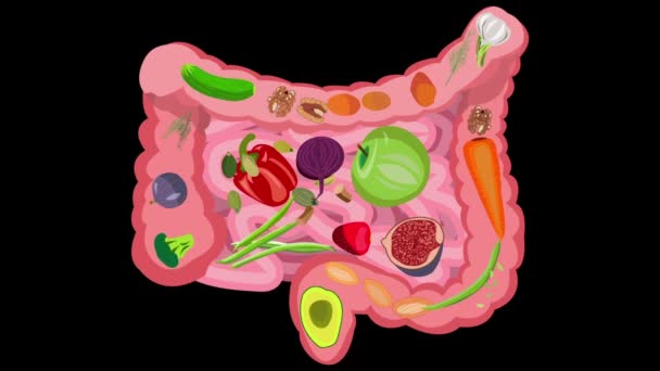
Healthy food for intestinal health. Healthy food for intestinal health. Fiber, vegetables, fruits, berries, bran, greens, prebiotics. Video on a black background
Healthy food for intestinal health. Video on a black background — Stock Video
Some organs contain much more water than others. The brain and kidneys possess the highest percentage of water; the bones and teeth contain the lowest proportion.
Some Organs Contain Much More Water Others Brain Kidneys Possess — Stock Video
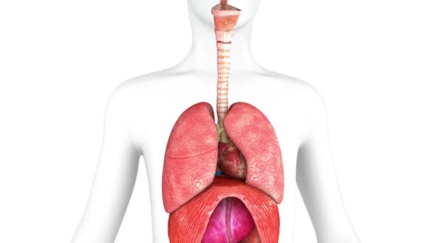
The lung is the essential respiration organ in many air-breathing animals, including most tetrapods, a few fish and a few snails. In mammals and the more complex life forms, the two lungs are located near the backbone on either side of the heart.
Lungs Anatomy — Stock Video
Breathing in and out with the expansion and contraction of the lungs.
Breathing Out Expansion Contraction Lungs — Stock Video
Animation illustrating the various stages of kidney cancer.
Animation illustrating the various stages of kidney cancer. — Stock Video
Pains ankle and wrist rheumatology disease animation ,4k video animated
Pains ankle and wrist rheumatology disease animation — Stock Video
Stomach and small intestine view
Stomach and small intestine — Stock Video
Footage of Human heart. Isolated on red background.
Footage Human Heart Isolated Red Background — Stock Video
Human ear structure footage
Human Ear Structure Footage — Stock Video

Usage Information

You can use this royalty-free vector image "Inflamation of the bronchus causing asthma" for personal and commercial purposes according to the Standard or Extended License. The Standard License covers most use cases, including advertising, UI designs, and product packaging, and allows up to 500,000 print copies. The Extended License permits all use cases under the Standard License with unlimited print rights and allows you to use the downloaded vector files for merchandise, product resale, or free distribution.

This stock vector image is scalable to any size. You can buy and download it in high resolution up to 4980x4109. Upload Date: Nov 3, 2013