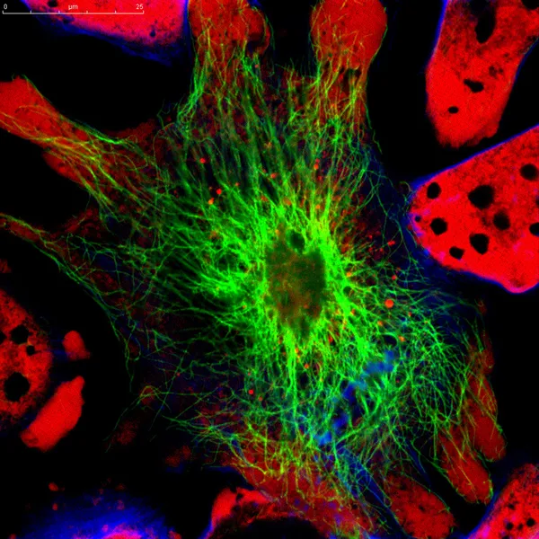
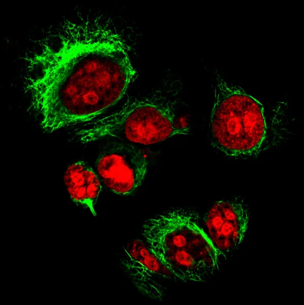
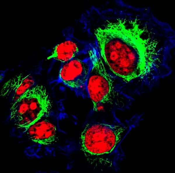
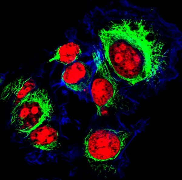
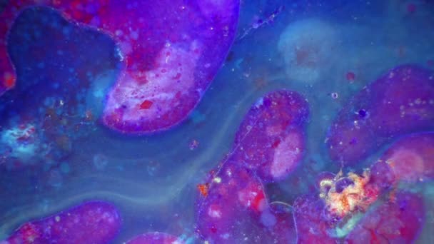
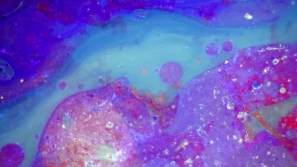
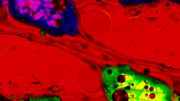
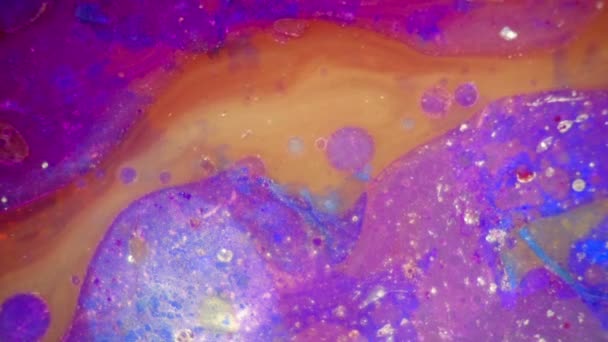
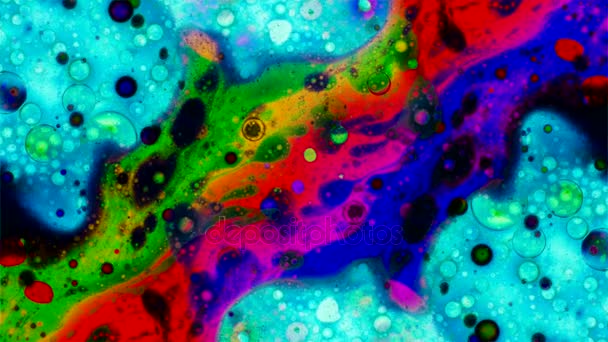
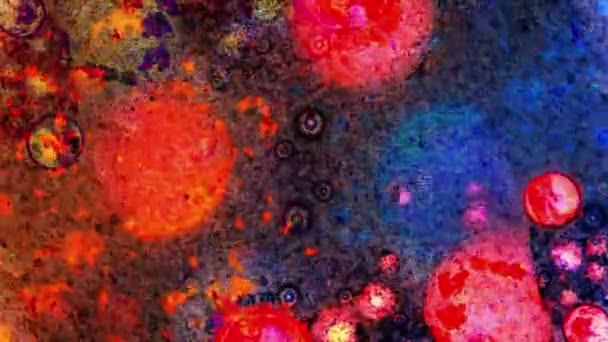
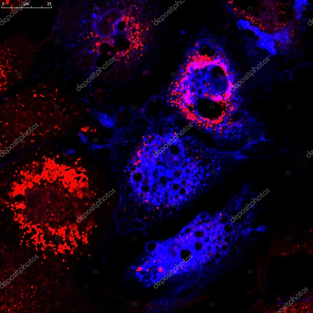
Mesenchymal stem cells labeled with fluorescent molecules — Stock Photo, Image

Mesenchymal stem cells labeled with fluorescent molecules — Photo

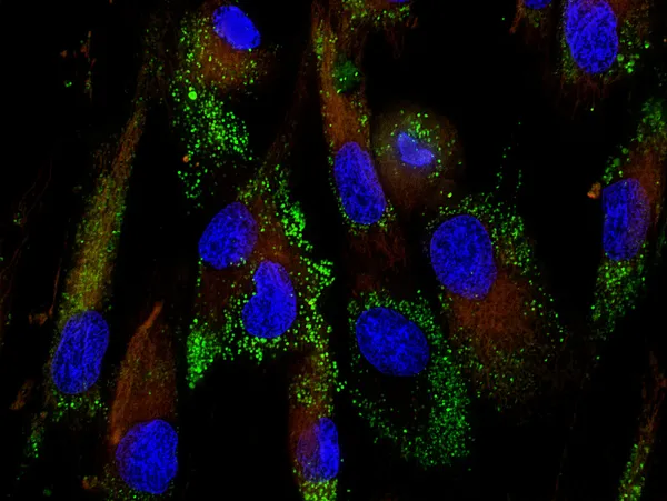
Mesenchymal stem cells stained with monoclonal antibodies for different molecules

 — Photo by vshivkova

Same Series:

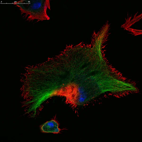
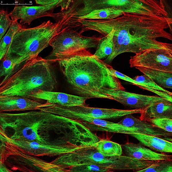
Real fluorescence microscopic view of human skin cells in culture. Nucleus are in blue, actin filaments are in green, tubulin was labeled with red
Confocal microscopy of fibroblast cells — Stock Photo, Image
This image was taken with a digital camera on a confocal microscope. Here is real skin cells from patients, was grown in culture, then stained and photographed by myself.
Fibroblasts (skin  cells) labeled with fluorescent dyes — Stock Photo, Image
Mesenchymal stem cells stained with monoclonal antibodies for different molecules
Mesenchymal stem cells labeled with fluorescent molecules — Stock Photo, Image
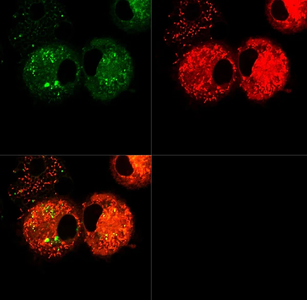
Real fluorescence microscopic view of human neuroblastoma cells
Real fluorescence microscopic view of human neuroblastoma cells — Stock Photo, Image
This image was taken with a digital camera on a confocal microscope
Dendritic cells labeled with fluorescent dyes — Stock Photo, Image
This image was taken with a digital camera on a confocal microscope. Here is real skin cells from patients, was grown in culture, then stained and photographed by myself.
Fibroblasts (skin  cells) labeled with fluorescent dyes — Stock Photo, Image
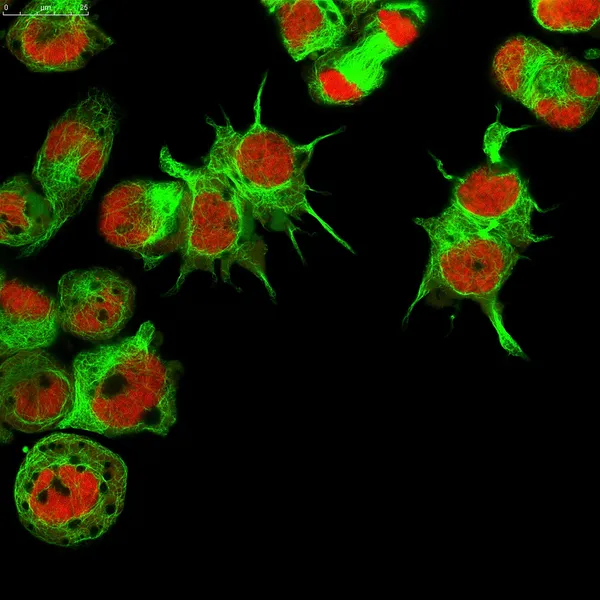
Human neuroblastoma cells: nuclei are stained in red, microfilaments are in green
Real fluorescence microscopic view of human neuroblastoma cells — Stock Photo, Image
This image was taken with a digital camera on a confocal microscope
Dendritic cells labeled with fluorescent dyes — Stock Photo, Image
This image was taken with a digital camera on a confocal microscope. Here is real skin cells from patients, was grown in culture, then stained and photographed by myself.
Fibroblasts (skin  cells) labeled with fluorescent dyes — Stock Photo, Image
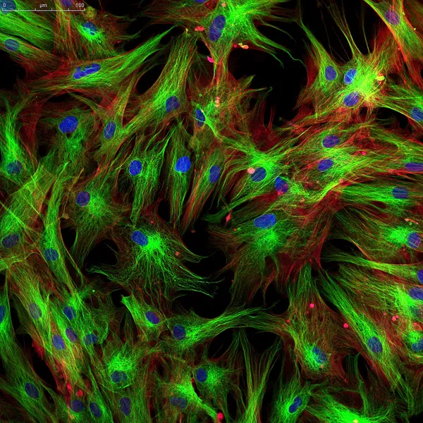
Real fluorescence microscopic view of human skin cells in culture. Nucleus are in blue, actin filaments are in red, tubulin was labeled with green
Confocal microscopy of fibroblast cells — Stock Photo, Image
This image was taken with a digital camera on a confocal microscope. Here is real skin cells from patients, was grown in culture, then stained and photographed by myself.
Fibroblasts (skin  cells) labeled with fluorescent dyes — Stock Photo, Image
Human neuroblastoma cells: nuclei are stained in red, microfilaments are in green
Real fluorescence microscopic view of human neuroblastoma cells — Stock Photo, Image
This image was taken with a digital camera on a confocal microscope
Background with fluorescent cancer cells — Stock Photo, Image
Tumor cells under microscope labeled with fluorescent molecules
Epithelial tumor cells labeled with fluorescent molecules — Stock Photo, Image
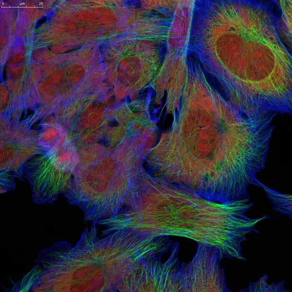
HeLa cells labeled with fluorescent molecules
Ovarian cancer cells labeled with fluorescent molecules — Stock Photo, Image
Tumor cells under microscope labeled with fluorescent molecules
Epithelial tumor cells labeled with fluorescent molecules — Stock Photo, Image

Usage Information

You can use this royalty-free photo "Mesenchymal stem cells labeled with fluorescent molecules" for personal and commercial purposes according to the Standard or Extended License. The Standard License covers most use cases, including advertising, UI designs, and product packaging, and allows up to 500,000 print copies. The Extended License permits all use cases under the Standard License with unlimited print rights and allows you to use the downloaded stock images for merchandise, product resale, or free distribution.

You can buy this stock photo and download it in high resolution up to 3200x3200. Upload Date: Jan 25, 2014