Structure of a nematocyst — Stock Vector

Structure of a nematocyst — Vector

A Cnidarian's cell that contains a nematocyst. A stinging cell. Nematocyst is a hollow tube that exists inside the cell in an inside out condition.

 — Vector by edesignua

Same Series:

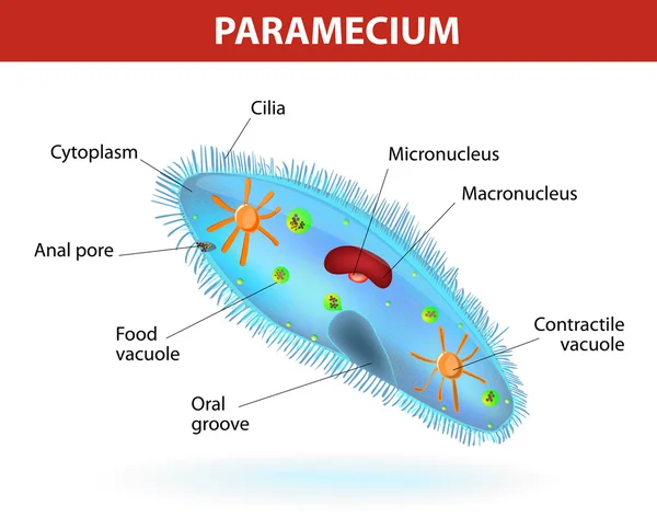
Anatomy of a paramecium. Vector diagram. Ciliate protozoan that lives in stagnant freshwater. Paramecium covered with cilia, which allow it to move about and to feed on bacteria.
Structure of a paramecium — Stock Vector
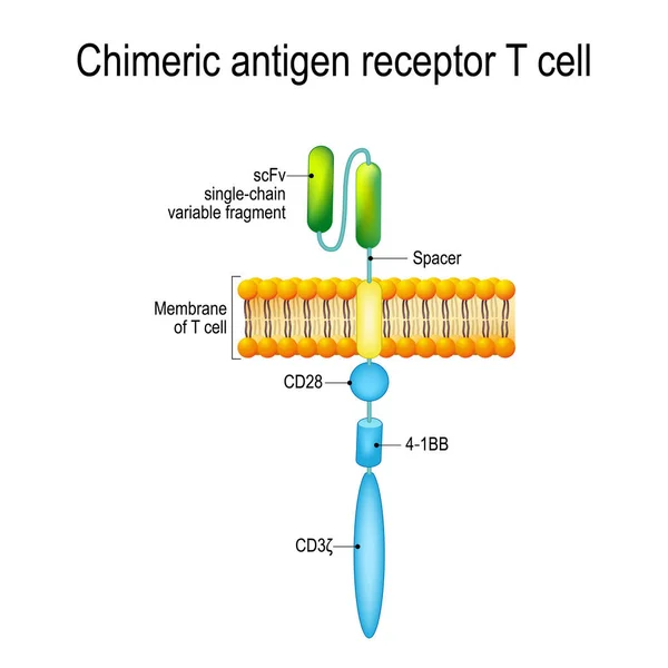
Chimeric antigen receptor T cell (CAR). Artificial T cell receptors are proteins that have been engineered for cancer therapy (killing of tumor cells). genetically engineered. Vector diagram for medical, educational and science use
Chimeric antigen receptor T cell (CAR) — Stock Vector
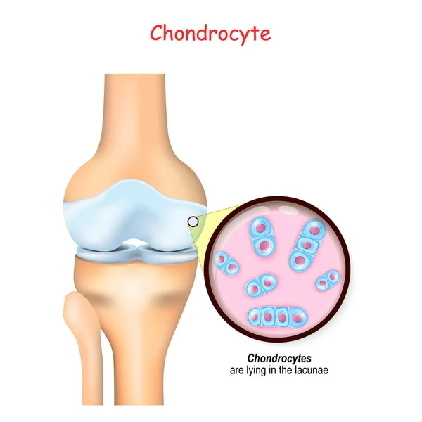
Knee and close up of cells of a cartilage. chondrocytes are lying in the lacunae and produce and maintain the cartilaginous matrix, and collagen.
Knee Close Cells Cartilage Chondrocytes Lying Lacunae Produce Maintain Cartilaginous — Stock Vector
Echinococcus granulosus. causes cystic echinococcosis (hydatid disease). Vector illustration for biological, science, and medical use.
Echinococcus Granulosus Causes Cystic Echinococcosis Hydatid Disease Vector Illustration Biological — Stock Vector
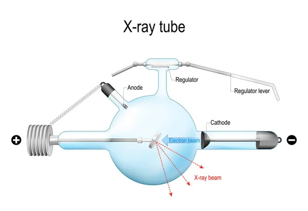
Crookes X-ray tube. structural schematic diagram of an x ray tube that used for radiation therapy, medical radiography and airport security. Diagnostic radiology. Realistic vector illustration. isolated on white background.
Crookes Ray Tube Structural Schematic Diagram Ray Tube Used Radiation — Stock Vector
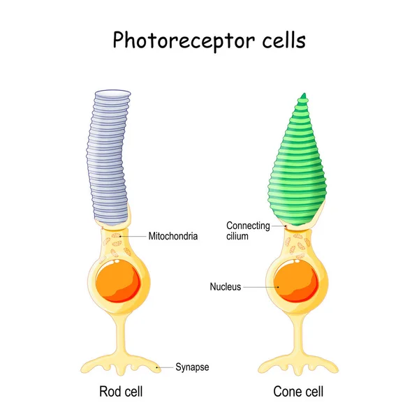
Anatomy of Photoreceptor. cell of a retina in the eye. Cone cells in respond to color vision and send signals to brain. Rod cells are used in peripheral vision
Anatomy Photoreceptor Cell Retina Eye Cone Cells Respond Color Vision — Stock Vector
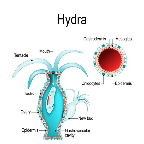
Hydra anatomy. Cnidaria. Vector illustration for biological, science and educational use
Hydra Anatomy Cnidaria Vector Illustration Biological Science Educational Use — Stock Vector
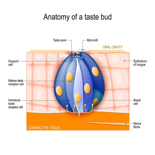
Taste bud. Mature and Immature taste Receptor, Support and Basal Cells, Epithelium Of tongue. Human Anatomy. Vector diagram for educational, biological, science and medical use
Taste Bud Mature Immature Taste Receptor Support Basal Cells Epithelium — Stock Vector
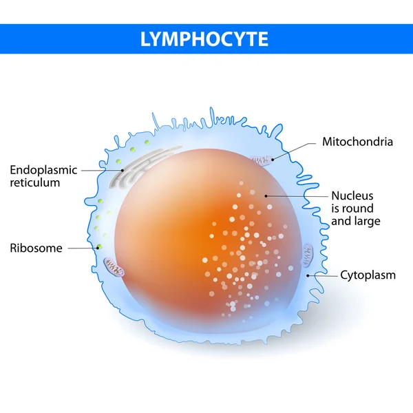
Lymphocyte. Structure cell. Lymphocyte - type of white blood cell
Lymphocyte. Vector illustration — Stock Vector
Enterocyte. intestinal absorptive epithelial cell with microvilli. Structure of the enterocyte. Infographics. Vector illustration on white background.
Enterocyte. Structure of the intestinal absorptive epithelial ce — Stock Vector
Plant cell anatomy. cross section. structure of a Eukaryotic cell. Vector diagram for your design, educational, medical, biological and science use
Plant Cell Anatomy Cross Section Structure Eukaryotic Cell Vector Diagram — Stock Vector
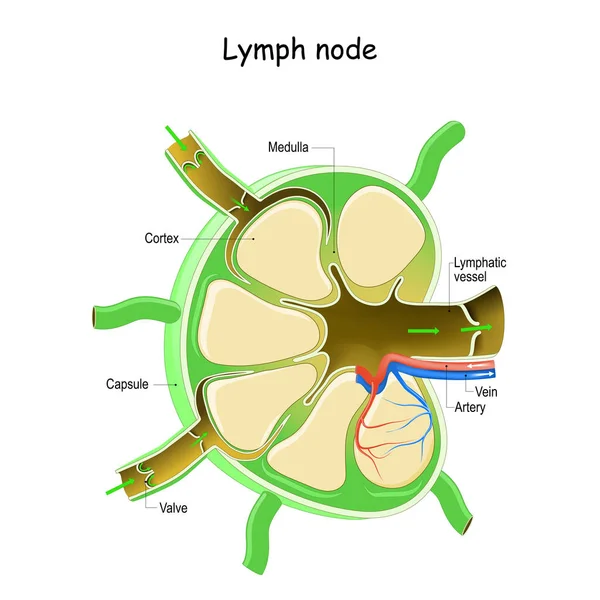
Lymph Node Anatomy. Labeled diagram showing the flow of lymph. Afferent and efferent vessels. Vector illustration
Lymph Node Anatomy Labeled Diagram Showing Flow Lymph Afferent Efferent — Stock Vector
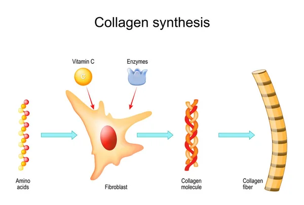
Collagen synthesis with Vitamin C and Enzymes. From Fibroblast and Amino acids to Collagen fiber that comprises molecules of protein. Vector
Collagen Synthesis Vitamin Enzymes Fibroblast Amino Acids Collagen Fiber Comprises — Stock Vector
Epidermophyton floccosum. fungal infections of the skin. Vector illustration
Epidermophyton Floccosum Fungal Infections Skin Vector Illustration — Stock Vector
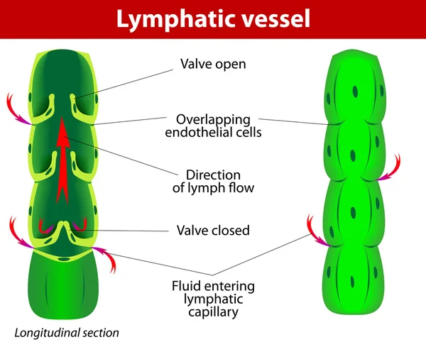
The internal structure of a lymph vessel
Lymphatic vessel — Stock Vector
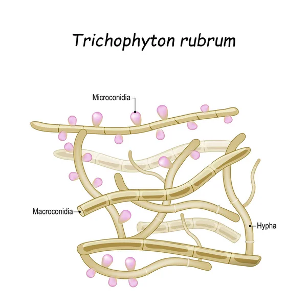
Trichophyton rubrum is a dermatophytic fungus that colonizes the upper layers of skin, and cause of athlete's foot, fungal infection of nail, and ringworm. Close-up of fungi with fertile hyphae, Macroconidia and Microconidia
Trichophyton rubrum. Close-up of fungi with fertile hyphae — Stock Vector

Similar Stock Videos:

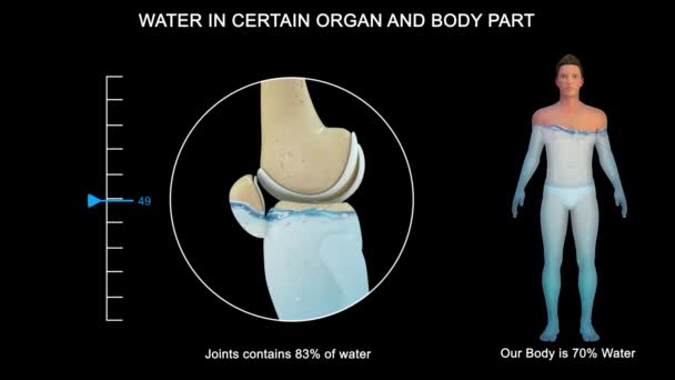
Some organs contain much more water than others. The brain and kidneys possess the highest percentage of water; the bones and teeth contain the lowest proportion.
Some Organs Contain Much More Water Others Brain Kidneys Possess — Stock Video
Graphic animation showing the structure and composition of a coronavirus.
Graphic Animation Showing Structure Composition Coronavirus — Stock Video
Human Organs Animation on white background
Human Organs Animation — Stock Video
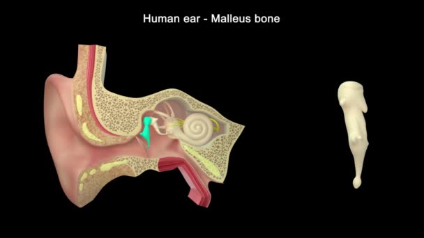
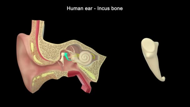
Human ear structure footage
Human Ear Structure Footage — Stock Video
Human Organs Animation on white background
Human Organs Animation — Stock Video
3d rendered medically illustration of male Internal organs
Rendered Medically Illustration Male Internal Organs — Stock Video
Digestive system on human silhouette cartoon High definition colorful scenes animation
Digestive system concept HD animation — Stock Video
Anatomy Diagram Ear Structure
Anatomy Diagram Ear Structure — Stock Video
The stomach lies between the esophagus and the duodenum (the first part of the small intestine). It is on the left upper part of the abdominal cavity. The top of the stomach lies against the diaphragm.
Stomach Layers — Stock Video
Animated endocannabinoid system on red background. High quality 4k footage
Animated endocannabinoid system on red background — Stock Video
The human digestive system consists of the gastrointestinal tract plus the accessory organs of digestion. Digestion involves the breakdown of food into smaller and smaller components until they can be absorbed and assimilated into the body.
Human Digestive System Consists Gastrointestinal Tract Accessory Organs Digestion Digestion — Stock Video
Stomach upset animated line icon. stomach upset sign. isolated on white background
Stomach upset line icon animation — Stock Video
Black line Virus cells in lung icon isolated on white background. Infected lungs. Coronavirus, COVID-19. 2019-nCoV. 4K Video motion graphic animation.
Black line Virus cells in lung icon isolated on white background. Infected lungs. Coronavirus, COVID-19. 2019-nCoV. 4K Video motion graphic animation — Stock Video
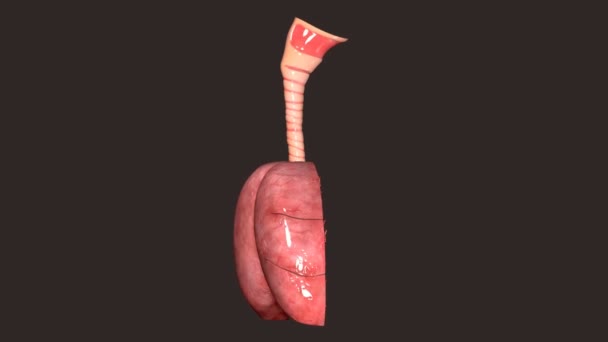
The lung is the essential respiration organ in many air-breathing animals, including most tetrapods, a few fish and a few snails. In mammals and the more complex life forms, the two lungs are located near the backbone on either side of the heart.
Lungs Anatomy — Stock Video
Human Ear Anatomy. 3d illustration
Human Ear Anatomy Illustration — Stock Video
Indigestion stomach animated glyph icon. indigestion stomach sign. isolated on white background
Indigestion stomach glyph icon animation — Stock Video
Animation illustrating the various stages of kidney cancer.
Animation illustrating the various stages of kidney cancer. — Stock Video

Usage Information

You can use this royalty-free vector image "Structure of a nematocyst" for personal and commercial purposes according to the Standard or Extended License. The Standard License covers most use cases, including advertising, UI designs, and product packaging, and allows up to 500,000 print copies. The Extended License permits all use cases under the Standard License with unlimited print rights and allows you to use the downloaded vector files for merchandise, product resale, or free distribution.

This stock vector image is scalable to any size. You can buy and download it in high resolution up to 5019x3910. Upload Date: Feb 11, 2014