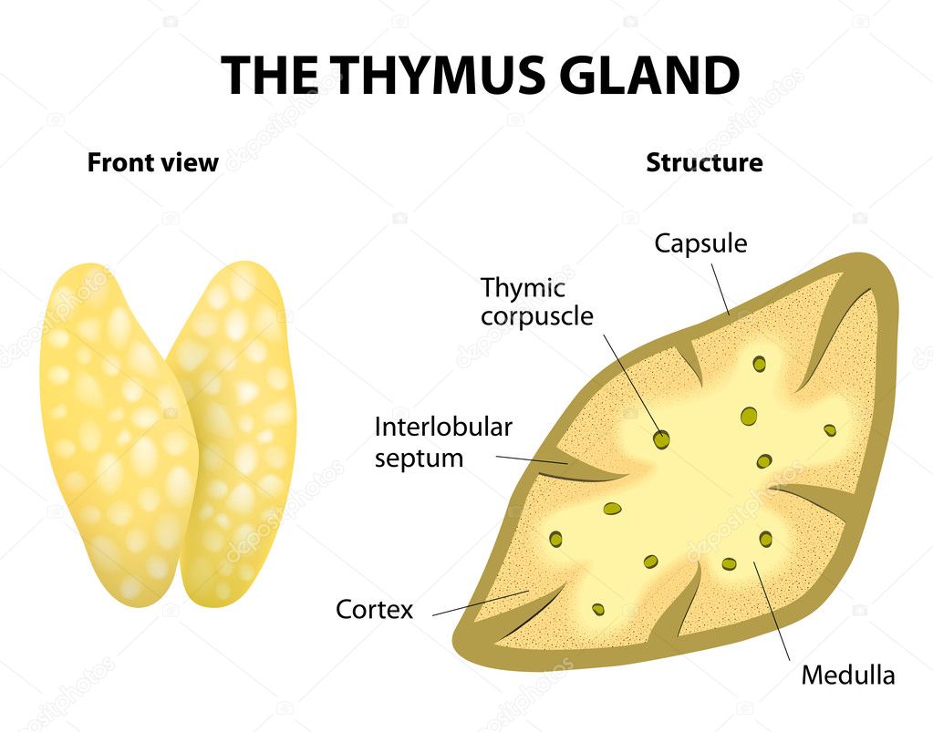
Thymus gland anatomy — Stock Vector

Thymus gland anatomy — Vector

Thymus structure. Vector diagram. Gland lies in the thoracic cavity, just above the heart. It secretes thymosin.

 — Vector by edesignua

Same Series:

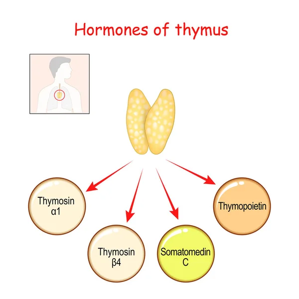
Hormones produced by the Thymus gland. Thymopoietin, Insulin-like growth factor (IGF-1), or somatomedin C, thymosin alpha and Thymosin beta. Vector illustration for medical, science and education use.
Hormones Produced Thymus Gland Thymopoietin Insulin Growth Factor Igf Somatomedin — Stock Vector
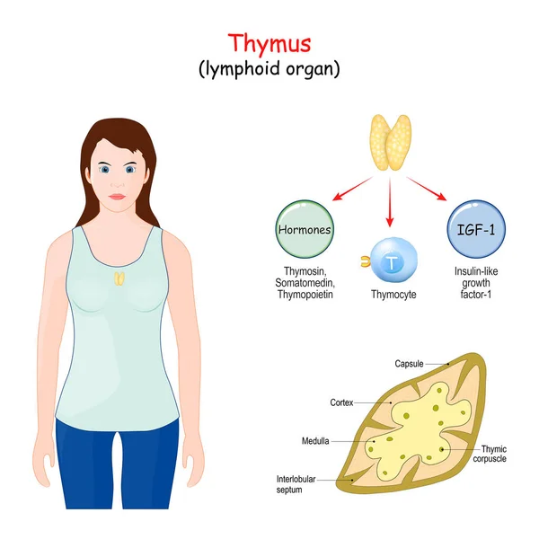
Thymus. lymphoid organ of the human immune system. Structure, Location, and function of the thymus. Woman with highlight of thymus gland
Thymus Lymphoid Organ Human Immune System Structure Location Function Thymus — Stock Vector
Anatomy of a thyroid gland. Close-up of thyroid Follicle structure with Follicular cell, C-cell, and Connective tissue. Vector illustration
Anatomy Thyroid Gland Close Thyroid Follicle Structure Follicular Cell Cell — Stock Vector
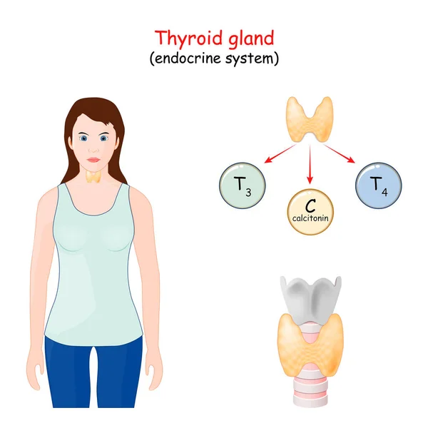
Thyroid gland hormones. Human endocrine system. infographics. Vector diagram
Thyroid Gland Hormones Human Endocrine System Infographics Vector Diagram — Stock Vector
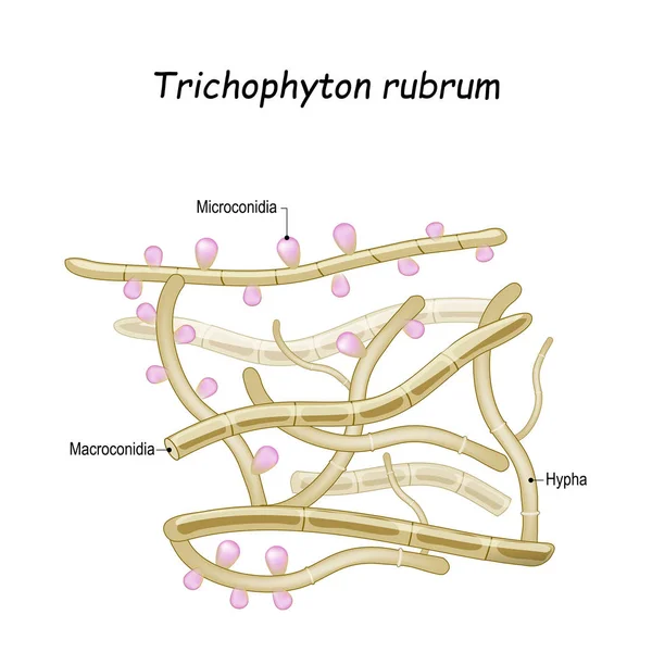
Trichophyton rubrum is a dermatophytic fungus that colonizes the upper layers of skin, and cause of athlete's foot, fungal infection of nail, and ringworm. Close-up of fungi with fertile hyphae, Macroconidia and Microconidia
Trichophyton rubrum. Close-up of fungi with fertile hyphae — Stock Vector
Echinococcus granulosus. causes cystic echinococcosis (hydatid disease). Vector illustration for biological, science, and medical use.
Echinococcus Granulosus Causes Cystic Echinococcosis Hydatid Disease Vector Illustration Biological — Stock Vector
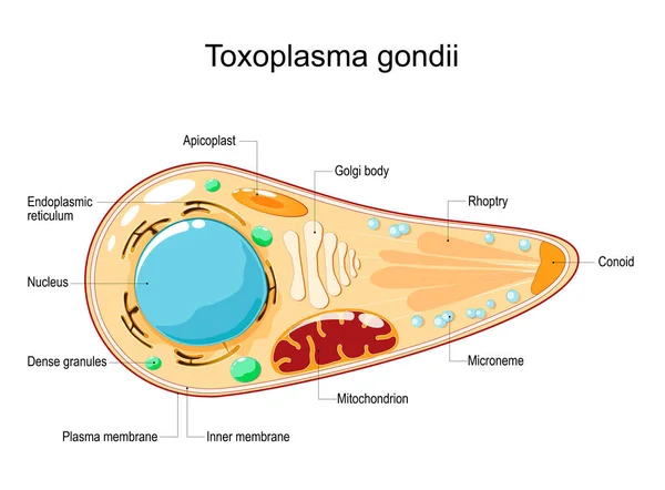
Toxoplasma gondii. Cell Structure and anatomy. Vector illustration. flat style. Toxoplasma is an obligate intracellular, parasitic protozoan that causes the disease toxoplasmosis
Toxoplasma Gondii Cell Structure Anatomy Vector Illustration Flat Style Toxoplasma — Stock Vector
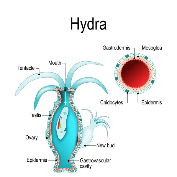
Hydra anatomy. Cnidaria. Vector illustration for biological, science and educational use
Hydra Anatomy Cnidaria Vector Illustration Biological Science Educational Use — Stock Vector
Hyperthyroidism and Hypothyroidism comparisons. disorders of the endocrine system. Female with Signs and symptoms of different thyroid gland diseases
Hyperthyroidism Hypothyroidism Comparisons Disorders Endocrine System Female Signs Symptoms Different — Stock Vector
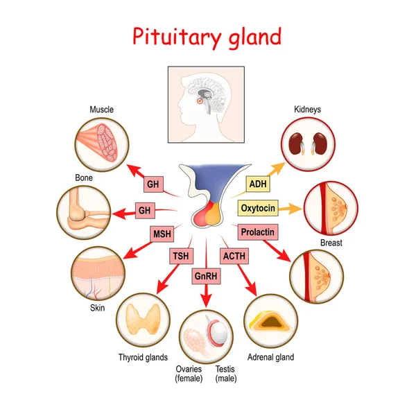
Hormones of pituitary gland. The two lobes, anterior and posterior, function as independent glands. Anatomy of Pituitary gland. vector diagram
Hormones Pituitary Gland Two Lobes Anterior Posterior Function Independent Glands — Stock Vector
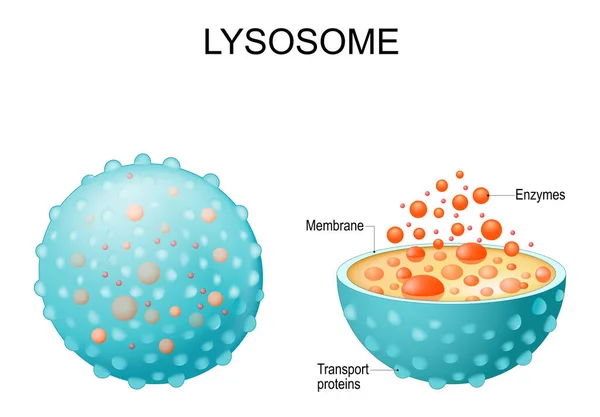
Lysosome. appearance, exterior and interior view. Cross section and Anatomy of the Lysosome: Hydrolytic enzymes, Membrane and transport proteins. Vector illustration
Lysosome Appearance Exterior Interior View Cross Section Anatomy Lysosome Hydrolytic — Stock Vector
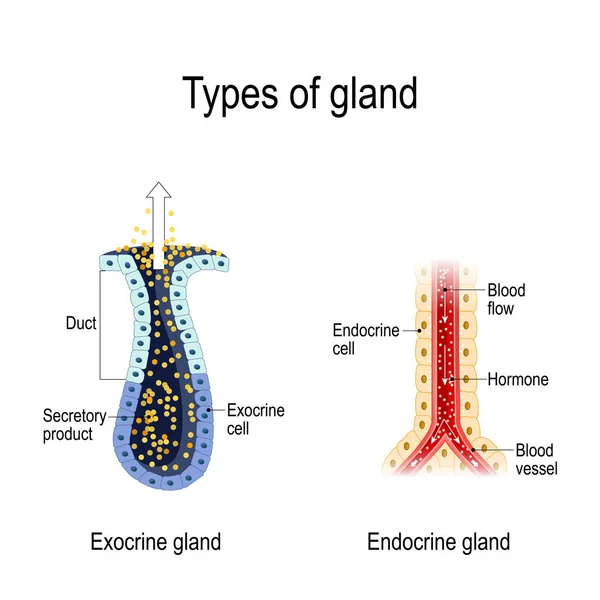
Types of gland. Anatomy of an Endocrine and exocrine glands. different of glands secretion. cross-section. Vector diagram for educational, medical, biological and science use
Types Gland Anatomy Endocrine Exocrine Glands Different Glands Secretion Cross — Stock Vector
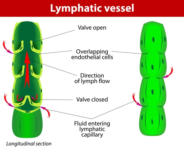
The internal structure of a lymph vessel
Lymphatic vessel — Stock Vector
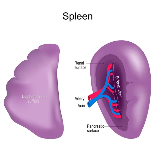
Spleen. Visceral and Diaphragmatic surfaces of the spleen. front and side view. Healthy Internal organ of the human lymphatic system. Realistic vector illustration. isolated on white background.
Spleen Visceral Diaphragmatic Surfaces Spleen Front Side View Healthy Internal — Stock Vector
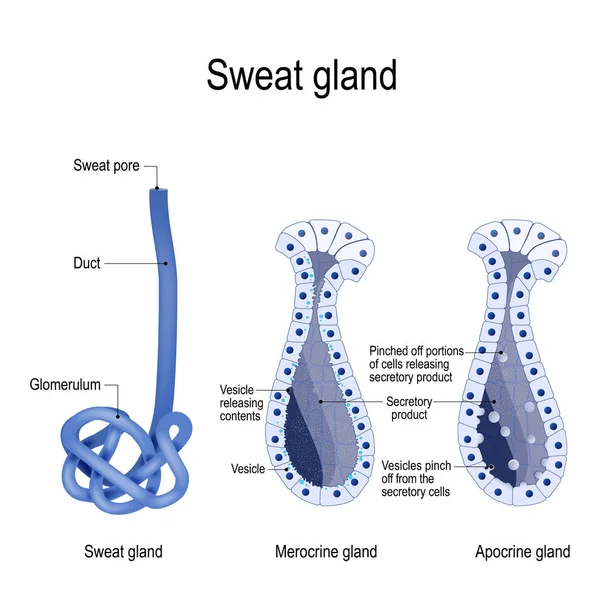
Sweat gland. Merocrine and apocrine. different of manner of secretion. cross-section of the human skin, with the sweat gland. Close-up of dark and clear cells, lumen, sweat duct, glomerulum and pore. labeled Vector diagram for educational, medical,
Sweat Gland Merocrine Apocrine Different Manner Secretion Cross Section Human — Stock Vector
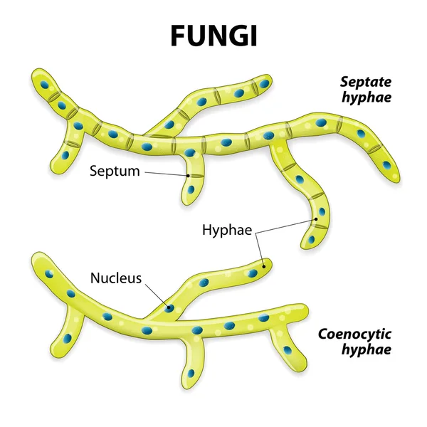
Fungi. Classification based on cell division. Septate hyphae (with septa) and aseptate hyphae (coenocytic or without septa).
Fungi. Classification based on cell division — Stock Vector

Similar Stock Videos:

Graphic animation showing the structure and composition of a coronavirus.
Graphic Animation Showing Structure Composition Coronavirus — Stock Video
Human Organs Animation on white background
Human Organs Animation — Stock Video
The stomach lies between the esophagus and the duodenum (the first part of the small intestine). It is on the left upper part of the abdominal cavity. The top of the stomach lies against the diaphragm.
Stomach Layers — Stock Video
White Human stomach icon isolated on purple background. 4K Video motion graphic animation.
White Human stomach icon isolated on purple background. 4K Video motion graphic animation — Stock Video
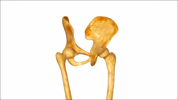
The hip bone (innominate bone, pelvic bone or coxal bone) is a large flat bone, constricted in the center and expanded above and below.
Pelvic Hip — Stock Video
3d rendered medically illustration of male Internal organs
Rendered Medically Illustration Male Internal Organs — Stock Video
Animation of human organs internal diagram, Body of human internal organs, brain, heart, lungs, liver, stomach, intestine
Animation Human Organs Internal Diagram Body Human Internal Organs Brain — Stock Video
Black line Virus cells in lung icon isolated on white background. Infected lungs. Coronavirus, COVID-19. 2019-nCoV. 4K Video motion graphic animation.
Black line Virus cells in lung icon isolated on white background. Infected lungs. Coronavirus, COVID-19. 2019-nCoV. 4K Video motion graphic animation — Stock Video
Prolapse of intervertebral disc
Prolapse of intervertebral disc — Stock Video
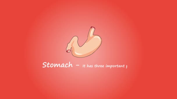
Footage of Human Stomach. Isolated on red background.
Footage Human Stomach Isolated Red Background — Stock Video
Human Organs Animation on white background
Human Organs Animation — Stock Video
Stomach upset animated line icon. stomach upset sign. isolated on white background
Stomach upset line icon animation — Stock Video
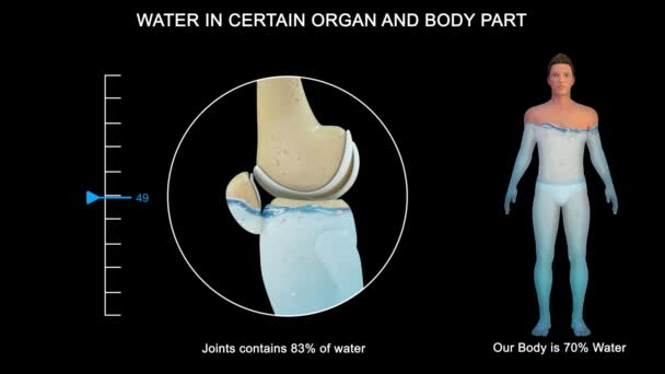
Some organs contain much more water than others. The brain and kidneys possess the highest percentage of water; the bones and teeth contain the lowest proportion.
Some Organs Contain Much More Water Others Brain Kidneys Possess — Stock Video
Indigestion stomach animated glyph icon. indigestion stomach sign. isolated on white background
Indigestion stomach glyph icon animation — Stock Video
An handmade, traditional, colored, animation of a hearth pumping
Handmade Traditional Colored Animation Hearth Pumping — Stock Video
Heart, Lungs and Diaphragm in Skeleton Body on dark background
Heart, Lungs and Diaphragm — Stock Video
Red blood cells, blood vessel, inside the blood vessel, white blood cells, white blood cells inside the blood vessel, High quality 3d render of blood cells, pulsing down artery, Red and white blood cells in artery
Red Blood Cells Blood Vessel Blood Vessel White Blood Cells — Stock Video
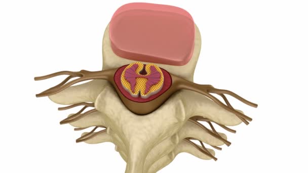
Spinal cord under pressure of bulging disc
Spinal cord under pressure of bulging disc — Stock Video

Usage Information

You can use this royalty-free vector image "Thymus gland anatomy" for personal and commercial purposes according to the Standard or Extended License. The Standard License covers most use cases, including advertising, UI designs, and product packaging, and allows up to 500,000 print copies. The Extended License permits all use cases under the Standard License with unlimited print rights and allows you to use the downloaded vector files for merchandise, product resale, or free distribution.

This stock vector image is scalable to any size. You can buy and download it in high resolution up to 4968x3898. Upload Date: Apr 9, 2014