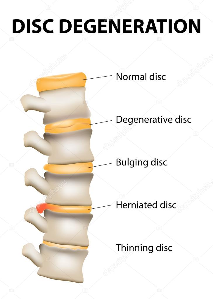
Disc degeneration — Stock Vector

Disc degeneration — Vector

Disc degeneration it's the normal wear and tear process of aging spine. intervertebral discs lose their flexibility, elasticity, and shock-absorbing characteristics.

 — Vector by edesignua

Same Series:

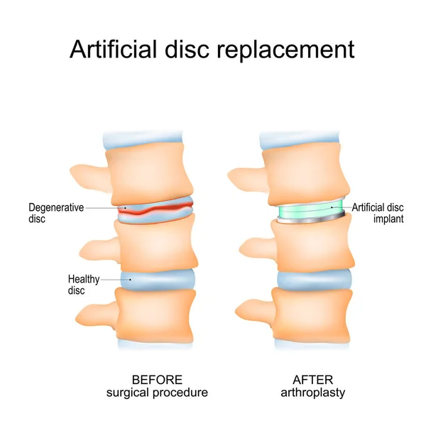
Intervertebral disc arthroplasty. Close-up of a human spinal column before Surgical procedure and after Arthroplasty. Degenerative Disc and Artificial disc replacement. Vector poster
Intervertebral Disc Arthroplasty Close Human Spinal Column Surgical Procedure Arthroplasty — Stock Vector
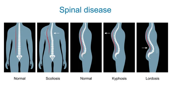
Spinal disease. types of spine defects. Human Skeleton and bones. Vertebral column disorders. Normal spine and Spinal deformity from Scoliosis to Lordosis and Kyphosis. Vector illustration like x-Ray image. Anterior and lateral view. infographics
Spinal Disease Types Spine Defects Human Skeleton Bones Vertebral Column — Stock Vector
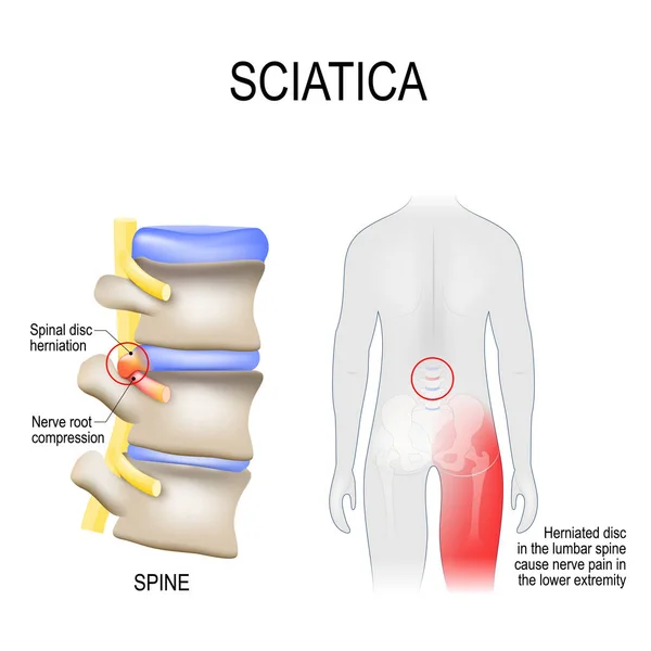
Sciatica. scheme with vertebrae, disks and nerves. Human body from back. Disc degeneration. normal wear and tear process of aging spine. Vector illustration for medical, biological, educational and science use
Sciatica. scheme with vertebrae, disks and nerves. Human body fr — Stock Vector
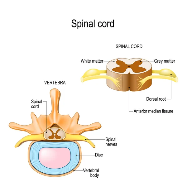
Section of the human vertebral column and cross-section of spinal cord. Central nervous system. Vector illustration for medical, biological, and educational use
Section Human Vertebral Column Cross Section Spinal Cord Central Nervous — Stock Vector
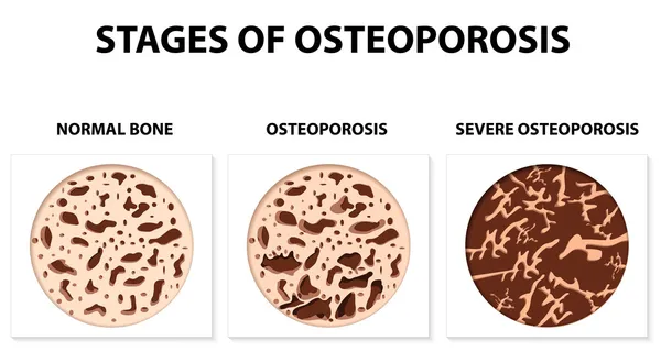
Osteoporosis is a disease characterized by low bone mass and deterioration of bone tissue, leading to fragility and fractures.
Stages of Osteoporosis — Stock Vector
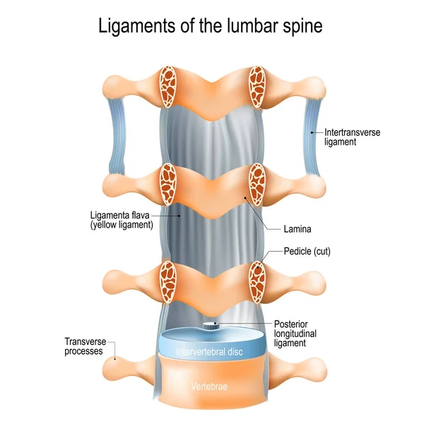
Ligaments of the lumbar spine: Ligamenta flava (yellow ligament), Intertransverse, and Posterior longitudinal ligaments. vector illustration for medical, educational and science use
Ligaments Lumbar Spine Ligamenta Flava Yellow Ligament Intertransverse Posterior Longitudinal — Stock Vector
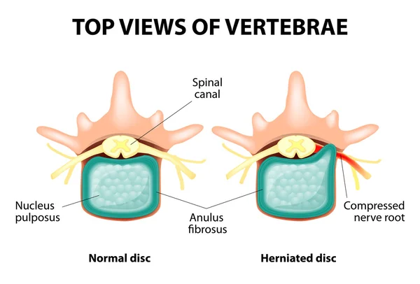
Herniated Disc. Prolapse of intervertebral disc closeup.
Herniated Disc — Stock Vector
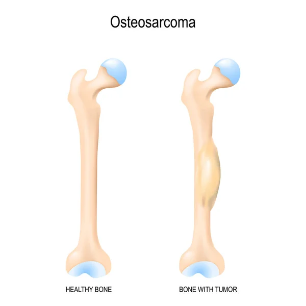
Osteosarcoma. Osteogenic sarcoma is a cancerous tumor in a bone. malignant neoplasm. Two human bones: healthy femur and bone with osteosarcoma. Vector illustration for biological, science, medical use.
Osteosarcoma Osteogenic Sarcoma Cancerous Tumor Bone Malignant Neoplasm Two Human — Stock Vector
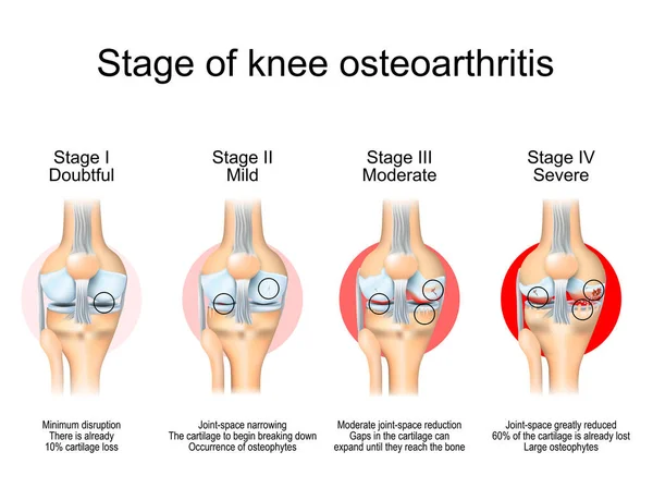
Stages of knee osteoarthritis. From Minimum disruption to lost a cartilage and joint-space reduction. Anatomy of knee joint. Vector illustration
Stages Knee Osteoarthritis Minimum Disruption Lost Cartilage Joint Space Reduction — Stock Vector
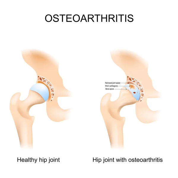
Hip joint with osteoarthritis with femoral head and the acetabulum of the pelvis. A comparison between a healthy hip joint and one with osteoarthritis, highlighting the key differences in structure. This can help patients understand diagnosis. Vector
Hip Joint Osteoarthritis Femoral Head Acetabulum Pelvis Comparison Healthy Hip — Stock Vector
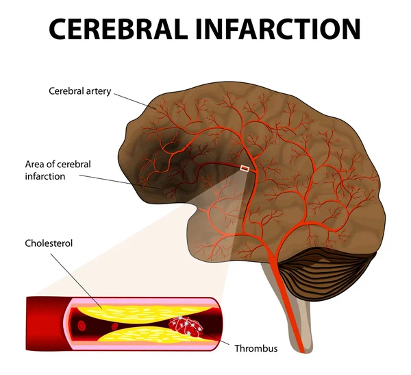
Brain stroke. Cerebral infarction. the obstruction is caused by a blood clot that forms in a cerebral artery, at an atheromatous plaque.
Cerebral infarction or ischemic stroke — Stock Vector
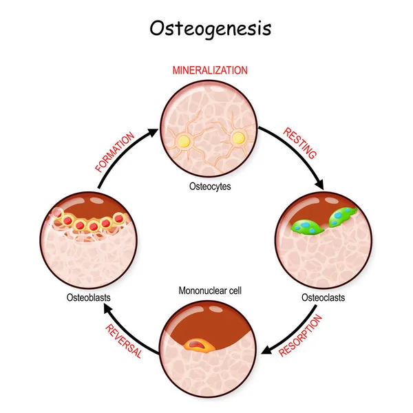
Bone remodeling (resting, resorption, reversal, mineralization, formation). Describe a process of Ossification. osteogenesis. Bone is broken down by osteoclasts, and rebuilt by osteoblasts
Bone Remodeling Resting Resorption Reversal Mineralization Formation Describe Process Ossification — Stock Vector
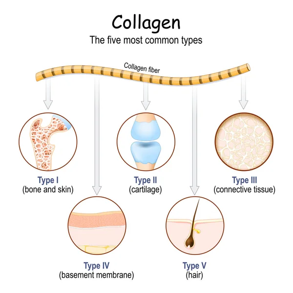
Collagen fibers. The five most common types of collagen protein. Vector
Collagen Fibers Five Most Common Types Collagen Protein Vector — Stock Vector
Fibular collateral ligament injury. joint anatomy. Vector illustration for biological, medical, science and educational use
Fibular Collateral Ligament Injury Joint Anatomy Vector Illustration Biological Medical — Stock Vector
Osteoporosis vs Osteoarthritis. comparison and difference between Healthy joint and degenerative joint disease, and joint with low bone mass. Cross section of skeletal bone
Osteoporosis Osteoarthritis Comparison Difference Healthy Joint Degenerative Joint Disease Joint — Stock Vector
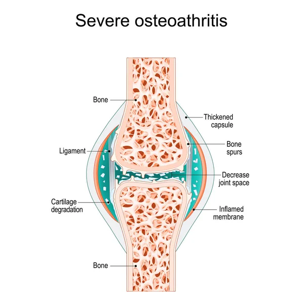
Severe osteoarthritis. Synovial joint with bone Spurs, Inflamed membrane, Decreased joint space, and Cartilage degradation. vector illustration
Severe Osteoarthritis Synovial Joint Bone Spurs Inflamed Membrane Decreased Joint — Stock Vector

Similar Stock Videos:

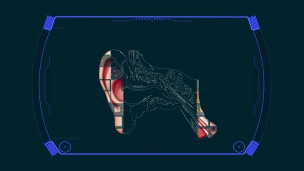
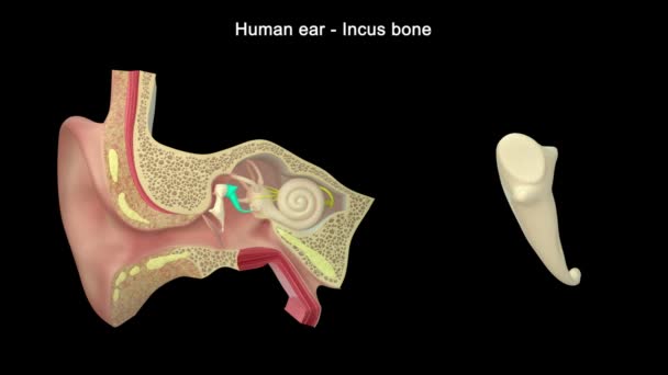
Anatomy Diagram Ear Structure
Anatomy Diagram Ear Structure — Stock Video
Animated endocannabinoid system on red background. High quality 4k footage
Animated endocannabinoid system on red background — Stock Video
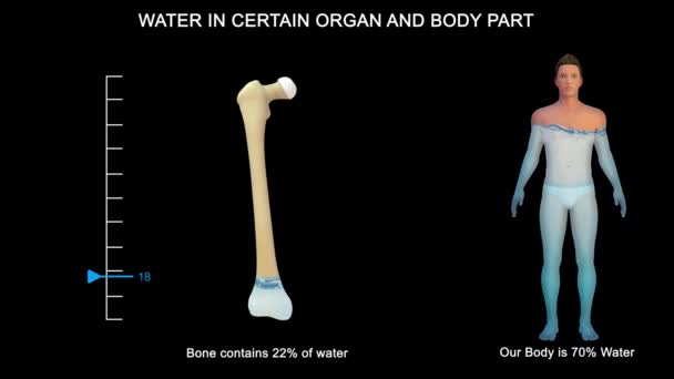
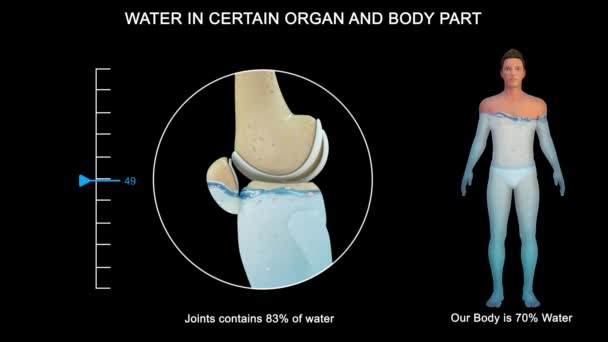
Some organs contain much more water than others. The brain and kidneys possess the highest percentage of water; the bones and teeth contain the lowest proportion.
Some Organs Contain Much More Water Others Brain Kidneys Possess — Stock Video
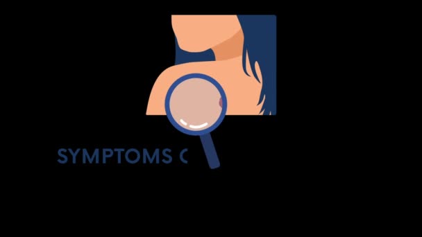
Skin cancer symptoms 2D animation on transparent background. Melanoma concept. Can be used for topics like oncology, tumor, awareness, healthcare
Skin Cancer Symptoms Animation Transparent Background Melanoma Concept Can Used — Stock Video
Human Organs Animation on white background
Human Organs Animation — Stock Video
Graphic animation showing the structure and composition of a coronavirus.
Graphic Animation Showing Structure Composition Coronavirus — Stock Video
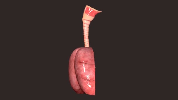
The lung is the essential respiration organ in many air-breathing animals, including most tetrapods, a few fish and a few snails. In mammals and the more complex life forms, the two lungs are located near the backbone on either side of the heart.
Lungs Anatomy — Stock Video
Some organs contain much more water than others. The brain and kidneys possess the highest percentage of water; the bones and teeth contain the lowest proportion.
Some Organs Contain Much More Water Others Brain Kidneys Possess — Stock Video
The heart is a muscular organ in humans and many other animals, which pumps blood through the blood vessels of the circulatory system. The blood provides the body with oxygen and nutrients, as well as removing metabolic wastes.
Human Heart — Stock Video
The human digestive system consists of the gastrointestinal tract plus the accessory organs of digestion. Digestion involves the breakdown of food into smaller and smaller components until they can be absorbed and assimilated into the body.
Human Digestive System Consists Gastrointestinal Tract Accessory Organs Digestion Digestion — Stock Video
Human Organs Animation on white background
Human Organs Animation — Stock Video
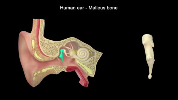
Human ear structure footage
Human Ear Structure Footage — Stock Video
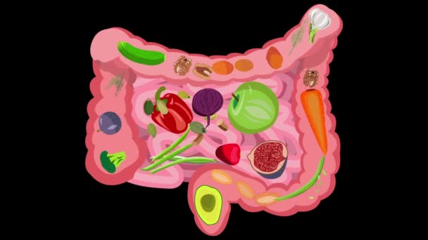
Healthy food for intestinal health. Healthy food for intestinal health. Fiber, vegetables, fruits, berries, bran, greens, prebiotics. Video on a black background
Healthy food for intestinal health. Video on a black background — Stock Video
The stomach lies between the esophagus and the duodenum (the first part of the small intestine). It is on the left upper part of the abdominal cavity. The top of the stomach lies against the diaphragm.
Stomach Layers — Stock Video
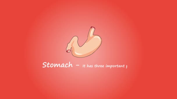
Footage of Human Stomach. Isolated on red background.
Footage Human Stomach Isolated Red Background — Stock Video
Human Ear Anatomy. 3d illustration
Human Ear Anatomy Illustration — Stock Video
Digestive system on human silhouette cartoon High definition colorful scenes animation
Digestive system concept HD animation — Stock Video

Usage Information

You can use this royalty-free vector image "Disc degeneration" for personal and commercial purposes according to the Standard or Extended License. The Standard License covers most use cases, including advertising, UI designs, and product packaging, and allows up to 500,000 print copies. The Extended License permits all use cases under the Standard License with unlimited print rights and allows you to use the downloaded vector files for merchandise, product resale, or free distribution.

This stock vector image is scalable to any size. You can buy and download it in high resolution up to 3159x4434. Upload Date: May 23, 2014