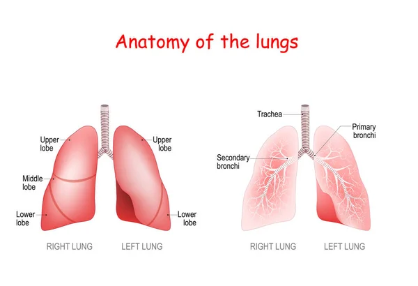
Anatomy of the human lungs — Stock Vector

Anatomy of the human lungs — Vector

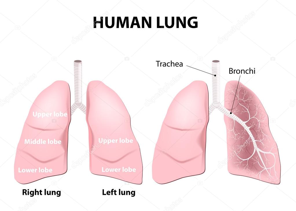
Detailed diagram of the human lungs.

 — Vector by edesignua

Same Series:

Anatomy of the human lungs. lobes, trachea and bronchi. Vector illustration
Anatomy Human Lungs Lobes Trachea Bronchi Vector Illustration — Stock Vector
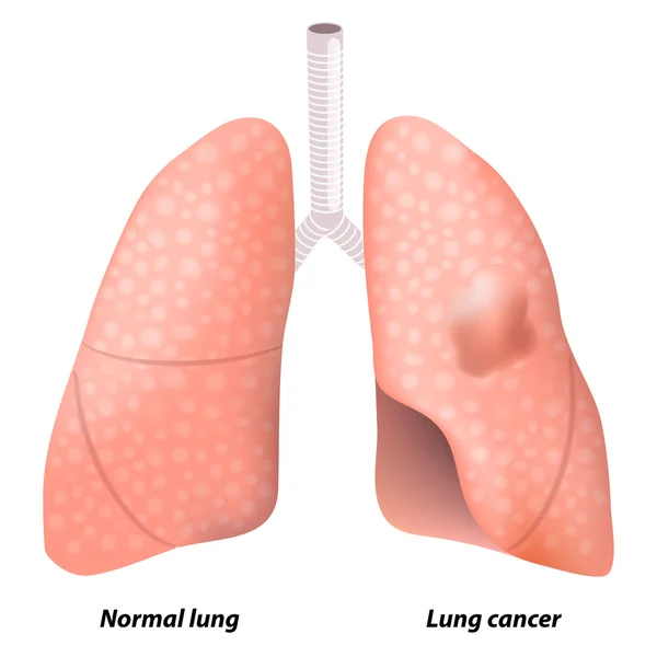
Lung cancer. This illustration shows malignant tumor in the upper lobe of the person's left lung. Human anatomy
Lung cancer — Stock Vector
Pulmonary alveolus. alveoli, trachea, and bronchiole in the lungs. Vector illustration
Pulmonary Alveolus Alveoli Trachea Bronchiole Lungs Vector Illustration — Stock Vector
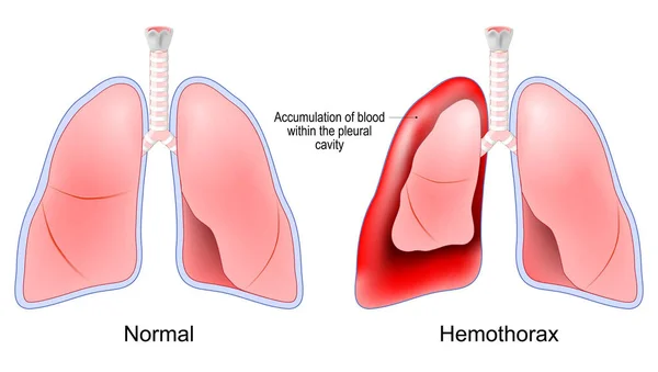
Hemothorax. Healthy human lungs and red lungs after accumulation of blood within the pleural cavity. Chest trauma. pulmonary embolism treatment. Vector poster
Hemothorax Healthy Human Lungs Red Lungs Accumulation Blood Pleural Cavity — Stock Vector
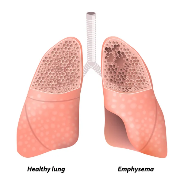
Emphysema. Chronic obstructive pulmonary disease. diagram showing a cross-section of normal lung and lungs damaged by COPD. Human anatomy
Chronic obstructive pulmonary disease — Stock Vector
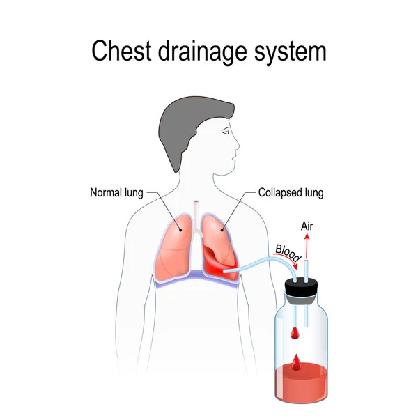
Hemothorax. Collapsed lung. abnormal collection of blood in the pleural space between the lung and the chest wall. Chest drainage system. Fluid collected in the chamber. Vector illustration for medical, biological, science and educational use
Hemothorax Collapsed Lung Abnormal Collection Blood Pleural Space Lung Chest — Stock Vector
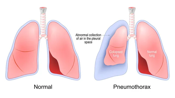
Pneumothorax. accumulation of air between the lung and the chest cavity. Normal, and collapsed lungs. Human Respiratory system. Vector illustration
Pneumothorax Accumulation Air Lung Chest Cavity Normal Collapsed Lungs Human — Stock Vector
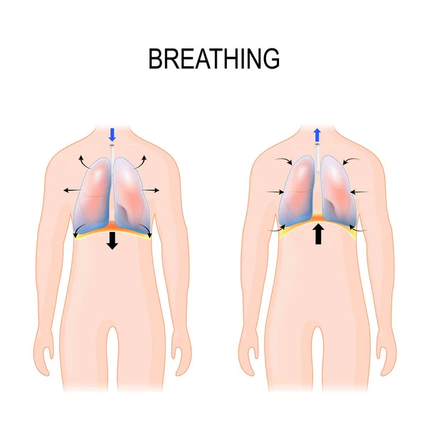
Breathing. Movement of ribcage during inspiration and expiration. diaphragm functions. enlarging the cavity creates suction that draws air into the lungs. illustration for medical, and educational use
Breathing. Movement of ribcage during inspiration and expiration — Stock Vector
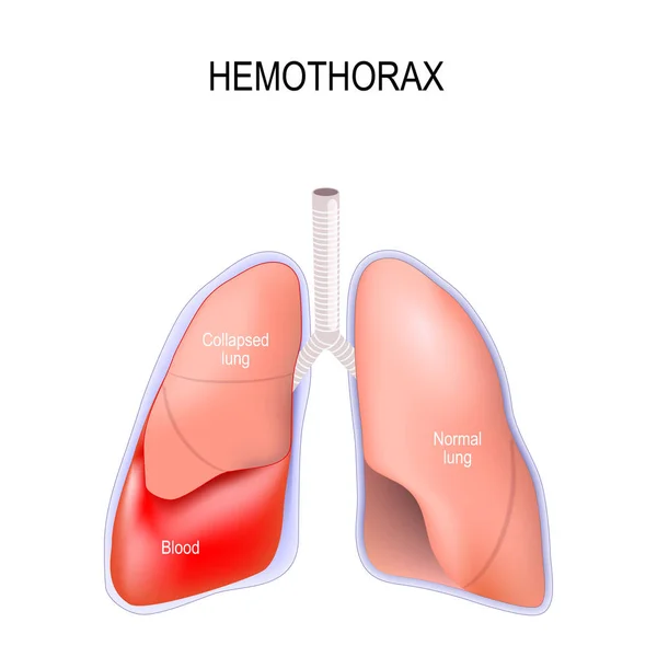
Hemothorax. Collapsed lung. abnormal collection of blood in the pleural space between the lung and the chest wall. Vector illustration for medical, biological, science and educational use
Hemothorax Collapsed Lung Abnormal Collection Blood Pleural Space Lung Chest — Stock Vector
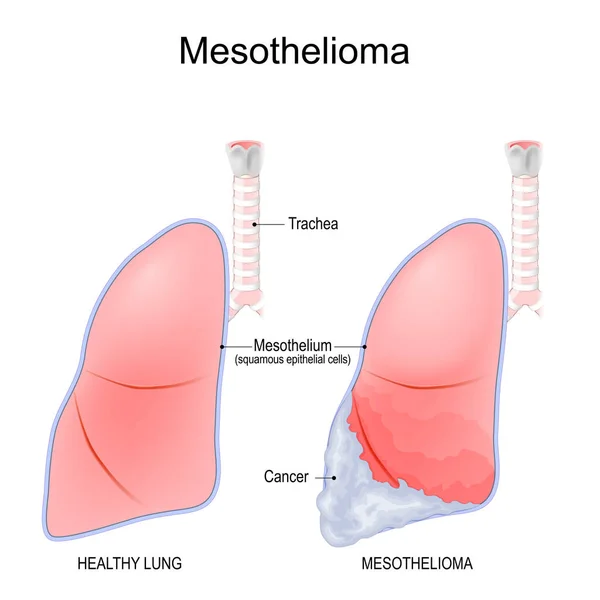
Mesothelioma. cancer that develops from the thin layer of lungs tissue - mesothelium. Malignant tumor that caused by exposure to asbestos, genetics or viral infection. Differences and comparisons of a healthy human lung, and lung with Mesothelioma. V
Mesothelioma Cancer Develops Thin Layer Lungs Tissue Mesothelium Malignant Tumor — Stock Vector
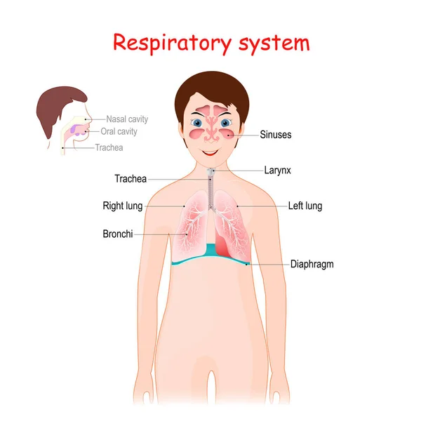
Human respiratory system for kids. Cute smiling girl with lungs, bronchi, sinuses, diaphragm, and trachea. poster for educational use. Vector illustration
Human Respiratory System Kids Cute Smiling Girl Lungs Bronchi Sinuses — Stock Vector
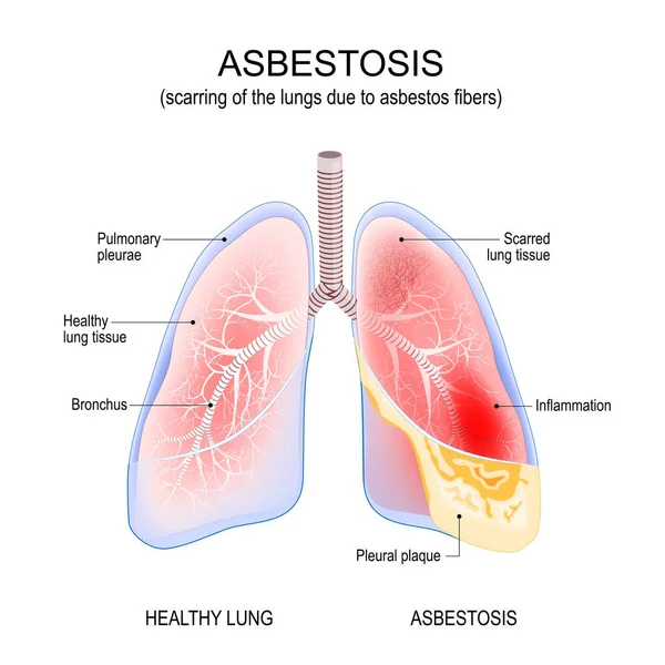
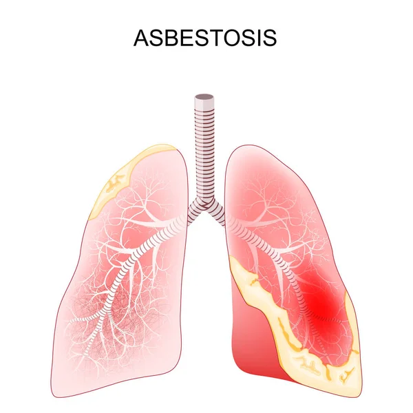
Asbestosis lungs. Inflammation and scarring of the lungs due to asbestos fibers. cancer. Vector illustration
Asbestosis Lungs Inflammation Scarring Lungs Due Asbestos Fibers Cancer Vector — Stock Vector
Lung cancer. carcinoma is a malignant lung tumor. vector illustration for medical use
Lung Cancer Carcinoma Malignant Lung Tumor Vector Illustration Medical Use — Stock Vector
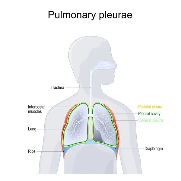
Pulmonary pleurae. Anatomy of a Respiratory system. Structure of Chest cavity. Parietal and Visceral pleura, lungs, ribs, trachea, Pleural cavity, bronchi and diaphragm on a grey human silhouette. Vector illustration
Pulmonary Pleurae Anatomy Respiratory System Structure Chest Cavity Parietal Visceral — Stock Vector
Asbestosis. lung disease as a result of exposure to asbestos fibers. Close-up of a cross section of human lungs with fibrosis and scarring. Vector illustration
Asbestosis Lung Disease Result Exposure Asbestos Fibers Close Cross Section — Stock Vector
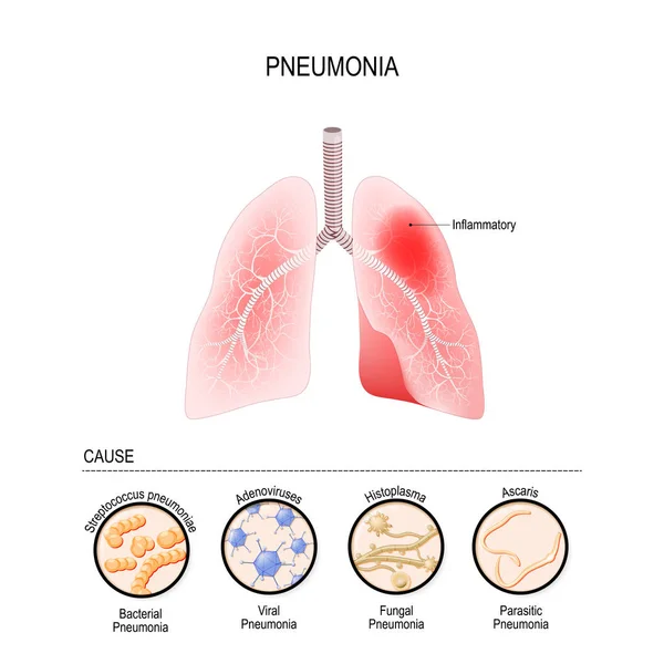
Pneumonia is caused by infection with viruses, bacteria, fungi and other microorganisms (for example, ascaris). closeup lungs, and bronchioles. human respiratory system. Vector illustration for medical, biological, educational and science use
Pneumonia is caused by infection with viruses, bacteria, fungi a — Stock Vector

Similar Stock Videos:

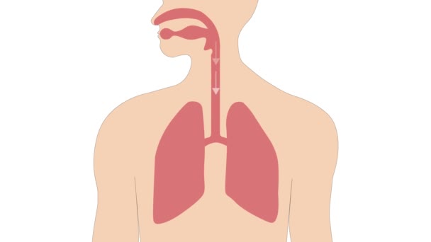
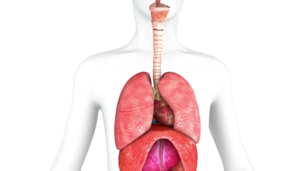
Breathing in and out with the expansion and contraction of the lungs.
Breathing Out Expansion Contraction Lungs — Stock Video
Polycystic ovarian syndrome 2D animation. Uterus, infertility, gynecology. For topics like pcos, disease, pelvic floor
Polycystic Ovarian Syndrome Animation Uterus Infertility Gynecology Topics Pcos Disease — Stock Video
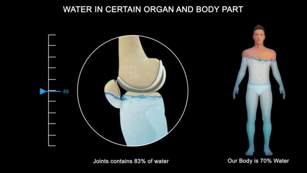
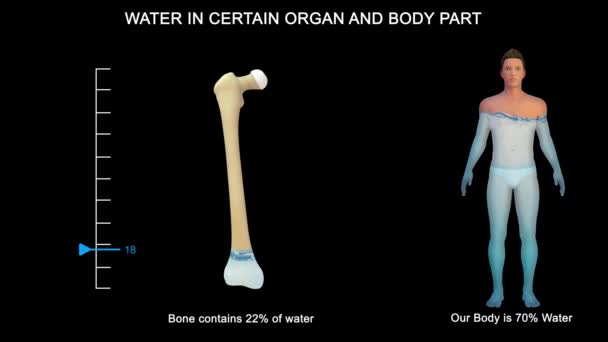
Some organs contain much more water than others. The brain and kidneys possess the highest percentage of water; the bones and teeth contain the lowest proportion.
Some Organs Contain Much More Water Others Brain Kidneys Possess — Stock Video
Breathing in and out with the expansion and contraction of the lungs.
Breathing Out Expansion Contraction Lungs — Stock Video
Animated endocannabinoid system on red background. High quality 4k footage
Animated endocannabinoid system on red background — Stock Video
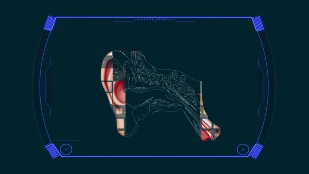
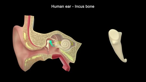
Anatomy Diagram Ear Structure
Anatomy Diagram Ear Structure — Stock Video
Some organs contain much more water than others. The brain and kidneys possess the highest percentage of water; the bones and teeth contain the lowest proportion.
Some Organs Contain Much More Water Others Brain Kidneys Possess — Stock Video
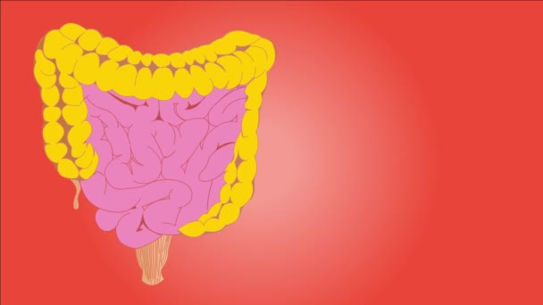
The human digestive system consists of the gastrointestinal tract plus the accessory organs of digestion. Digestion involves the breakdown of food into smaller and smaller components until they can be absorbed and assimilated into the body.
Human Digestive System Consists Gastrointestinal Tract Accessory Organs Digestion Digestion — Stock Video
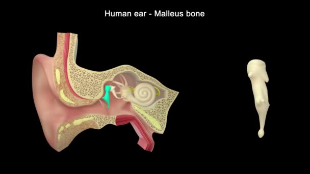
Human ear structure footage
Human Ear Structure Footage — Stock Video
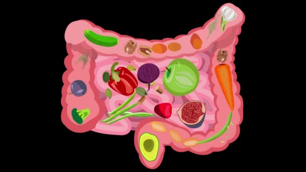
Healthy food for intestinal health. Healthy food for intestinal health. Fiber, vegetables, fruits, berries, bran, greens, prebiotics. Video on a black background
Healthy food for intestinal health. Video on a black background — Stock Video
Abstract futuristic motion background with graphic effects
Abstract Futuristic Motion Background Graphic Effects — Stock Video
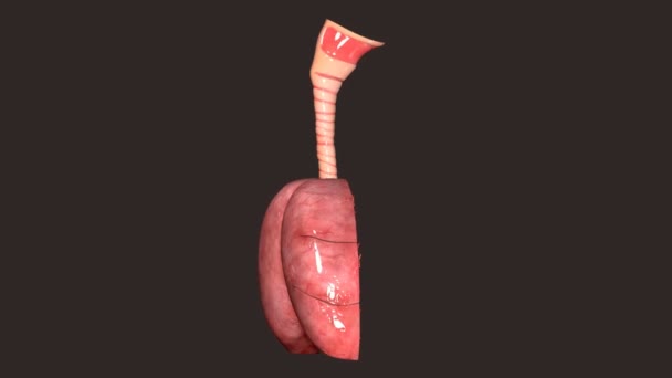
This video shows 3d lungs breathing
Video Shows Lungs Breathing — Stock Video
The lung is the essential respiration organ in many air-breathing animals, including most tetrapods, a few fish and a few snails. In mammals and the more complex life forms, the two lungs are located near the backbone on either side of the heart.
Lungs Anatomy — Stock Video
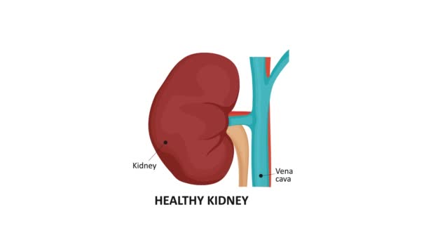
Animation illustrating the various stages of kidney cancer.
Animation illustrating the various stages of kidney cancer. — Stock Video
Digestive system on human silhouette cartoon High definition colorful scenes animation
Digestive system concept HD animation — Stock Video
The heart is a muscular organ in humans and many other animals, which pumps blood through the blood vessels of the circulatory system. The heart is located in the middle compartment (mediastinum) of the chest.
Human Heart — Stock Video
Graphic animation showing the structure and composition of a coronavirus.
Graphic Animation Showing Structure Composition Coronavirus — Stock Video
Human Ear Anatomy. 3d illustration
Human Ear Anatomy Illustration — Stock Video

Usage Information

You can use this royalty-free vector image "Anatomy of the human lungs" for personal and commercial purposes according to the Standard or Extended License. The Standard License covers most use cases, including advertising, UI designs, and product packaging, and allows up to 500,000 print copies. The Extended License permits all use cases under the Standard License with unlimited print rights and allows you to use the downloaded vector files for merchandise, product resale, or free distribution.

This stock vector image is scalable to any size. You can buy and download it in high resolution up to 5280x3767. Upload Date: Oct 29, 2014