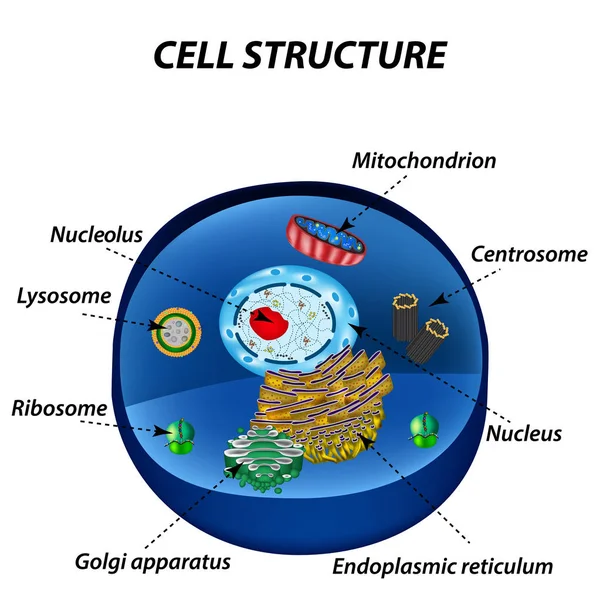
The internal structure of an animal cell — Stock Vector

The internal structure of an animal cell — Vector

The internal structure of an animal cell. Illustration

 — Vector by mikrostoker

Same Series:

Structure of human cells. Organelles. The core nucleus, endoplasmic reticulum, Golgi apparatus, lysosomes, ribosomes, mitochondria centriole Vector illustration
Structure of human cells. Organelles. The core nucleus, endoplasmic reticulum, Golgi apparatus, lysosomes, ribosomes, mitochondria, centriole. Vector illustration — Stock Vector
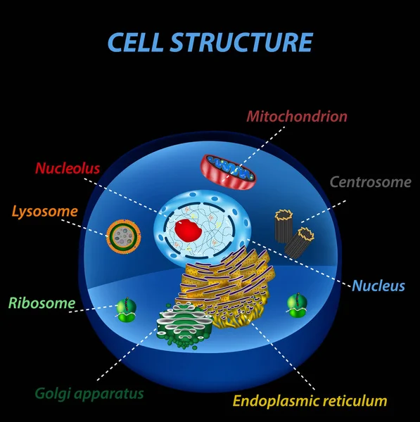
Structure of human cells. Organelles. The core nucleus, endoplasmic reticulum, Golgi apparatus, lysosomes, ribosomes, mitochondria, centriole Vector illustration on a black background
Structure of human cells. Organelles. The core nucleus, endoplasmic reticulum, Golgi apparatus, lysosomes, ribosomes, mitochondria, centriole. Vector illustration on a black background — Stock Vector
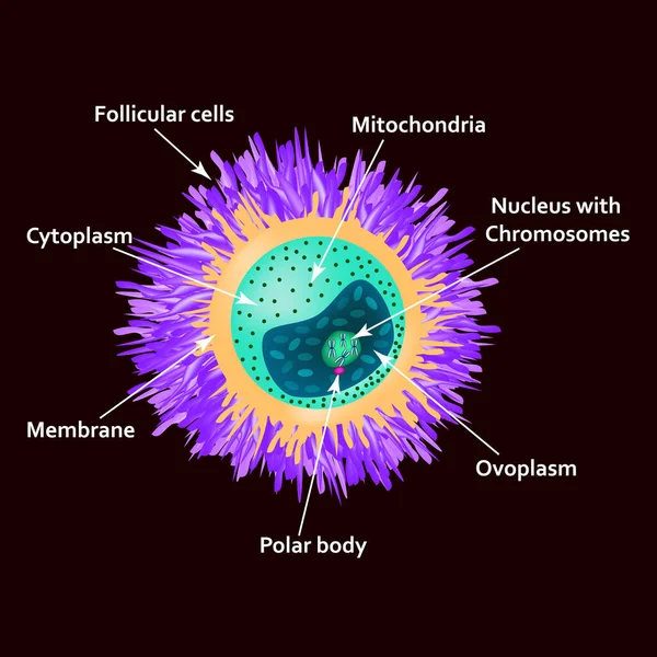
The structure of the egg. Ovum anatomy. Vector illustration on isolated background.
The structure of the egg. Ovum anatomy. Vector illustration on isolated background — Stock Vector
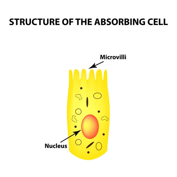
Structure of the enterocyte. absorptive cells intestine. Infographics. Vector illustration on isolated background
Structure of the enterocyte. absorptive cells intestine. Infographics. Vector illustration on isolated background. — Stock Vector
The structure of the egg. Ovum anatomy. Vector illustration on isolated background.
The structure of the egg. Ovum anatomy. Vector illustration on isolated background — Stock Vector
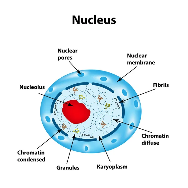
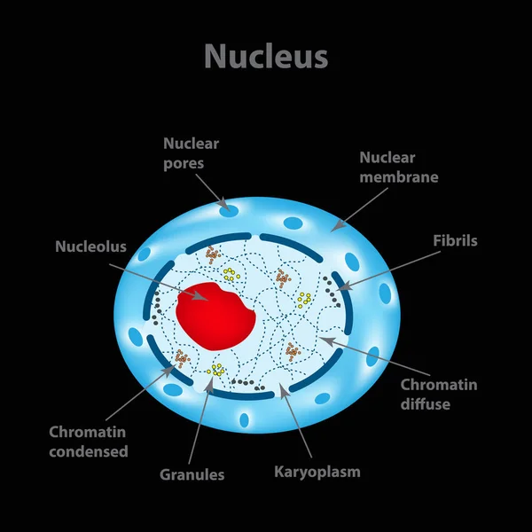
The structure of the human cell nucleus. Infographics. Vector illustration on isolated background.
The structure of the human cell nucleus. Infographics. Vector illustration on isolated background — Stock Vector
The structure of the human cell nucleus. Infographics. Vector illustration.
The structure of the human cell nucleus. Infographics. Vector illustration — Stock Vector
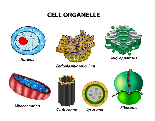
Set the cell organelles. Nucleus, endoplasmic reticulum, Golgi apparatus, mitochondria, centrosome, lysosome, the ribosome. Infographics. Vector illustration on isolated background.
Set the cell organelles. Nucleus, endoplasmic reticulum, Golgi apparatus, mitochondria, centrosome, lysosome, the ribosome. Infographics. Vector illustration on isolated background — Stock Vector
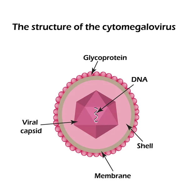
The structure of the cytomegalovirus. Infographics. Vector
The structure of the cytomegalovirus. Infographics. Vector illustration — Stock Vector
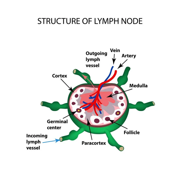
The anatomical structure of the lymph node. Infographics. Vector illustration on isolated background.
The anatomical structure of the lymph node. Infographics. Vector illustration on isolated background — Stock Vector
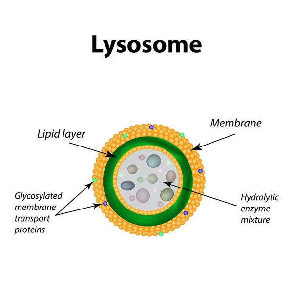
The structure of lysosomes. Infographics. Vector illustration on isolated background.
The structure of lysosomes. Infographics. Vector illustration on isolated background — Stock Vector
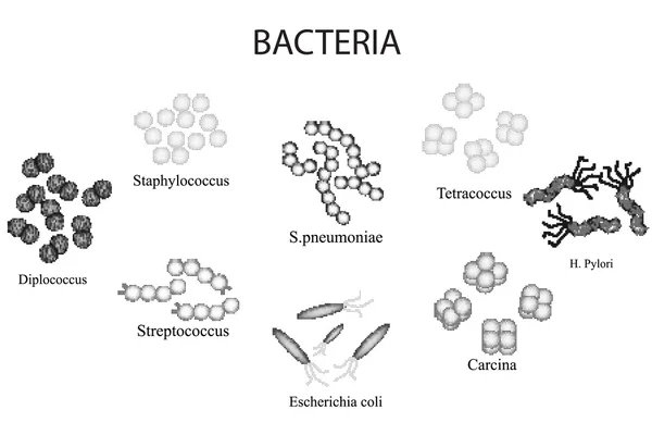
Set of bacteria. Black and white. Staphylococcus, Streptococcus, Streptococcus pneumoniae, Escherichia, coli, Helicobacter pylori, sartsiny, Diplococci, Tetrakokki. Hand draw. Vector illustration
Set of bacteria. Black and white. Staphylococcus, Streptococcus, Streptococcus pneumoniae, Escherichia, coli, Helicobacter pylori, sartsiny, Diplococci, Tetrakokki. Hand draw. — Stock Vector
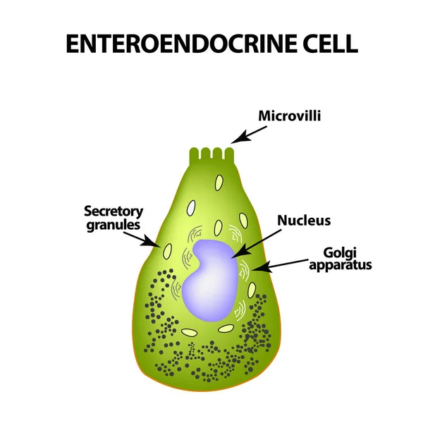
Enteroendocrine cell. Cell of the intestines. Vector illustration on isolated background.
Enteroendocrine cell. Cell of the intestines. Vector illustration on isolated background — Stock Vector
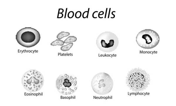
Blood cells. Set of monochrome cells. Red blood cells, platelets, leukocytes, lymphocytes, eosinophils, neutrophils, basophils monocytes Infographics Vector illustration on isolated background
Blood cells. Set of monochrome cells. Red blood cells, platelets, leukocytes, lymphocytes, eosinophils, neutrophils, basophils, monocytes. Infographics. Vector illustration on isolated background — Stock Vector
Blood cells. Set of monochrome cells. Red blood cells, platelets, leukocytes, lymphocytes, eosinophils, neutrophils, basophils monocytes Infographics illustration on isolated background
Blood cells. Set of monochrome cells. Red blood cells, platelets, leukocytes, lymphocytes, eosinophils, neutrophils, basophils, monocytes. Infographics. illustration on isolated background — Stock Photo, Image
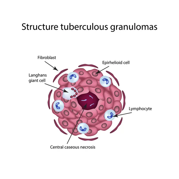
The structure of tuberculous granulomas. Infographics. Vector illustration on isolated background.
The structure of tuberculous granulomas. Vector illustration on isolated background — Stock Vector

Similar Stock Videos:
Graphic animation showing the structure and composition of a coronavirus.
Graphic Animation Showing Structure Composition Coronavirus — Stock Video
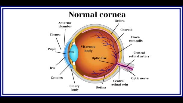
Keratoconus. Dystrophic disease of the cornea. Animation.
Keratoconus Dystrophic Disease Cornea Animation — Stock Video
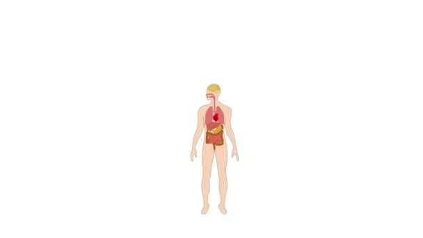
Human Organs Animation on white background
Human Organs Animation — Stock Video
Black line Virus cells in lung icon isolated on white background. Infected lungs. Coronavirus, COVID-19. 2019-nCoV. 4K Video motion graphic animation.
Black line Virus cells in lung icon isolated on white background. Infected lungs. Coronavirus, COVID-19. 2019-nCoV. 4K Video motion graphic animation — Stock Video
Abstract futuristic motion background with graphic effects
Abstract Futuristic Motion Background Graphic Effects — Stock Video
Human Organs Animation on white background
Human Organs Animation — Stock Video
Stomach upset animated line icon. stomach upset sign. isolated on white background
Stomach upset line icon animation — Stock Video
Animation of human organs internal diagram, Body of human internal organs, brain, heart, lungs, liver, stomach, intestine
Animation Human Organs Internal Diagram Body Human Internal Organs Brain — Stock Video
3d rendered medically illustration of male Internal organs
Rendered Medically Illustration Male Internal Organs — Stock Video
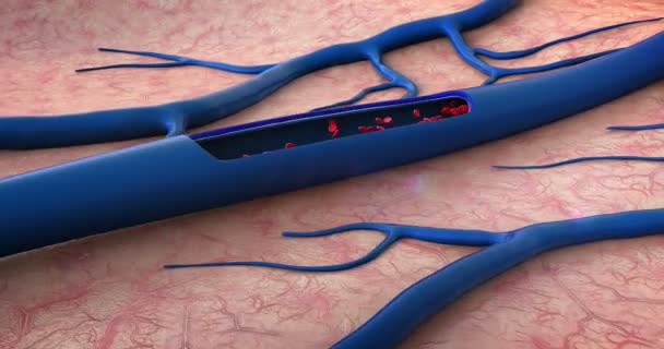
Red blood cells, blood vessel, inside the blood vessel, white blood cells, white blood cells inside the blood vessel, High quality 3d render of blood cells, pulsing down artery, Red and white blood cells in artery
Red Blood Cells Blood Vessel Blood Vessel White Blood Cells — Stock Video
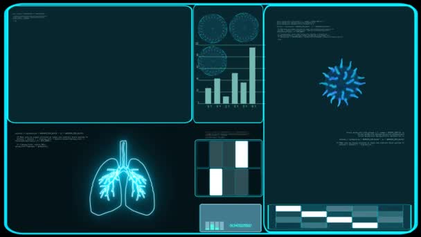
Covid virus spread all world analysis lung and digital rotate blue monitor zoom to america
Covid virus spread all world analysis lung and digital rotate blue monitor zoom to america — Stock Video
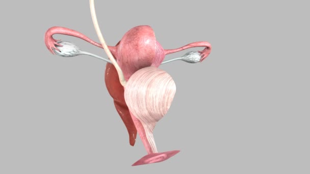
Female reproductive system medicine science animation concept
Female reproductive system — Stock Video
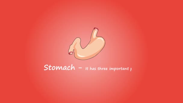
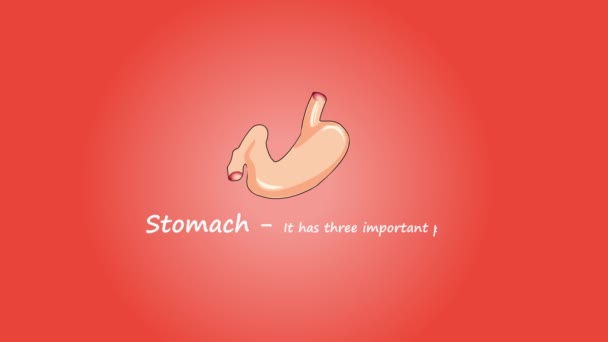
Footage of Human Stomach. Isolated on red background.
Footage Human Stomach Isolated Red Background — Stock Video
The stomach lies between the esophagus and the duodenum (the first part of the small intestine). It is on the left upper part of the abdominal cavity. The top of the stomach lies against the diaphragm.
Stomach Layers — Stock Video
Abstract futuristic motion background with graphic effects
Abstract Futuristic Motion Background Graphic Effects — Stock Video
Covid virus spread all world analysis and digital graph bar blue monitor zoom to america
Covid virus spread all world analysis and digital graph bar blue monitor zoom to america — Stock Video
Human Organs Animation on white background
Human Organs Animation — Stock Video
An handmade, traditional, colored, animation of a hearth pumping
Handmade Traditional Colored Animation Hearth Pumping — Stock Video

Usage Information

You can use this royalty-free vector image "The internal structure of an animal cell" for personal and commercial purposes according to the Standard or Extended License. The Standard License covers most use cases, including advertising, UI designs, and product packaging, and allows up to 500,000 print copies. The Extended License permits all use cases under the Standard License with unlimited print rights and allows you to use the downloaded vector files for merchandise, product resale, or free distribution.

This stock vector image is scalable to any size. You can buy and download it in high resolution up to 5000x5000. Upload Date: Mar 27, 2015